Ultrasound-guided truncal blocks: A new frontier in regional anaesthesia
- PMID: 27761032
- PMCID: PMC5064693
- DOI: 10.4103/0019-5049.191665
Ultrasound-guided truncal blocks: A new frontier in regional anaesthesia
Abstract
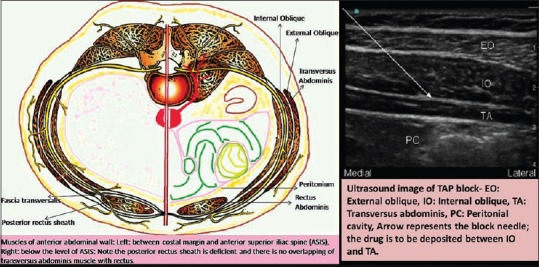
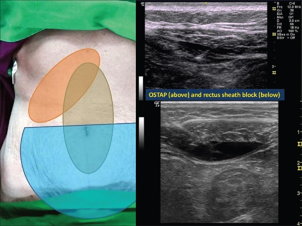
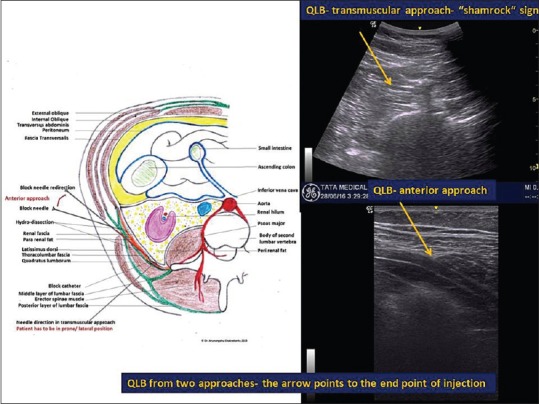
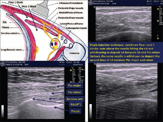
The practice of regional anaesthesia is rapidly changing with the introduction of ultrasound into the working domain of the anaesthesiologist. New techniques are being pioneered. Among the recent techniques, notable are the truncal blocks, for example, the transversus abdominis plane block, rectus sheath block, hernia block and quadratus lumborum block in the abdomen and the pectoral nerves (Pecs) block 1 and 2, serratus anterior plane block and intercostal nerve block. This narrative review covers the brief anatomical discourse along with technical description of the ultrasound-guided truncal blocks.
Keywords: Pecs block; transversus abdominis plane block; truncal blocks; ultrasound-guided regional anaesthesia.
Figures
References
-
- Hebbard P, Fujiwara Y, Shibata Y, Royse C. Ultrasound-guided transversus abdominis plane (TAP) block. Anaesth Intensive Care. 2007;35:616–7. - PubMed
-
- Rozen WM, Tran TM, Ashton MW, Barrington MJ, Ivanusic JJ, Taylor GI. Refining the course of the thoracolumbar nerves: A new understanding of the innervation of the anterior abdominal wall. Clin Anat. 2008;21:325–33. - PubMed
-
- Rafi AN. Abdominal field block: A new approach via the lumbar triangle. Anaesthesia. 2001;56:1024–6. - PubMed
-
- McDonnell JG, O'Donnell B, Curley G, Heffernan A, Power C, Laffey JG. The analgesic efficacy of transversus abdominis plane block after abdominal surgery: A prospective randomized controlled trial. Anesth Analg. 2007;104:193–7. - PubMed
-
- Carney J, Finnerty O, Rauf J, Bergin D, Laffey JG, Mc Donnell JG. Studies on the spread of local anaesthetic solution in transversus abdominis plane blocks. Anaesthesia. 2011;66:1023–30. - PubMed
Publication types
LinkOut - more resources
Full Text Sources
Other Literature Sources
Medical