Review
. 2016 Jun 30;10(6):1-8.
doi: 10.3941/jrcr.v10i6.1618. eCollection 2016 Jun.

Carotid Stent Fracture from Stylocarotid Syndrome

Affiliations
Review

Carotid Stent Fracture from Stylocarotid Syndrome

Jeffrey D Hooker et al. J Radiol Case Rep. .

Abstract

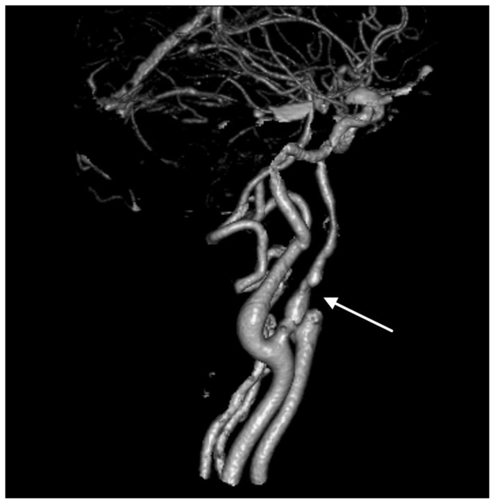
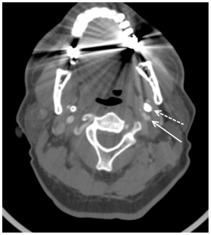
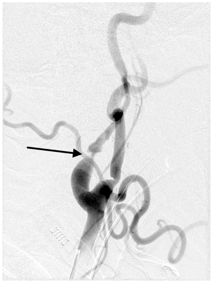
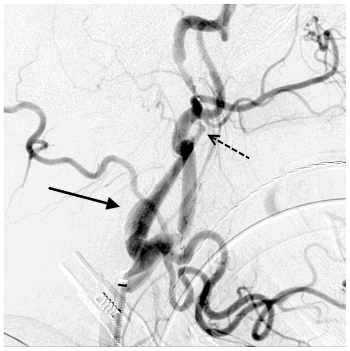
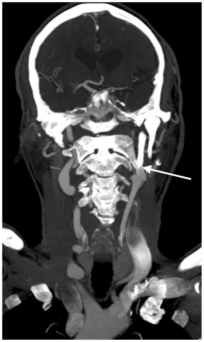
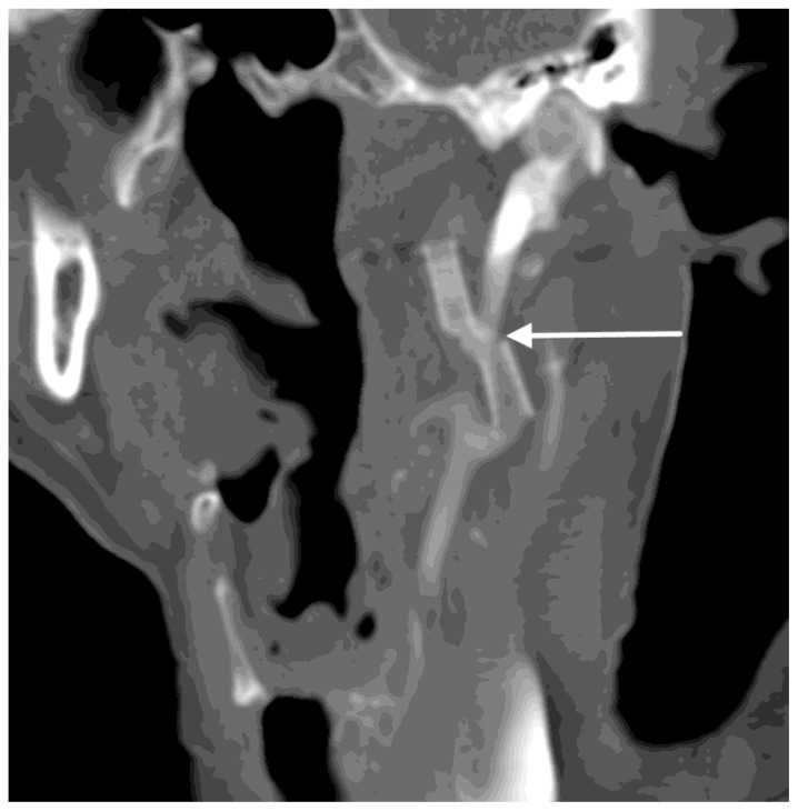
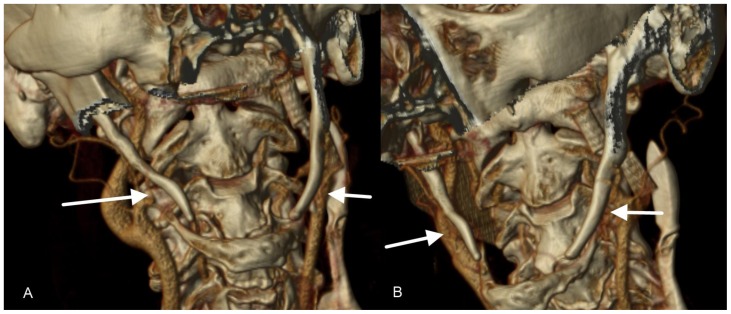
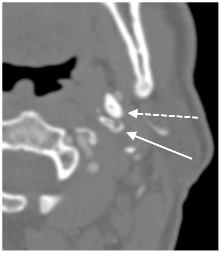
Eagle syndrome is a rare condition resulting from elongation of the temporal styloid process or calcification of its associated stylohyoid ligaments. Although usually presenting with pain or odynophagia, Eagle syndrome has been reported to cause a multitude of neurologic symptoms or vascular complications, some of which can be life-threatening. We present a case in which an endovascularly placed internal carotid artery stent in close proximity to a calcified stylohyoid ligament resulted in stent fracture with subsequent stent and vessel occlusion. We review and discuss the presentation, diagnosis, etiology, complications and treatment options of the syndrome.

Keywords: CT; Eagle syndrome; carotid stent; stent fracture; stylocarotid syndrome.


Figures

Figure 1
Figure 1
64-year-old male with left internal carotid artery stenosis presumed to be due to stylocarotid syndrome. FINDINGS: 3D reconstruction from CTA of the head and neck demonstrates a focal high-grade stenosis (arrow) in the cervical segment of the internal carotid artery with irregularity of the vessel proximal and distal to the stenosis. TECHNIQUE: Volumetric 3D reconstruction from CTA created in Philips Brilliance, 120 kVp, 498 mAs, 512×512 matrix, FOV 21.6 cm.
Figure 2
Figure 2
64-year-old male with left internal carotid artery stenosis presumed to be due to stylocarotid syndrome. FINDINGS: Short-segment irregularity of the left cervical internal carotid artery with focal severe stenosis (solid arrow) adjacent to the thickened, elongated left styloid process (dashed arrow). The elongated styloid process measured approximately 8 cm. TECHNIQUE: Axial image from CTA of the neck, slice thickness 2.5mm, 120 kVp, 498 mAs, 512×512 matrix, FOV 21.6 cm.
Figure 3
Figure 3
64-year-old male with left internal carotid artery stenosis presumed to be due to stylocarotid syndrome. FINDINGS: Focal severe stenosis (arrow) in the proximal cervical segment of the left internal carotid artery. TECHNIQUE: Digital subtraction angiography of the neck, lateral view, through a 5-Fr catheter in the left common carotid artery.
Figure 4
Figure 4
64-year-old male with left internal carotid artery stenosis presumed to be due to stylocarotid syndrome. FINDINGS: Resolution of focal internal carotid artery stenosis after stent placement (solid arrow). Incidental vasospasm distal to the stent (dashed arrow) which resolved during the procedure. TECHNIQUE: Digital subtraction angiography of the neck, lateral projection, from a 5-Fr catheter in the left common carotid artery.
Figure 5
Figure 5
64-year-old male with occluded, fractured left internal carotid artery stent presumed to be due to stylocarotid syndrome. FINDINGS: Occlusion of the fractured left internal carotid artery stent (arrow), which abuts the elongated, ossified left styloid process. The elongated styloid process measured approximately 8 cm. TECHNIQUE: Coronal MIP reconstruction from CT of the head and neck in the arterial phase, 120 kVp, 531 mAs, 512×512 matrix, FOV 20.6 cm with 75mL of intravenous ioversal (Optiray 320).
Figure 6
Figure 6
64-year-old male with occluded, fractured left internal carotid artery stent presumed to be due to stylocarotid syndrome. FINDINGS: Fractured left internal carotid artery stent (arrow) which abuts the elongated, ossified left styloid process. Nonopacification of the stent consistent with stent occlusion. The elongated styloid process measured approximately 8 cm. TECHNIQUE: Coronal reconstruction from CT of the head and neck in the arterial phase, 120 kVp, 531 mAs, 512×512 matrix, FOV 20.6 cm with 75 mL intravenous ioversal (Optiray 320).
Figure 7
Figure 7
64-year-old male with bilateral elongated styloid processes and fractured left internal carotid artery stent presumed to be due to stylocarotid syndrome. FINDINGS: Bilateral elongated styloid processes (arrows). The fractured left internal carotid stent is seen next to the elongated left styloid process. The elongated styloid process measured approximately 8 cm. TECHNIQUE: 3D reconstruction performed in Philips Brilliance from CT of the head and neck in the arterial phase, 120 kVp, 531 mAs, 512×512 matrix, FOV 20.6 cm with 75mL intravenous ioversal (Optiray 320).
Figure 8
Figure 8
64-year-old male with elongated styloid processes and fractured, occluded left internal carotid artery stent presumed to be due to stylocarotid syndrome. FINDINGS: Fractured, severely stenotic left internal carotid artery stent (solid arrow) adjacent to the elongated left styloid process (dashed arrow). Nonopacification of the stent consistent with thrombus. The elongated styloid process measured approximately 8 cm. TECHNIQUE: Axial image from CTA of the head and neck, slice thickness 0.75mm, 120 kVp, 531 mAs, 512×512 matrix, FOV 20.6 cm, with 75mL intravenous ioversal (Optiray 320).

References

    1. Mupparapu M, Robinson MD. The mineralized and elongated styloid process: a review of current diagnostic criteria and evaluation strategies. Gen Dent. 2005;53(1):54–59. - PubMed
    1. Eagle WW. Elongated styloid process: Report of two cases. Arch Otolaryngol. 1937;25:584–587. - PubMed
    1. Martin TJ, Friedland DR, Merati AL. Transcervical resection of the styloid process in Eagle syndrome. Ear Nose Throat J. 2008;87(7):399–401. - PubMed
    1. Dao A, Karnezis S, Lane J, III, Fujitani RM, Saremi F. Eagle syndrome presenting with external carotid artery pseudoaneurysm. Emerg Radiol. 2011;18(3):263–265. - PMC - PubMed
    1. Beder E, Ozgursoy OB, Ozgursoy SK, Anadolu Y. Three-dimensional computed tomography and surgical treatment for Eagle’s syndrome. Ear Nose Throat J. 2006;85(7):443–445. - PubMed

MeSH terms

Supplementary concepts

LinkOut - more resources