Case Reports
. 2016 Aug 31;10(8):1-11.
doi: 10.3941/jrcr.v10i8.2683. eCollection 2016 Aug.

Intracranial Migration of Silicone Delaying Life Saving Surgical Management: A Mimicker of Hemorrhage

Affiliations
Case Reports

Intracranial Migration of Silicone Delaying Life Saving Surgical Management: A Mimicker of Hemorrhage

Dani Sarohia et al. J Radiol Case Rep. .

Abstract

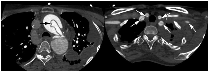
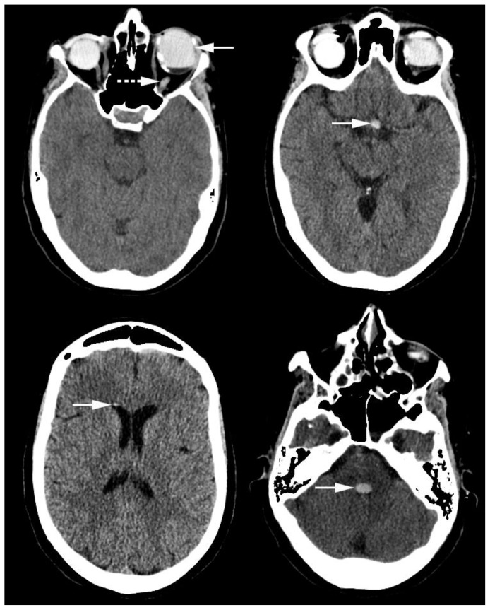
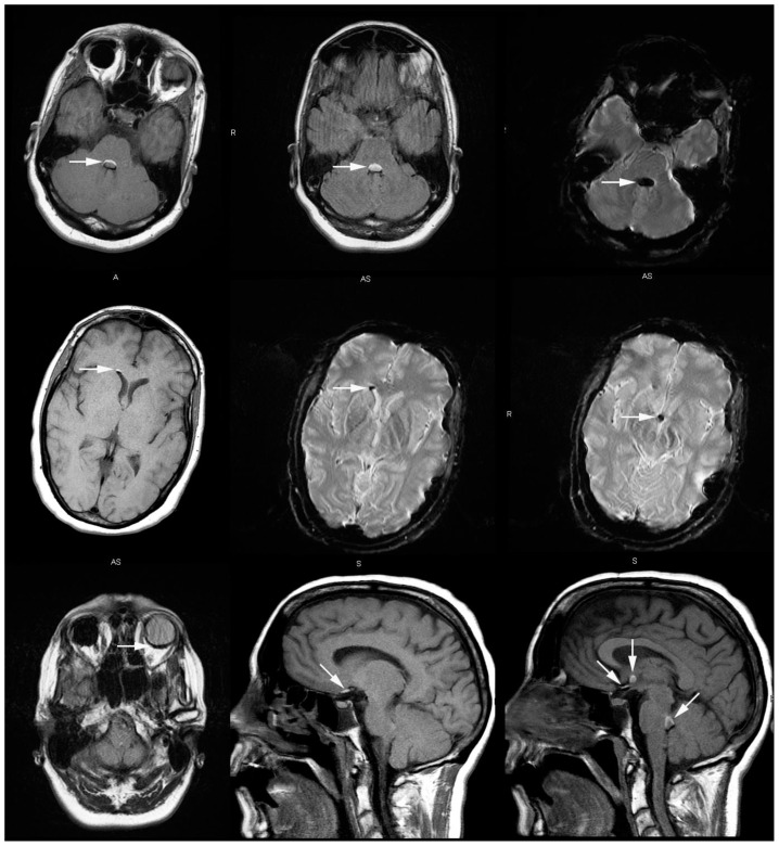
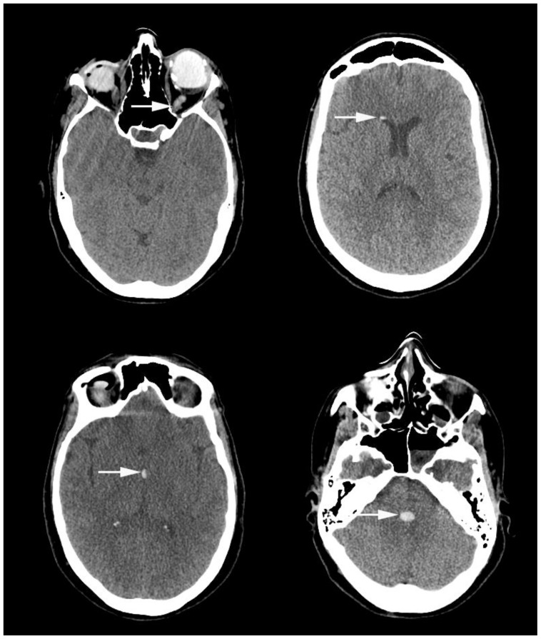
We present a case in which intraocular silicone injection for complex retinal detachment resulted in migration and distribution of silicone along the intracranial visual pathway, and ultimately throughout the ventricular system. Misinterpretation of this material as intracranial hemorrhage on outside computed tomography imaging delayed emergent repair of a Type A aortic dissection until the diagnosis was made on repeat imaging. A discussion of this case and salient computed tomography and magnetic resonance imaging characteristics of silicone is provided.

Keywords: Intraocular silicone; Marfan syndrome; intracranial hemorrhage; intracranial silicone; intraventricular hemorrhage; retinal detachment.


Figures

Figure 1
Figure 1
51-year-old female with Marfan syndrome and blindness secondary to bilateral retinal detachment. FINDINGS: A. Axial contrast enhanced CT through the chest in the arterial phase demonstrates Type A dissection involving the aortic arch (black arrow). B. Type A dissection extending into the bilateral common carotid and subclavian arteries (white arrows). TECHNIQUE: GE (GE lightspeed VCT) 64 slice multidetector CT 646 mAs, 120 kV, 1.3 mm slice thickness.
Figure 2
Figure 2
51-year-old Female with Marfan syndrome and blindness secondary to bilateral retinal detachment. FINDINGS: Axial CT slices through the brain showing hyperdense silicone mimicking intraventricular hemorrhage in A. The left globe (arrow) tracking in the left optic nerve sheath (dotted arrow), which was not observed on the initial outside report. B. The optic chiasm (white arrow). C. The frontal horn of the right lateral ventricle; the non-dependent nature was not observed on the initial outside report (white arrow). D. The fourth ventricle. HU = 100 (white arrow). TECHNIQUE: Toshiba CT scanner 250 mAs, 120 kV, 3mm slice thickness.
Figure 3
Figure 3
51-year-old Female with Marfan syndrome and blindness secondary to bilateral retinal detachment. FINDINGS: A. Axial FLAIR MR image showing chemical shift artifact at the interface between silicone and adjacent brain parenchyma in the fourth ventricle (white arrow). B. Axial FLAIR MR image showing hyperintense silicone mimicking intraventricular hemorrhage filling the fourth ventricle (white arrow). C. Axial susceptibility weighted MR image showing susceptibility artifact caused by silicone mimicking intraventricular hemorrhage in the fourth ventricle (white arrow). D. E. Axial diffusion weighted image (DWI) and Apparent Diffusion Coefficient (ADC) map at the level of the cerebellum showing lack of restricted diffusion in the silicone filling the fourth ventricle (white arrow). F. Non-contrast axial T1 MR image showing T1 hyperintense silicone within the left globe and along the optic nerve (white arrow). G Non-contrast axial T1 image through the lateral ventricles with T1 hyperintense silicone situated non-dependently in the right frontal horn. H. Axial Gradient Recall Echo (GRE) image through the lateral ventricles showing susceptibility artifact corresponding with T1 hyperintense silicone (white arrow). I. Non-contrast sagittal T1 MR image with hyperintense silicone in the optic chiasm, the third ventricle, and the fourth ventricle (white arrows) mimicking intraventricular hemorrhage. TECHNIQUE: 1.5T GE MR Scanner (GE discovery 450) Axial T2-FLAIR: 5 mm slice thickness, TR = 8000 ms, TE = 124.3 ms, Flip Angle 160. Axial SWI 2.2 mm slice thickness, TR = 5500 ms, TE = 25 ms, Flip Angle 90. Axial Diffusion Weighted Image 5 mm slice thickness, TR = 8000, TE = 83.7, Flip Angle 90. Axial Apparent Diffusion Coefficient Map: 5 mm slice thickness, TR = 8000, TE = 83.7, Flip Angle 90. Axial T1: 5 mm slice thickness, TR = 500 ms, TE = 12 ms, Flip Angle 90. Axial Gradient Recall Echo: 5 mm slice thickness, TR = 575 ms, TE = 15 ms, Flip Angle 20. Sagittal T1: 5 mm slice thickness, TR = 466.668 ms, TE = 12 ms, Flip Angle 90.
Figure 4
Figure 4
51-year-old Female with Marfan syndrome and blindness secondary to bilateral retinal detachment. FINDINGS: Axial CT images taken 4 days after the initial outside CT scan showing A. Hyperdense silicone in the left globe tracking along the left optic sheath (white arrow). B. Hyperdense silicone layering non-dependently in the frontal horn of the right lateral ventricle (white arrow). C. Hyperdense silicone in the third ventricle, identical in position compared with the CT performed 4 days prior (white arrow). D. Hyperdense silicone in the fourth ventricle, also identical in position compared with the prior CT (white arrow). Silicone in the discussed locations was mistaken for hemorrhage. TECHNIQUE: GE (GE lightspeed VCT) 64 slice multidetector CT 226 mAs, 100 kV, 2.5 mm slice thickness.

Similar articles

Cited by

References

    1. Atlas SW, DuBois P, Singer MB, Lu D. Diffusion measurements in intracranial hematomas: implications for MR imaging of acute stroke. AJNR Am J Neuroradiol. 2000;21(7):1190–4. - PMC - PubMed
    1. Silvera S, Oppenheim C, Touze E, Ducreux D, Page P, Domigo V, et al. Spontaneous intracerebral hematoma on diffusion-weighted images: influence of T2-shine-through and T2-blackout effects. AJNR Am J Neuroradiol. 2005;26(2):236–41. - PMC - PubMed
    1. La Heij EC, Hendrikse F. Retinal detachments and retinal surgery. Ned Tijdschr Geneeskd. 1999;143(15):781–5. - PubMed
    1. Eller AW, Friberg TR, Mah F. Migration of silicone oil into the brain: a complication of intraocular silicone oil for retinal tamponade. Am J Ophthalmol. 2000;129(5):685–8. - PubMed
    1. Chen JX, NA, Aygun N, Gujar SK, Gandhi D. Intravitreal silicone oil migration into the subarachnoid space and ventricles: A case report and review of literature. European Journal of Radiology Extra. 2011;78:e81–e83. Not referenced.

Publication types

MeSH terms