Left Anterior Descending Coronary Artery and Multiple Peripheral Mycotic Aneurysms Due to Mycobacterium Bovis Following Intravesical Bacillus Calmette-Guerin Therapy: A Case Report
- PMID: 27761190
- PMCID: PMC5065280
- DOI: 10.3941/jrcr.v10i8.2697
Left Anterior Descending Coronary Artery and Multiple Peripheral Mycotic Aneurysms Due to Mycobacterium Bovis Following Intravesical Bacillus Calmette-Guerin Therapy: A Case Report
Abstract
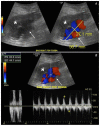
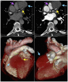
The use of live attenuated intravesicular Bacillus Calmette-Guerin (BCG) therapy is a generally accepted safe and effective method for the treatment of superficial transitional cell carcinoma (TCC) of the bladder. Although rare, < 5% of patient's treated with intravesicular BCG therapy may develop potentially serious complications, including localized infections to the genitourinary tract, mycotic aneurysms and osteomyelitis. We present here a case of a 63-year-old male who developed left coronary and multiple peripheral M. Bovis mycotic aneurysms as a late complication of intravesicular BCG therapy for superficial bladder cancer. The patient initially presented with acute onset pain and swelling in the left knee > 2 years following initial therapy, and initial workup revealed a ruptured saccular aneurysm of the left popliteal artery as well as incidental bilateral common femoral artery aneurysms. Following endovascular treatment and additional workup, the patient was discovered to have additional aneurysms in the right popliteal artery and left anterior descending artery (LAD). Surgical pathology and bacterial cultures obtained from the excised femoral aneurysms and surgical groin wounds were positive for Mycobacterium Bovis, and the patient was initiated on a nine-month antimycobacterial course of isoniazid, rifampin and ethambutol. Including the present case, there has been a total of 32 reported cases of mycotic aneurysms as a complication from intravesicular BCG therapy, which we will review here. The majority of reported cases involve the abdominal aorta; however, this represents the first known reported case of a coronary aneurysm.
Keywords: BCG; Bacillus Calmette-Guerin; Mycobacterium bovis; Mycotic aneurysm; intravesicular.
Figures


Similar articles
-
Mycobacterium bovis abdominal aortic and femoral artery aneurysms following intravesical bacillus Calmette-Guérin therapy for bladder cancer.Cardiovasc Pathol. 2010 Mar-Apr;19(2):e29-32. doi: 10.1016/j.carpath.2008.09.003. Epub 2008 Nov 20. Cardiovasc Pathol. 2010. PMID: 19026573
-
Ruptured mycotic abdominal aortic aneurysm secondary to Mycobacterium bovis after intravesical treatment with bacillus Calmette-Guérin.J Vasc Surg. 2007 Jul;46(1):131-4. doi: 10.1016/j.jvs.2007.01.054. J Vasc Surg. 2007. PMID: 17606130 Review.
-
Disseminated Mycotic Aneurysms following Intravesical Bacillus Calmette-Guérin Therapy for Bladder Cancer: Case Discussion and Systematic Treatment Algorithm.Ann Vasc Surg. 2017 Feb;39:284.e5-284.e10. doi: 10.1016/j.avsg.2016.05.120. Epub 2016 Aug 13. Ann Vasc Surg. 2017. PMID: 27531091
-
Arteriocutaneous Fistula Associated with Bilateral Femoral Pseudoaneurysms Caused by Bacillus Calmette-Guérin. Apropos of a Case and Review of Literature.Ann Vasc Surg. 2017 Feb;39:291.e1-291.e6. doi: 10.1016/j.avsg.2016.07.094. Epub 2016 Nov 27. Ann Vasc Surg. 2017. PMID: 27903467 Review.
-
Abdominal aortic aneurysmal and endovascular device infection with iliopsoas abscess caused by Mycobacterium bovis as a complication of intravesical bacillus Calmette-Guérin therapy.Ann Vasc Surg. 2013 Nov;27(8):1186.e1-5. doi: 10.1016/j.avsg.2012.12.004. Epub 2013 Aug 21. Ann Vasc Surg. 2013. PMID: 23972639
Cited by
-
Failed endovascular abdominal aortic aneurysm repair due to Mycobacterium bovis infection following intravesical bacillus Calmette-Guérin therapy.J Vasc Surg Cases Innov Tech. 2022 Nov 8;8(4):807-812. doi: 10.1016/j.jvscit.2022.10.020. eCollection 2022 Dec. J Vasc Surg Cases Innov Tech. 2022. PMID: 36507086 Free PMC article.
-
Coronary artery aneurysm combined with other multiple aneurysms at multiple locations: A case report and systematic review.Medicine (Baltimore). 2017 Dec;96(50):e9230. doi: 10.1097/MD.0000000000009230. Medicine (Baltimore). 2017. PMID: 29390352 Free PMC article.
-
Fluorescence in situ hybridization microscopic detection of Bacilli Calmette Guérin mycobacteria in aortic lesions: A case report.Medicine (Baltimore). 2018 Jul;97(30):e11321. doi: 10.1097/MD.0000000000011321. Medicine (Baltimore). 2018. PMID: 30045257 Free PMC article.
-
Mycotic aneurysm formation after bacillus Calmette-Guérin instillation for recurrent bladder cancer.CMAJ. 2018 Apr 16;190(15):E467-E471. doi: 10.1503/cmaj.171214. CMAJ. 2018. PMID: 29661816 Free PMC article. No abstract available.
-
Case Report with Systematic Literature Review on Vascular Complications of BCG Intravesical Therapy for Bladder Cancer.J Clin Med. 2022 Oct 21;11(20):6226. doi: 10.3390/jcm11206226. J Clin Med. 2022. PMID: 36294547 Free PMC article.
References
-
- Schellhammer PF, Ladaga LE, Fillion MB. Bacillus Calmette-Guerin for superficial transitional cell carcinoma of the bladder. J Urol. 1986;135:261–4. - PubMed
-
- Safdar N, Abad CL, Kaul DR, Jarrard D, Saint S. An unintended consequence. N Engl J Med. 2008;358:1496–501. - PubMed
-
- Lamm DL, van der Meijden PM, Morales A, Brosman SA, Catalona WJ, Herr HW, Soloway MS, Steg A, Debruyne FM. Incidence and treatment of complications of bacillus Calmette-Guerin intravesical therapy in superficial bladder cancer. J Urol. 1992;147:596–600. - PubMed
-
- Gonzalez OY, Musher DM, Brar I, Furgeson S, Boktour MR, Septimus EJ, Hamill RJ, Graviss EA. Spectrum of bacilli Calmette-Guerin (BCG) infection after intravesical BCG immunotherapy. Clin Infect Dis. 2003;36:140–8. - PubMed
-
- Coscas R, Arlet JB, Belhomme D, Fabiani JN, Pouchot J. Multiple mycotic aneurysms due to Mycobacterium bovis after intravesical bacillus Calmette-Guerin therapy. J Vasc Surg. 2009;50:1185–90. - PubMed
Publication types
MeSH terms
Substances
LinkOut - more resources
Full Text Sources
Other Literature Sources
Medical

