

Impact of Isometric Contraction of Anterior Cervical Muscles on Cervical Lordosis
- PMID: 27761195
- PMCID: PMC5065270
- DOI: 10.3941/jrcr.v10i9.2885
Impact of Isometric Contraction of Anterior Cervical Muscles on Cervical Lordosis
Abstract
Objective: This study investigates the impact of isometric contraction of anterior cervical muscles on cervical lordosis.
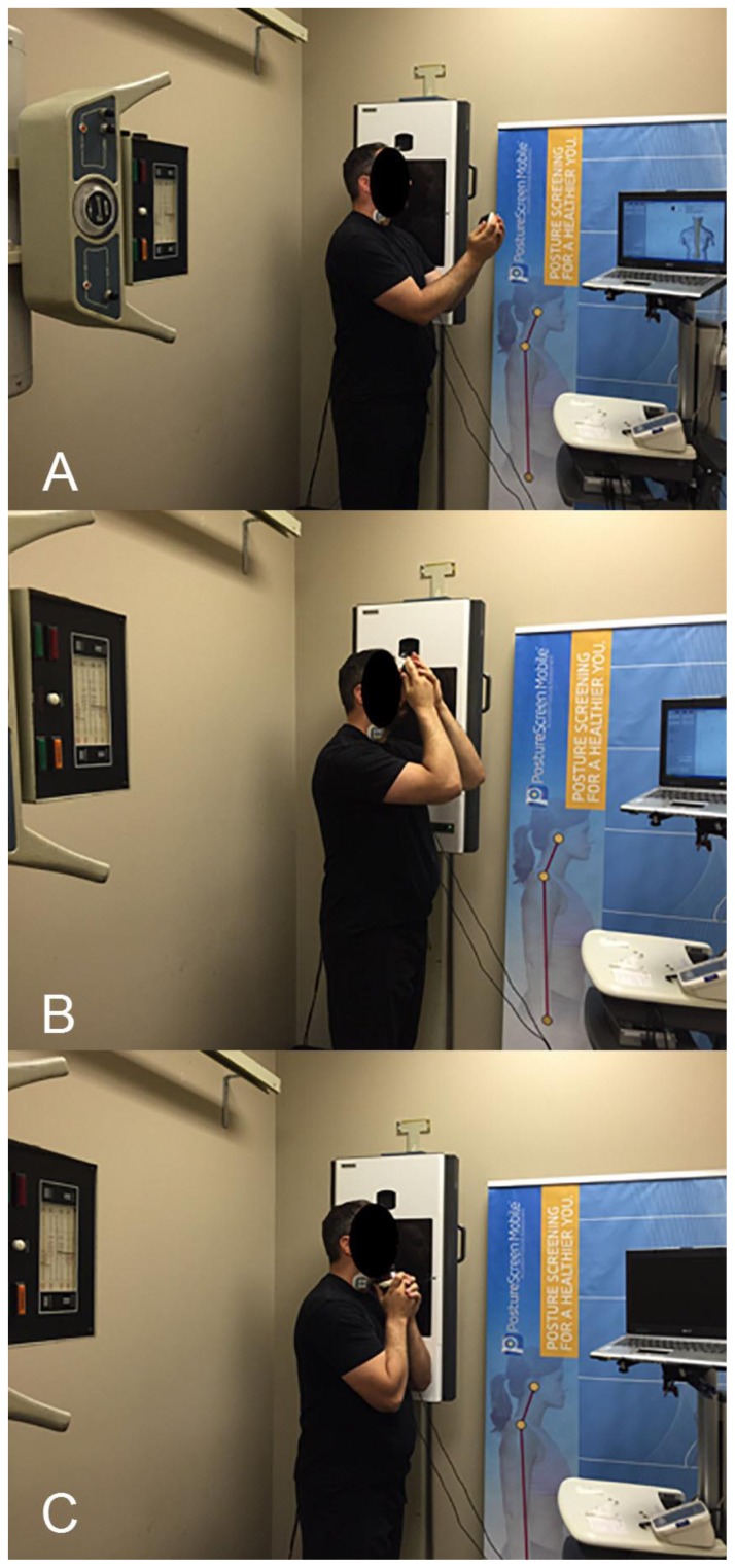
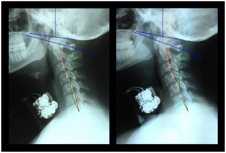
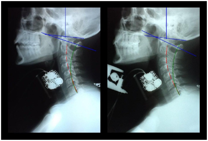
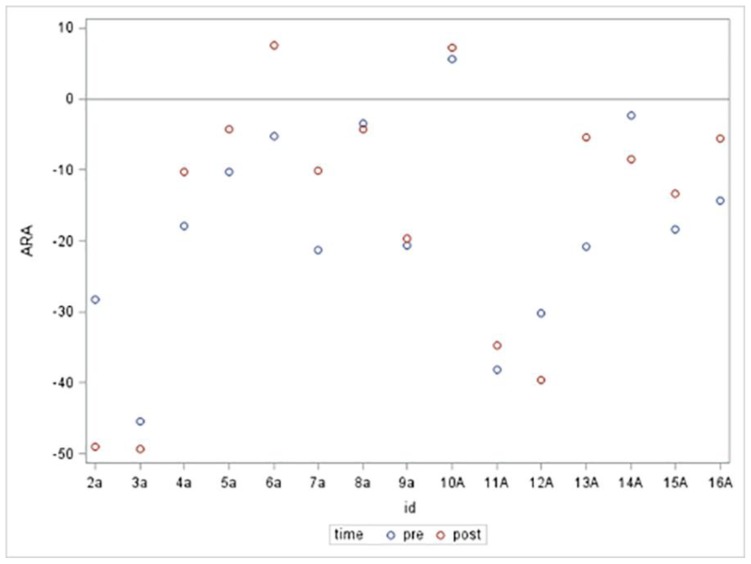
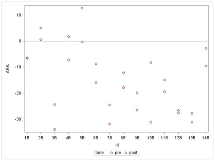
Methods: 29 volunteers were randomly assigned to an anterior head translation (n=15) or anterior head flexion (n=14) group. Resting neutral lateral cervical x-rays were compared to x-rays of sustained isometric contraction of the anterior cervical muscles producing anterior head translation or anterior head flexion.
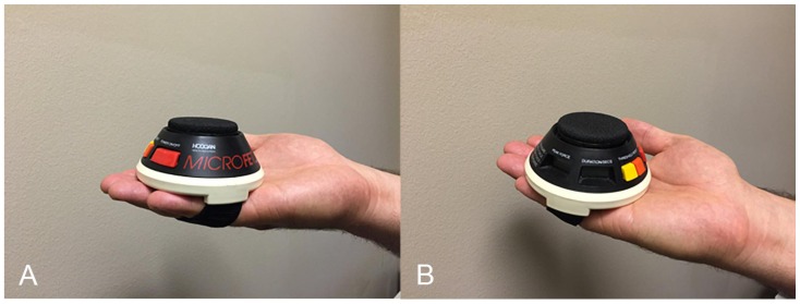
Results: Paired sample t-tests indicate no significant difference between pre and post anterior head translation or anterior head flexion. Analysis of variance suggests that gender and peak force were not associated with change in cervical lordosis. Chamberlain's to atlas plane line angle difference was significantly associated with cervical lordosis difference during anterior head translation (p=0.01).
Conclusion: This study shows no evidence that hypertonicity, as seen in muscle spasms, of the muscles responsible for anterior head translation and anterior head flexion have a significant impact on cervical lordosis.
Keywords: anterior cervical muscles; anterior head flexion; anterior head translation; cervical biomechanics; forward head posture; hypertonicity; hypolordosis; muscle spasm; neutral lateral cervical radiograph.
Figures
Similar articles
-
EMG power spectra of cervical muscles in lateral flexion and comparison with sagittal and oblique plane activities.Eur J Appl Physiol. 2003 May;89(3-4):367-76. doi: 10.1007/s00421-003-0797-3. Epub 2003 Mar 25. Eur J Appl Physiol. 2003. PMID: 12736847
-
Effects of suboccipital release with craniocervical flexion exercise on craniocervical alignment and extrinsic cervical muscle activity in subjects with forward head posture.J Electromyogr Kinesiol. 2016 Oct;30:31-7. doi: 10.1016/j.jelekin.2016.05.007. Epub 2016 May 24. J Electromyogr Kinesiol. 2016. PMID: 27261928
-
Further evaluation of an EMG technique for assessment of the deep cervical flexor muscles.J Electromyogr Kinesiol. 2006 Dec;16(6):621-8. doi: 10.1016/j.jelekin.2005.10.003. Epub 2005 Dec 15. J Electromyogr Kinesiol. 2006. PMID: 16359872
-
Postural Consequences of Cervical Sagittal Imbalance: A Novel Laboratory Model.Spine (Phila Pa 1976). 2015 Jun 1;40(11):783-92. doi: 10.1097/BRS.0000000000000877. Spine (Phila Pa 1976). 2015. PMID: 25768685
-
Concordance and Reliability of Photogrammetric Protocols for Measuring the Cervical Lordosis Angle: A Systematic Review of the Literature.J Manipulative Physiol Ther. 2018 Jan;41(1):71-80. doi: 10.1016/j.jmpt.2017.08.004. J Manipulative Physiol Ther. 2018. PMID: 29366490
Cited by
-
Restoring cervical lordosis by cervical extension traction methods in the treatment of cervical spine disorders: a systematic review of controlled trials.J Phys Ther Sci. 2021 Oct;33(10):784-794. doi: 10.1589/jpts.33.784. Epub 2021 Oct 13. J Phys Ther Sci. 2021. PMID: 34658525 Free PMC article. Review.
-
The Efficacy of Cervical Lordosis Rehabilitation for Nerve Root Function and Pain in Cervical Spondylotic Radiculopathy: A Randomized Trial with 2-Year Follow-Up.J Clin Med. 2022 Nov 2;11(21):6515. doi: 10.3390/jcm11216515. J Clin Med. 2022. PMID: 36362743 Free PMC article.
-
Abnormal Static Sagittal Cervical Curvatures following Motor Vehicle Collisions: A Retrospective Case Series of 41 Patients before and after a Crash Exposure.Diagnostics (Basel). 2024 May 2;14(9):957. doi: 10.3390/diagnostics14090957. Diagnostics (Basel). 2024. PMID: 38732372 Free PMC article.
-
Does restoration of sagittal cervical alignment improve cervicogenic headache pain and disability: A 2-year pilot randomized controlled trial.Heliyon. 2021 Mar 15;7(3):e06467. doi: 10.1016/j.heliyon.2021.e06467. eCollection 2021 Mar. Heliyon. 2021. PMID: 33786392 Free PMC article.
-
Restoring lumbar lordosis: a systematic review of controlled trials utilizing Chiropractic Bio Physics® (CBP®) non-surgical approach to increasing lumbar lordosis in the treatment of low back disorders.J Phys Ther Sci. 2020 Sep;32(9):601-610. doi: 10.1589/jpts.32.601. Epub 2020 Sep 1. J Phys Ther Sci. 2020. PMID: 32982058 Free PMC article. Review.
References
-
- Jones AMM, Stringer EA, Wong DA. Plated cervical fusions yield better outcomes. Trans Orthop Res Soc. 1998;22:524.
-
- Kawakami M, Tamaki T, Yoshida M, et al. Axial symptoms and cervical alignments after cervical anterior spinal fusion for patients with cervical myelopathy. J Spinal Disord. 1999;2:50–6. - PubMed
-
- Matsunaga S, Sakou T, Sunahara N, et al. Biomechanical analysis of buckling alignment of the cervical spines. Predictive value for subaxial subluxation after occipitocervical fusion. Spine. 1997;22:765–71. - PubMed
-
- Lowery G. Three-dimensional screw divergence and sagittal balance: a personal philosophy relative to cervical biomechanics. Spine: State of the Art Reviews. 1996;10:343–56.
-
- Katsuura A, Hukuda S, Imanaka T, et al. Anterior cervical plate used in degenerative disease can maintain cervical Lordosis. J Spinal Disorders. 1996;9:470–6. - PubMed
Publication types
MeSH terms
LinkOut - more resources
Full Text Sources
Other Literature Sources
Medical