Case Reports
. 2016 Aug 23;4(10):935-943.
doi: 10.1002/ccr3.639. eCollection 2016 Oct.

Idiopathic sclerosing orbital inflammation mimicking a malignant spindle cell tumor in a dog

Affiliations
Case Reports

Idiopathic sclerosing orbital inflammation mimicking a malignant spindle cell tumor in a dog

Nina Marie Rzechorzek et al. Clin Case Rep. .

Abstract

A dog presented with a retrobulbar mass, diagnosed histopathologically as malignant spindle cell neoplasia. Emergence of analogous findings in the contralateral orbit prompted extended immunohistochemistry of the original mass and reassignment to idiopathic sclerosing orbital inflammation. Early incisional biopsy with extended immunohistochemical analysis should be considered for canine orbital tumors.

Keywords: Case report; IgG4; exophthalmos; idiopathic orbital inflammation; lymphoplasmacytic infiltration; magnetic resonance imaging; radiation therapy; retrobulbar.


Figures

Figure 1
Figure 1
MRI of the head highlighting the mass in the left retrobulbar and periorbital region. (A) Mildly hyperintense signal on transverse T2w sequence (yellow arrow). (B) Moderate to marked contrast‐enhancing signal on transverse T1w postgadolinium sequence (yellow arrow). The medial aspect of the left nasal cavity is partially filled with noncontrast‐enhancing material. (C) Moderate to marked contrast‐enhancing signal on dorsal T1w postgadolinium sequence (yellow arrow). A poorly circumscribed, extra‐axial contrast‐enhancing mass appears to breach through the ethmoid bone, compressing the ventrolateral aspect of the left olfactory bulb (red arrow); this mass appeared to be contiguous with the retrobulbar mass and the nasal hyperintensity. (D) Marked meningeal enhancement surrounding the lateral aspect of the left olfactory bulb (yellow arrow) on a more caudal slice of transverse T1w postgadolinium sequence.
Figure 2
Figure 2
Photomicrographs capturing key histological features of the left retrobulbar mass (hematoxylin and eosin stain). Spindle cell proliferation with lymphoplasmacytic inflammatory nodules (yellow arrows; lacking germinal follicular architecture) and diffuse inflammation infiltrating muscle and adipose tissue. (A) Scale bar = 200 μm (B) scale bar = 50 μm.
Figure 3
Figure 3
Computed tomography images of the head, postresection of left retrobulbar mass; soft tissue window postcontrast (iohexol). (A) Immediately postsurgery for radiation therapy planning, showing local inflammation around the surgical site. (B) 20 weeks postsurgery. Thickened subcutaneous tissue can be seen within the irradiated field with mild contrast enhancement (yellow arrows) but no gross tumor recurrence. The regional lymph nodes, left eye socket, left orbital bone lysis, seroma, and soft tissue lesion within the left nasal cavity are static compared to (A). There is evidence of reduced local inflammatory reaction (lack of contrast enhancement) in the left retro‐orbital region. Note, however, mild contrast uptake in the right orbital rim – potentially a sign of early inflammation developing on this side (red arrows).
Figure 4
Figure 4
Contralateral emergence of identical retrobulbar lesion 26 weeks postsurgery, identified with advanced imaging. (A) CT (soft tissue window postcontrast) shows right retrobulbar soft tissue mass (yellow arrow), lacking fat intensity, with right temporal and pterygoid myositis. (B) Concordant findings on MRI (dorsal T1w postgadolinium sequence; yellow arrow). The following additional sequences were also performed in transverse (T1w, T2w, T1w postgadolinium, T2w*, fluid‐attenuated inversion recovery), sagittal (T2w), and dorsal (T2w) planes (data not shown).
Figure 5
Figure 5
Photomicrographs capturing key immunohistochemical features of the left retrobulbar mass. (A) The inflammatory infiltrate (brown‐stained cells) was predominantly composed of T lymphocytes (CD3 immunolabel; scale bar = 50 μm). (B) Small numbers of IgG4‐expressing plasma cells were noted (brown‐stained cells highlighted by yellow arrows; IgG4 immunolabel; scale bar = 50 μm).
Figure 6
Figure 6
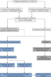
Proposed diagnostic approach to progressive exophthalmos in the dog. Early referral to ophthalmology (and/or neurology) is indicated for progressive exophthalmos. Ultrasonography should include both orbits; upon observation of a retrobulbar mass, ultrasound‐guided FNA for cytology and culture and sensitivity (aerobic/anaerobic bacteria, mycoplasma, and fungi) should be performed. In some geographical locations, a latex cryptococcal antigen agglutination test might be warranted. For infectious inflammatory lesions, blood culture and targeted antimicrobial therapy with or without surgical exploration should be considered. Lack of evidence for an infectious cause should prompt advanced imaging including MRI with contrast and CSF analysis (if MRI indicates intracranial and/or meningeal involvement). At this stage, an incisional biopsy of any soft tissue mass should be performed followed by histopathology, extensive immunohistochemistry, and culture. Incisional biopsy was not considered prior to enucleation and debulking in this case because of (a) the rapid progression of clinical signs and pain that were compromising welfare (b) the consideration that biopsy would also require general anesthesia and (based on our top differential) we anticipated that debulking of the mass would still be required in due course, and (c) that incisional biopsy would have provided less information than excision with respect to diagnosis and staging. In the case of a positive culture, the diagnosis should be revised (dotted arrow). For confirmed neoplasms, three‐view thoracic radiographs and FNA of regional lymph nodes are required for staging. Note that a bilateral presentation should rouse suspicion of a noninfectious inflammatory pathogenesis. For these lesions, the subtype should be determined and IgG4/IgG ratio should be investigated by both serology and immunohistochemical analysis. Monitoring for pain and ulcerative keratitis is paramount throughout the diagnostic work‐up and should be treated promptly. *Signs of ocular pain in the dog include photophobia, blepharospasm, self‐trauma, increased lacrimation, acute aversive response on palpation of the globe or upon mouth opening and globe retraction (note that globe retraction may be prohibited by a retrobulbar mass). **Enucleation may be required at later disease stages on welfare grounds. IMT, inflammatory myofibroblastic tumor; NGE, nodular granulomatous episcleritis; IgG4‐RD, IgG4‐related disease; IOI, idiopathic orbital inflammation; ISOI, idiopathic sclerosing orbital inflammation.

References

    1. Billson, F. M. , Miller‐Michau T., and Mould J. R.. 2006. Idiopathic sclerosing orbital pseudotumor in seven cats. Vet. Ophthalmol. 9:45–51. - PMC - PubMed
    1. Knight, C. , Fan E., and Riis R.. 2009. Inflammatory myofibroblastic tumors in two dogs. Vet. Pathol. 46:273–276. - PubMed
    1. Miller, S. A. , Van der Woerdt A., and Bartick T. E.. 2000. Retrobulbar pseudotumor of the orbit in a cat. J. Am. Vet. Med. Assoc. 216:356–358. - PubMed
    1. LaDue, T. , Klein M. K., and Veterinary Radiation Therapy Oncology Group . 2001. Toxicity criteria of the veterinary radiation therapy oncology group. Vet. Radiol. Ultrasound. 42:475–476. - PubMed
    1. Chow, S. P. , Nastri A., and Hardy T.. 2010. Infratemporal inflammatory myofibroblastic tumour with orbital extension. Clin. Experiment Ophthalmol. 38:727–730. - PubMed

Publication types