. 2016 Oct 11:4:e2494.
doi: 10.7717/peerj.2494. eCollection 2016.

Comparative functional characterization of novel non-syndromic GJB2 gene variant p.Gly45Arg and lethal syndromic variant p.Gly45Glu

Affiliations

Comparative functional characterization of novel non-syndromic GJB2 gene variant p.Gly45Arg and lethal syndromic variant p.Gly45Glu

Juan Rodriguez-Paris et al. PeerJ. .

Abstract

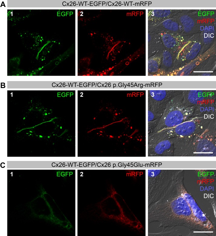
We characterized a novel GJB2 missense variant, c.133G>A, p.Gly45Arg, and compared it with the only other variant at the same amino acid position of the connexin 26 protein (Cx26) reported to date: c.134G>A, p.Gly45Glu. Whereas both variants are associated with hearing loss and are dominantly inherited, p.Gly45Glu has been implicated in the rare fatal keratitis-ichthyosis-deafness (KID) syndrome, which results in cutaneous infections and septicemia with premature demise in the first year of life. In contrast, p.Gly45Arg appears to be non-syndromic. Subcellular localization experiments in transiently co-transfected HeLa cells demonstrated that Cx26-WT (wild-type) and p.Gly45Arg form gap junctions, whereas Cx26-WT with p.Gly45Glu protein does not. The substitution of a nonpolar amino acid glycine in wildtype Cx26 at position 45 with a negatively charged glutamic acid (acidic) has previously been shown to interfere with Ca2+ regulation of hemichannel gating and to inhibit the formation of gap junctions, resulting in cell death. The novel variant p.Gly45Arg, however, changes this glycine to a positively charged arginine (basic), resulting in the formation of dysfunctional gap junctions that selectively affect the permeation of negatively charged inositol 1,4,5-trisphosphate (IP3) and contribute to hearing loss. Cx26 p.Gly45Arg transfected cells, unlike cells transfected with p.Gly45Glu, thrived at physiologic Ca2+ concentrations, suggesting that Ca2+ regulation of hemichannel gating is unaffected in Cx26 p.Gly45Arg transfected cells. Thus, the two oppositely charged amino acids that replace the highly conserved uncharged glycine in p.Gly45Glu and p.Gly45Arg, respectively, produce strikingly different effects on the structure and function of the Cx26 protein.

Keywords: Connexin 26; FRAP; GJB2; Hearing loss; IP3; p.Gly45Arg; p.Gly45Glu.


Conflict of interest statement

At the time of submission, Iris Schrijver was an Academic Editor for PeerJ.

Figures

Figure 1
Figure 1. Co-expression of Wild-type (WT) Cx26 with Cx26 variants in HeLa cells.
Green color corresponds to Enhanced Green Fluorescent Protein (EGFP) and red color corresponds to monomeric Red Fluorescent Protein (mRFP). Yellow color indicates areas of co-localization. DAPI staining is represented by the blue color. (A) Confocal images of transiently co-transfected HeLa cells that express Cx26-WT with C-terminal EGFP-tag (panel 1) and Cx26-WT with C-terminal mRFP-tag (panel 2). Co-localization of both proteins at the gap junctions is shown in yellow (panel 3). (B) Co-expression of Cx26-WT-EGFP (panel 1) and Cx26-p.Gly45Arg with C-terminal tagged mRFP (panel 2) results in co-localization of both proteins at the gap junctions (panel 3). (C) Cells co-transfected with Cx26-WT-EGFP (panel 1) and Cx26-p.Gly45Glu-mRFP (panel 2) do not form gap junctions as evidenced by the absence of plaques (panel 3). Scale bars: 20 µm.
Figure 2
Figure 2. Co-expression of Cx26 variants with p.Tyr136Stop in HeLa cells.
Green color corresponds to Enhanced Green Fluorescent Protein (EGFP) and red color corresponds to monomeric Red Fluorescent Protein (mRFP). Yellow color indicates areas of co-localization. DAPI staining is represented by the blue color. (A–B) Cells co-transfected with Cx26-WT-EGFP (panel 1) and, respectively, Cx26 p.Gly45Glu-p.Tyr136Stop-mRFP (panel 2) (A) and Cx26 p.Tyr136Stop-mRFP (panel 2) (B) form only Cx26-WT gap junctions (green color; panels 1 and 3).(C) Cells co-transfected with Cx26 p.Gly45Arg-EGFP (panel 1) and Cx26 p.Tyr136Stop-mRFP (panel 2) in trans only form Cx26 p.Gly45Arg gap junctions (green color; panels 1 and 3). Brightness and contrast were adjusted to show fluorescent signal indicating successful transfection of the cells. (D) Cells co-transfected with Cx26 p.Gly45Glu-EGFP (panel 1) and Cx26 p.Tyr136Stop-mRFP (panel 2) in trans do not form gap junctions (panels 1, 2 and 3). Brightness and contrast were adjusted to show fluorescent signal indicating successful transfection of the cells. Scale bars: 20 µm.
Figure 3
Figure 3. IP3-permeability and fluorescence recovery after photo bleaching (FRAP) in Cx26-G45R coupled cells.
(A) Cx26-WT-EGFP transfected HeLa cells. Cell 1 and cell 2 are connected by gap junctions expressing Cx26-WT-EGFP (panel 1). Cells are loaded with caged IP3, Rhod2 AM and Calcein violet AM. Upon focal irradiation of cell 1, functional IP3 permeability allows for increase in Calcium levels in cell 1 and 2 as measured by changes in relative Rhod2 fluorescence intensity levels (n = 4; mean ± SD) (panels 2, 3 and 4). Consecutive photo-bleaching of Calcein violet in cell 1 reduces detected fluorescence levels immediately after bleaching (panels 5, 6 and 8). Calcein violet dye flow through the gap junction recovers fluorescence values (n = 4; mean ± SD) (panels 7 and 8). (B) Cx26-WT-EGFP and Cx26-G45R co-transfected HeLa cells. Cell 1 and cell 2 are connected by gap junctions co-expressing Cx26-WT-EGFP and Cx26-G45R (panel 1). Cells are loaded with caged IP3, Rhod2 AM and Calcein violet AM. Upon focal irradiation of cell 1, impaired IP3 permeability results in the reduced release of Calcium in cell 2 in comparison to cell 1 (n = 4; mean ± SD) (panels 2, 3 and 4). Consecutive photo-bleaching of Calcein violet in cell 1 reduces detected fluorescence levels immediately after bleaching (panels 5, 6 and 8). Calcein violet dye flow through the Cx26-WT-EGFP/Cx26-G45R gap junction recovers fluorescence values (n = 3; mean ± SD) (panels 7 and 8). (C) Cx26-G45R-EGFP transfected HeLa cells. Cell 1 and cell 2 are connected by gap junctions expressing Cx26-G45R-EGFP (panel 1). Cells are loaded with caged IP3, Rhod2 AM and Calcein violet AM. Upon focal irradiation of cell 1, loss of IP3 permeability results in IP3-uncoupling of cell 1 and 2 as demonstrated by the absence of increased Rhod2 fluorescence levels in cell 2 (n = 4; mean ± SD) (panels 2, 3 and 4). Consecutive photo-bleaching of Calcein violet in cell 1 reduces detected fluorescence levels immediately after bleaching (panels 5, 6 and 8). Calcein violet dye flow through the Cx26-G45R-EGFP gap junction recovers fluorescence values (n = 3; mean ± SD) (panels 7 and 8). Scale bars: 10 µm.

References

    1. Adzhubei IA, Schmidt S, Peshkin L, Ramensky VE, Gerasimova A, Bork P, Kondrashov AS, Sunyaev SR. A method and server for predicting damaging missense mutations. Nature Methods. 2010;7:248–249. doi: 10.1038/nmeth0410-248. - DOI - PMC - PubMed
    1. Ambrosi C, Walker AE, DePriest AD, Cone AC, Lu C, Badger J, Skerrett IM, Sosinsky GE. Analysis of trafficking, stability and function of human connexin 26 gap junction channels with deafness-causing mutations in the fourth transmembrane helix. PLoS ONE. 2013;8:e70916–e70924. doi: 10.1371/journal.pone.0070916. - DOI - PMC - PubMed
    1. Beltramello M, Piazza V, Bukauskas FF, Pozzan T, Mammano F. Impaired permeability to Ins(1,4,5)P3 in a mutant connexin underlies recessive hereditary deafness. Nature Cell Biology. 2005;7:63–69. doi: 10.1038/ncb1205. - DOI - PubMed
    1. Bennett BC, Purdy MD, Baker KA, Acharya C, McIntire WE, Stevens RC, Zhang Q, Harris AL, Abagyan R, Yeager M. An electrostatic mechanism for Ca2+-mediated regulation of gap junction channels. Nature Communications. 2016;7:8770–8782. doi: 10.1038/ncomms9770. - DOI - PMC - PubMed
    1. Capriotti E, Calabrese R, Casadio R. Predicting the insurgence of human genetic diseases associated to single point protein mutations with support vector machines and evolutionary information. Bioinformatics. 2006;22:2729–2734. doi: 10.1093/bioinformatics/btl423. - DOI - PubMed