A new hynobiid-like salamander (Amphibia, Urodela) from Inner Mongolia, China, provides a rare case study of developmental features in an Early Cretaceous fossil urodele
- PMID: 27761316
- PMCID: PMC5068444
- DOI: 10.7717/peerj.2499
A new hynobiid-like salamander (Amphibia, Urodela) from Inner Mongolia, China, provides a rare case study of developmental features in an Early Cretaceous fossil urodele
Abstract
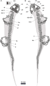
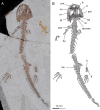
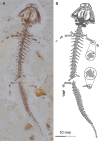
A new fossil salamander, Nuominerpeton aquilonaris (gen. et sp. nov.), is named and described based on specimens from the Lower Cretaceous Guanghua Formation of Inner Mongolia, China. The new discovery documents a far northern occurrence of Early Cretaceous salamanders in China, extending the geographic distribution for the Mesozoic fossil record of the group from the Jehol area (40th-45th parallel north) to near the 49th parallel north. The new salamander is characterized by having the orbitosphenoid semicircular in shape; coracoid plate of the scapulocoracoid greatly expanded with a convex ventral and posterior border; ossification of two centralia in carpus and tarsus; and first digit being about half the length of the second digit in both manus and pes. The new salamander appears to be closely related to hynobiids, although this inferred relationship awaits confirmation by research in progress by us on a morphological and molecular combined analysis of cryptobranchoid relationships. Comparison of adult with larval and postmetamorphic juvenile specimens provides insights into developmental patterns of cranial and postcranial skeletons in this fossil species, especially resorption of the palatine and anterior portions of the palatopterygoid in the palate and the coronoid in the mandible during metamorphosis, and postmetamorphic ossification of the mesopodium in both manus and pes. Thus, this study provides a rare case study of developmental features in a Mesozoic salamander.
Keywords: Developmental features; Early Cretaceous; Hynobiid-like salamander; Larval–juvenile–adult forms; New fossil taxon.
Conflict of interest statement
The authors declare there are no competing interests.
Figures


Similar articles
-
Comparative osteology of the hynobiid complex Liua-Protohynobius-Pseudohynobius (Amphibia, Urodela): Ⅰ. Cranial anatomy of Pseudohynobius.J Anat. 2021 Feb;238(2):219-248. doi: 10.1111/joa.13311. Epub 2020 Sep 22. J Anat. 2021. PMID: 32964448 Free PMC article.
-
Osteology of Batrachuperus londongensis (Urodela, Hynobiidae): study of bony anatomy of a facultatively neotenic salamander from Mount Emei, Sichuan Province, China.PeerJ. 2018 Mar 28;6:e4517. doi: 10.7717/peerj.4517. eCollection 2018. PeerJ. 2018. PMID: 29610705 Free PMC article.
-
A New Basal Salamandroid (Amphibia, Urodela) from the Late Jurassic of Qinglong, Hebei Province, China.PLoS One. 2016 May 4;11(5):e0153834. doi: 10.1371/journal.pone.0153834. eCollection 2016. PLoS One. 2016. PMID: 27144770 Free PMC article.
-
Morphology and ontogeny of carpus and tarsus in stereospondylomorph temnospondyls.PeerJ. 2023 Oct 26;11:e16182. doi: 10.7717/peerj.16182. eCollection 2023. PeerJ. 2023. PMID: 37904842 Free PMC article.
-
Repeated ecological and life cycle transitions make salamanders an ideal model for evolution and development.Dev Dyn. 2022 Jun;251(6):957-972. doi: 10.1002/dvdy.373. Epub 2021 Jun 2. Dev Dyn. 2022. PMID: 33991029 Review.
Cited by
-
Comparative osteology of the hynobiid complex Liua-Protohynobius-Pseudohynobius (Amphibia, Urodela): Ⅰ. Cranial anatomy of Pseudohynobius.J Anat. 2021 Feb;238(2):219-248. doi: 10.1111/joa.13311. Epub 2020 Sep 22. J Anat. 2021. PMID: 32964448 Free PMC article.
-
Middle Jurassic stem hynobiids from China shed light on the evolution of basal salamanders.iScience. 2021 Jun 17;24(7):102744. doi: 10.1016/j.isci.2021.102744. eCollection 2021 Jul 23. iScience. 2021. PMID: 34278256 Free PMC article.
-
Inter-amphibian predation in the Early Cretaceous of China.Sci Rep. 2019 May 23;9(1):7751. doi: 10.1038/s41598-019-44247-7. Sci Rep. 2019. PMID: 31123302 Free PMC article.
-
Cranial biomechanics in basal urodeles: the Siberian salamander (Salamandrella keyserlingii) and its evolutionary and developmental implications.Sci Rep. 2017 Aug 31;7(1):10174. doi: 10.1038/s41598-017-10553-1. Sci Rep. 2017. PMID: 28860600 Free PMC article.
-
Osteology of Batrachuperus yenyuanensis (Urodela, Hynobiidae), a high-altitude mountain stream salamander from western China.PLoS One. 2019 Jan 25;14(1):e0211069. doi: 10.1371/journal.pone.0211069. eCollection 2019. PLoS One. 2019. PMID: 30682102 Free PMC article.
References
-
- Amiot R, Wang X, Zhou Z, Wang X, Buffetaut E, Lécuyer C, Ding Z, Fluteau F, Hibino T, Kusuhashi N, Mo J, Suteethorn V, Wang Y, Xu X, Zhang F. Oxygen isotopes of East Asian dinosaurs reveal exceptionally cold Early Cretaceous climates. Proceedings of the National Academy of Sciences of the United States of America. 2011;108:5179–5183. doi: 10.1073/pnas.1011369108. - DOI - PMC - PubMed
-
- AmphibiaTree “Batrachuperus persicus”. Digital Morphology. 2004. http://digimorph.org/specimens/Batrachuperus_persicus/ [30 August 2015]. http://digimorph.org/specimens/Batrachuperus_persicus/
-
- AmphibiaTree “Onychodactylus japonicus”. Digital Morphology. 2007a. http://digimorph.org/specimens/Onychodactylus_japonicus/head/ [29 February 2016]. http://digimorph.org/specimens/Onychodactylus_japonicus/head/
-
- AmphibiaTree “Rhyacotriton variegatus”. Digital Morphology. 2007b. http://digimorph.org/specimens/Rhyacotriton_variegatus/head/ [30 August 2015]. http://digimorph.org/specimens/Rhyacotriton_variegatus/head/
-
- AmphibiaWeb . Information on amphibian biology and conservation. Berkeley: AmphibiaWeb; 2016. [10 August 2016].
LinkOut - more resources
Full Text Sources
Other Literature Sources

