

Histological variability in the limb bones of the Asiatic wild ass and its significance for life history inferences
- PMID: 27761353
- PMCID: PMC5068390
- DOI: 10.7717/peerj.2580
Histological variability in the limb bones of the Asiatic wild ass and its significance for life history inferences
Abstract
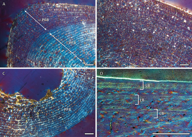
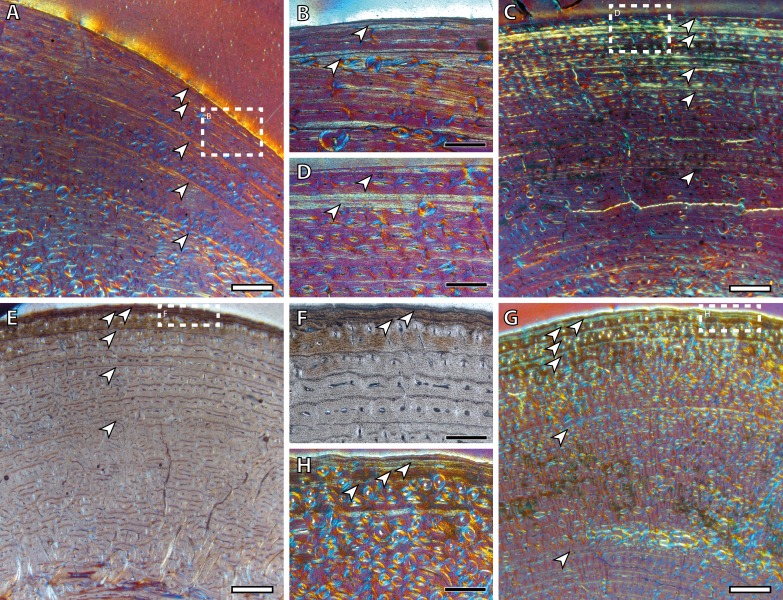
The study of bone growth marks (BGMs) and other histological traits of bone tissue provides insights into the life history of present and past organisms. Important life history traits like longevity or age at maturity, which could be inferred from the analysis of these features, form the basis for estimations of demographic parameters that are essential in ecological and evolutionary studies of vertebrates. Here, we study the intraskeletal histological variability in an ontogenetic series of Asiatic wild ass (Equus hemionus) in order to assess the suitability of several skeletal elements to reconstruct the life history strategy of the species. Bone tissue types, vascular canal orientation and BGMs have been analyzed in 35 cross-sections of femur, tibia and metapodial bones of 9 individuals of different sexes, ages and habitats. Our results show that the number of BGMs recorded by the different limb bones varies within the same specimen. Our study supports that the femur is the most reliable bone for skeletochronology, as already suggested. Our findings also challenge traditional beliefs with regard to the meaning of deposition of the external fundamental system (EFS). In the Asiatic wild ass, this bone tissue is deposited some time after skeletal maturity and, in the case of the femora, coinciding with the reproductive maturity of the species. The results obtained from this research are not only relevant for future studies in fossil Equus, but could also contribute to improve the conservation strategies of threatened equid species.
Keywords: Bone growth marks; Bone histology; Equus hemionus; External fundamental system; Intraskeletal histological variability; Life history; Limb bones; Longevity; Reproductive maturity; Skeletochronology.
Conflict of interest statement
The authors declare there are no competing interests.
Figures


Similar articles
-
Limb bone histology records birth in mammals.PLoS One. 2018 Jun 20;13(6):e0198511. doi: 10.1371/journal.pone.0198511. eCollection 2018. PLoS One. 2018. PMID: 29924818 Free PMC article.
-
Osteohistology and palaeobiology of giraffids from the Mio-Pliocene Langebaanweg (South Africa).J Anat. 2023 May;242(5):953-971. doi: 10.1111/joa.13825. Epub 2023 Feb 6. J Anat. 2023. PMID: 36748181 Free PMC article. Review.
-
Reassessing the evolutionary history of ass-like equids: insights from patterns of genetic variation in contemporary extant populations.Mol Phylogenet Evol. 2015 Apr;85:88-96. doi: 10.1016/j.ympev.2015.01.005. Epub 2015 Feb 12. Mol Phylogenet Evol. 2015. PMID: 25681678
-
Calibration of life history traits with epiphyseal closure, dental eruption and bone histology in captive and wild red deer.J Anat. 2019 Aug;235(2):205-216. doi: 10.1111/joa.13016. Epub 2019 May 30. J Anat. 2019. PMID: 31148188 Free PMC article.
-
Developmental palaeontology of Reptilia as revealed by histological studies.Semin Cell Dev Biol. 2010 Jun;21(4):462-70. doi: 10.1016/j.semcdb.2009.11.005. Epub 2009 Nov 11. Semin Cell Dev Biol. 2010. PMID: 19913103 Review.
Cited by
-
Insular giant leporid matured later than predicted by scaling.iScience. 2023 Aug 17;26(9):107654. doi: 10.1016/j.isci.2023.107654. eCollection 2023 Sep 15. iScience. 2023. PMID: 37694152 Free PMC article.
-
Osteohistology of Greererpeton provides insight into the life history of an early Carboniferous tetrapod.J Anat. 2021 Dec;239(6):1256-1272. doi: 10.1111/joa.13520. Epub 2021 Jul 26. J Anat. 2021. PMID: 34310687 Free PMC article. Review.
-
Identifying animal taxa used to manufacture bone tools during the Middle Stone Age at Sibudu, South Africa: Results of a CT-rendered histological analysis.PLoS One. 2018 Nov 29;13(11):e0208319. doi: 10.1371/journal.pone.0208319. eCollection 2018. PLoS One. 2018. PMID: 30496272 Free PMC article.
-
Limb bone histology records birth in mammals.PLoS One. 2018 Jun 20;13(6):e0198511. doi: 10.1371/journal.pone.0198511. eCollection 2018. PLoS One. 2018. PMID: 29924818 Free PMC article.
-
Osteohistology and palaeobiology of giraffids from the Mio-Pliocene Langebaanweg (South Africa).J Anat. 2023 May;242(5):953-971. doi: 10.1111/joa.13825. Epub 2023 Feb 6. J Anat. 2023. PMID: 36748181 Free PMC article. Review.
References
-
- Amprino R. La structure du tissu osseux envisagée comme expression de différences dans la vitesse de l’accroissement. Archives of Biology. 1947;58:315–330.
-
- Amson E, Kolb C, Scheyer TM, Sánchez-Villagra MR. Growth and life history of Middle Miocene deer (Mammalia, Cervidae) based on bone histology. Comptes Rendus Palevol. 2015;14:637–645. doi: 10.1016/j.crpv.2015.07.001. - DOI
-
- Asa CS. Reproductive physiology. In: Kleiman DG, Thompson KV, Kirk-Baer C, editors. Wild Mammals in Captivity. The University of Chicago Press; Chicago: 2010. pp. 219–252.
-
- Cambra-Moo O, Nacarino-Meneses C, Díaz-Güemes I, Enciso S, García Gil O, Llorente Rodríguez L, Rodríguez Barbero MA, De Aza AH, González Martín A. Multidisciplinary characterization of the long-bone cortex growth patterns through sheep’s ontogeny. Journal of Structural Biology. 2015;191:1–9. doi: 10.1016/j.jsb.2015.06.013. - DOI - PubMed
LinkOut - more resources
Full Text Sources
Other Literature Sources
Miscellaneous
