

A novel step osteotomy for correction of hemifacial microsomia - A case report
- PMID: 27761391
- PMCID: PMC5064979
- DOI: 10.1016/j.jobcr.2016.05.002
A novel step osteotomy for correction of hemifacial microsomia - A case report
Abstract
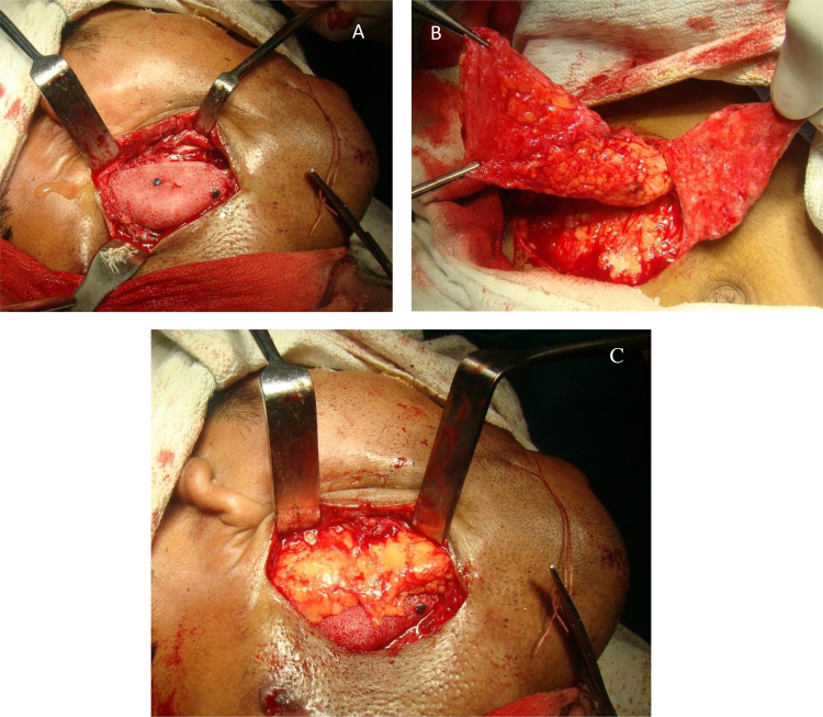
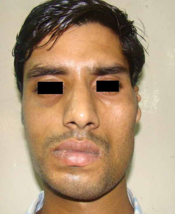
Facial asymmetry is one of the commonest facial anomalies, with reported incidence as high as 34%. Hemifacial microsomia (HFM) has an incidence of 1 in every 4000-5600 children and is one of the commonest causes of facial asymmetry. The standard treatment of HFM is orthognathic surgery by bilateral saggital split osteotomy (BSSO) or distraction osteogenesis (DO) of the mandible, both of which involve prolonged periods of occlusal adjustments by an orthodontist. Here, we present distraction of the mandible by means of a novel modified step osteotomy to correct the facial asymmetry in a case of hemifacial microsomia without disturbing the occlusion. This novel technique can prove to be a new tool in the maxillofacial surgeons armamentarium to treat facial asymmetry.
Keywords: Bilateral saggital split osteotomy (BSSO); Distraction osteogenesis (DO); Genioplasty; Hemifacial microsomia (HFM); Novel step osteotomy.
Figures


References
-
- Severt T.R., Proffit W.R. The prevalence of facial asymmetry in the dentofacial deformities population at the University of North Carolina. Int J Adult Orthodon Orthognath Surg. 1997;12(3):171–176. - PubMed
-
- Grabb W.C. The first and second branchial arch syndrome. Plast Reconstr Surg. 1965;36(4):85–508. - PubMed
-
- Poswillo D. The pathogenesis of the first and second branchial arch syndrome. Oral Surg Oral Med Oral Pathol. 1973;35:302–328. - PubMed
-
- Mehrotra D., Vishwakarma K., Chellapa A.L., Mahajan N. Pre-arthroplasty simultaneous maxillomandibular distraction osteogenesis for the correction of post-ankylotic dentofacial deformities. Int J Oral Maxillofac Surg. 2016 - PubMed
-
- Posnick J.C. Elsevier; China: 2014. Principles and Practice of Orthognathic Surgery.
LinkOut - more resources
Full Text Sources
Other Literature Sources
