Evaluate the growth and adhesion of osteoblast cells on nanocomposite scaffold of hydroxyapatite/titania coated with poly hydroxybutyrate
- PMID: 27761431
- PMCID: PMC5070039
- DOI: 10.4103/2277-9175.188486
Evaluate the growth and adhesion of osteoblast cells on nanocomposite scaffold of hydroxyapatite/titania coated with poly hydroxybutyrate
Abstract
Background: The generation of bioartificial bone tissues may help to overcome the problems related to donor site morbidity and size limitations.
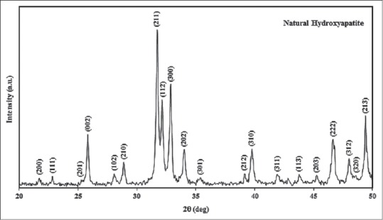
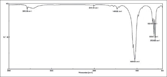
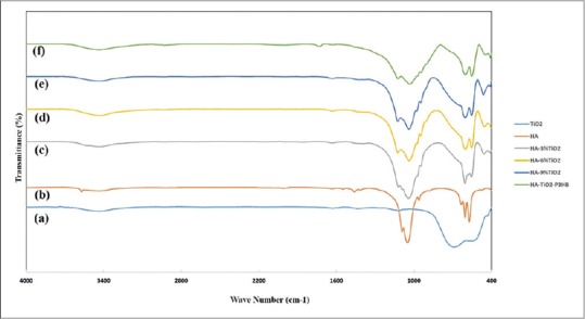
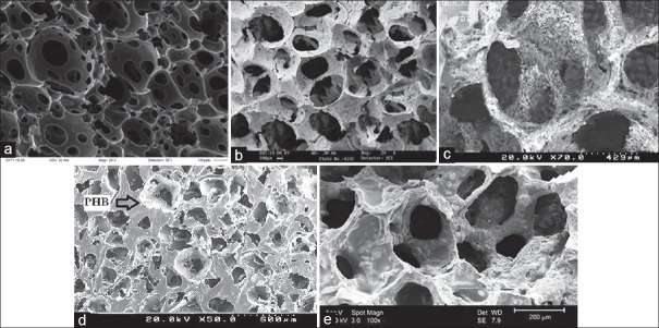
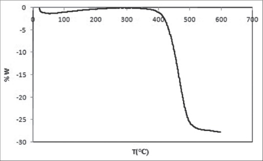
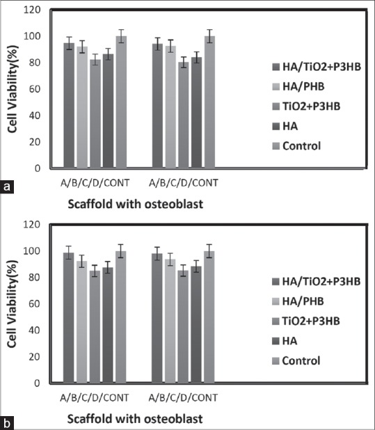
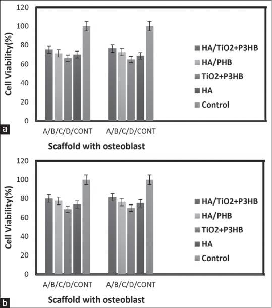
Materials and methods: In this paper, hydroxyapatite (HA) powder was made out of bovine bone by thermal analysis at 900°C and first, and then, porous HA (50 weight percentage) was produced by polyurethane sponge replication method. In order to improve the scaffold mechanical properties, they have been coated with poly hydroxybutyrate. In terms of phase studies, morphology, and specifying agent groups, the specific characterization devices such as X-ray diffraction and Fourier transform infrared, were employed. To compare the behavior of cellular scaffolds, they were divided into four groups of scaffolds. The osteoblast cells were cultured. To perform phase studies, analysis of Methylthiazole tetrazolium (MTT) and Trypan blue were carried out for the viability and attachment on the surface of the scaffold, and the specification of Scanning electron microscopy was employed for the morphology of the cells.
Results: The results of MTT analysis performed on four groups of scaffolds have shown that Titanium oxide (Tio2) had no effect on cell growth alone and HA was the main factor of growth and cell osteoblast adhesion on the scaffold. Moreover, the results showed that the use of coating with poly-3-hydroxybutyrate saved the factors and placed the osteoblasts within the pore. Since the main part of bone consists of HA, the TiO2 accelerates the formation of apatite crystals at the scaffold surface which is the evidence for bone tissue regeneration.
Conclusions: It is likely that the relation between HA and TiO2 leads to an increase in osteoblast adhesion and growth of cells on the scaffold surface.
Keywords: Osteoblast; poly- hydroxybutyrate; scaffold; tissue Engineering; titania.
Figures
Similar articles
-
Evaluation of the effects of nano-TiO2 on bioactivity and mechanical properties of nano bioglass-P3HB composite scaffold for bone tissue engineering.J Mater Sci Mater Med. 2016 Jan;27(1):2. doi: 10.1007/s10856-015-5613-1. Epub 2015 Nov 26. J Mater Sci Mater Med. 2016. PMID: 26610925
-
Novel hydroxyapatite/chitosan bilayered scaffold for osteochondral tissue-engineering applications: Scaffold design and its performance when seeded with goat bone marrow stromal cells.Biomaterials. 2006 Dec;27(36):6123-37. doi: 10.1016/j.biomaterials.2006.07.034. Epub 2006 Aug 30. Biomaterials. 2006. PMID: 16945410
-
A comparison study on the behavior of human endometrial stem cell-derived osteoblast cells on PLGA/HA nanocomposite scaffolds fabricated by electrospinning and freeze-drying methods.J Orthop Surg Res. 2018 Mar 27;13(1):63. doi: 10.1186/s13018-018-0754-9. J Orthop Surg Res. 2018. PMID: 29587806 Free PMC article.
-
Fabrication and characterization of highly porous barium titanate based scaffold coated by Gel/HA nanocomposite with high piezoelectric coefficient for bone tissue engineering applications.J Mech Behav Biomed Mater. 2018 Mar;79:195-202. doi: 10.1016/j.jmbbm.2017.12.034. Epub 2017 Dec 30. J Mech Behav Biomed Mater. 2018. PMID: 29306083
-
Natural hydroxyapatite-based nanobiocomposites and their biomaterials-to-cell interaction for bone tissue engineering.Discov Nano. 2024 Oct 7;19(1):169. doi: 10.1186/s11671-024-04119-0. Discov Nano. 2024. PMID: 39375246 Free PMC article. Review.
Cited by
-
Recent progress in nanocomposites of carbon dioxide fixation derived reproducible biomedical polymers.Front Chem. 2022 Oct 7;10:1035825. doi: 10.3389/fchem.2022.1035825. eCollection 2022. Front Chem. 2022. PMID: 36277338 Free PMC article. Review.
-
Applying extrusion-based 3D printing technique accelerates fabricating complex biphasic calcium phosphate-based scaffolds for bone tissue regeneration.J Adv Res. 2022 Sep;40:69-94. doi: 10.1016/j.jare.2021.12.012. Epub 2021 Dec 28. J Adv Res. 2022. PMID: 36100335 Free PMC article. Review.
-
Development and biological evaluation of Ti6Al7Nb scaffold implants coated with gentamycin-saturated bacterial cellulose biomaterial.PLoS One. 2018 Oct 24;13(10):e0205205. doi: 10.1371/journal.pone.0205205. eCollection 2018. PLoS One. 2018. PMID: 30356274 Free PMC article.
-
Biomimetic polyurethane/TiO2 nanocomposite scaffolds capable of promoting biomineralization and mesenchymal stem cell proliferation.Mater Sci Eng C Mater Biol Appl. 2018 Apr 1;85:79-87. doi: 10.1016/j.msec.2017.12.008. Epub 2017 Dec 18. Mater Sci Eng C Mater Biol Appl. 2018. PMID: 29407160 Free PMC article.
-
Bacterial-Derived Polyhydroxyalkanoate/Bioceramic Composites in Clinical Practice: State of the Art and Future Perspectives.ACS Biomater Sci Eng. 2025 Aug 11;11(8):4653-4670. doi: 10.1021/acsbiomaterials.5c00407. Epub 2025 Jul 21. ACS Biomater Sci Eng. 2025. PMID: 40690593 Free PMC article. Review.
References
-
- Langer R, Vacanti JP. Tissue engineering. Science. 1993;260:920–6. - PubMed
-
- Hollinger JO, Einhorn TA, Doll B, Sfeir C, editors. Bone tissue engineering. CRC press. 2004 Oct 14;
-
- Buckwalter JA, Glimcher MJ, Cooper RR, Recker R. Bone biology. II: Formation, form, modeling, remodeling, and regulation of cell function. Instr Course Lect. 1996;45:387–99. - PubMed
-
- Buckwalter JA, Glimcher MJ, Cooper RR, Recker R. Bone biology. I: Structure, blood supply, cells, matrix, and mineralization. Instr Course Lect. 1996;45:371–86. - PubMed
-
- Ackerman LV, Spjut HJ, Abell MR. Bones and Joints (Monographs in Pathology) Baltimore: Williams and Wilkins; 1976.
LinkOut - more resources
Full Text Sources
Other Literature Sources