Review
. 2016 Oct 7;3(6):e288.
doi: 10.1212/NXI.0000000000000288. eCollection 2016 Dec.

Immunology of neuromyelitis optica during pregnancy

Affiliations
Review

Immunology of neuromyelitis optica during pregnancy

Vahid Davoudi et al. Neurol Neuroimmunol Neuroinflamm. .

Abstract

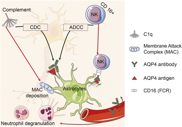
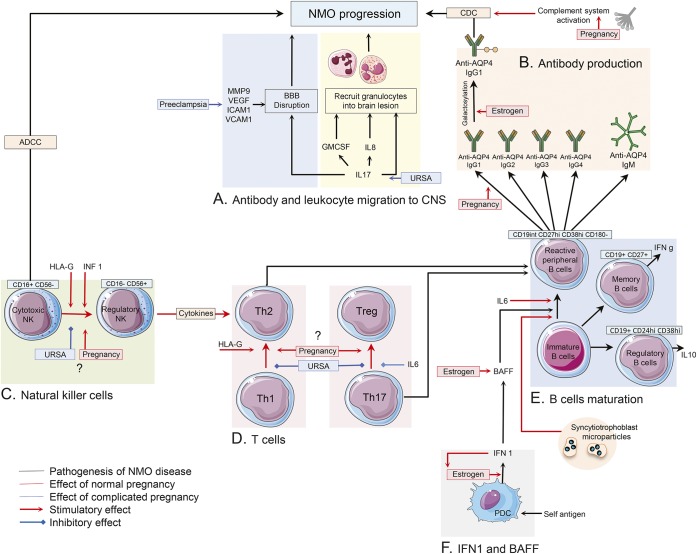
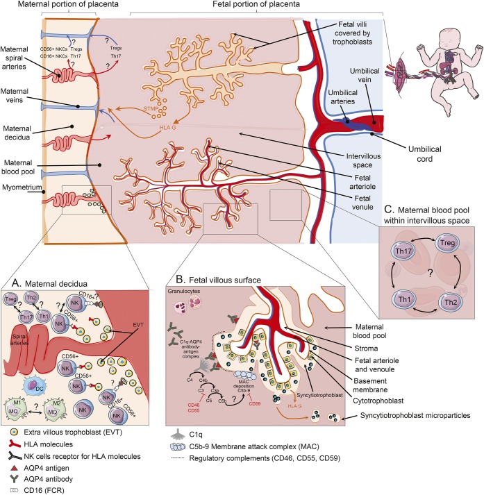
Anti-aquaporin-4 (AQP4) autoantibody plays a key role in the pathogenesis of neuromyelitis optica (NMO). Studies have shown increased relapse rates in patients with NMO during pregnancy and postpartum. High estrogen levels during pregnancy can increase activation-induced cytidine deaminase expression, which is responsible for immunoglobulin production. Additionally, sex hormones may influence antibody glycosylation, with effects on antibody function. Estrogen decreases apoptosis of self-reactive B cells, through upregulation of antiapoptotic molecules. Furthermore, high estrogen levels during pregnancy can boost B-cell activating factor and type 1 interferon (IFN) production, facilitating development of self-reactive peripheral B cells in association with increased disease activity. Elevated levels of estrogen during pregnancy decrease IFN-γ generation, which causes a shift toward T helper (Th) 2 immunity, thereby propagating NMO pathogenesis. Women with NMO have an elevated rate of pregnancy complications including miscarriage and preeclampsia, which are associated with increased Th17 cells and reduction of T-regulatory cells. These in turn can enhance inflammation in NMO. Increased regulatory natural killer cells (CD56-) during pregnancy can enhance Th2-mediated immunity, thereby increasing inflammation. In the placenta, trophoblasts express AQP4 antigen and are exposed to maternal blood containing anti-AQP4 antibodies. Animal models have shown that anti-AQP4 antibodies can bind to AQP4 antigen in placenta leading to complement deposition and placental necrosis. Reduction of regulatory complements has been associated with placental insufficiency, and it is unclear whether these are altered in NMO. Further studies are required to elucidate the specific mechanisms of disease worsening, as well as the increased rate of complications during pregnancy in women with NMO.


Figures

Figure 1
Figure 1. Aquaporin-4 (AQP4)–immunoglobulin G binds to AQP4 antigen located on the foot processes of astrocytes
This complex activates complement resulting in membrane attack complexes (MAC) deposition. Neutrophil degranulation leads to astrocyte death, which in turn causes oligodendrocyte death and axonal degeneration. AQP4-antibody-antigen complex leads to complement-dependent cytotoxicity (CDC) in the presence of complement system or antibody-dependent cell-mediated cytotoxicity (ADCC) using cytotoxic CD16+ natural killer (NK) cells.
Figure 2
Figure 2. Immune changes in peripheral blood of pregnant patients with NMO
(A) Interleukin (IL)–17, IL-8, and granulocyte colony-stimulating factor (G-CSF) are proposed as a signal for the recruitment of neutrophils and eosinophil into the perivascular spaces. Matrix metalloproteinase–9 (MMP-9), vascular endothelial growth factor A (VEGF-A), intercellular adhesion molecule–1 (ICAM-1), and vascular cell adhesion molecule–1 (VCAM-1) are considered to have roles in blood–brain barrier (BBB) disruption and lymphocyte recruitment into the CNS of patients with NMO. Serum level of most of these molecules was found to be elevated during preeclampsia. (B) Change in AQP4 antibody classes, subclasses, and glycosylation pattern under influence of sex hormones during and after pregnancy may cause NMO disease progression. (C) During normal pregnancy, NMO could subside due to decreased rate of cytotoxic natural killer cells (NKCs) (CD16+) in peripheral blood, though regulatory NKCs (CD56+) can enhance T-helper (Th2)–mediated immunity and cause NMO disease progression. In patients with unknown recurrent spontaneous abortion (URSA), a shift toward cytotoxic NKCs was reported. Interferon (IFN)–β and human leukocyte antigen G induce a decreased number of cytotoxic NKCs. (D, E) CD19int CD27high CD38high CD180 B-cell plasmablasts are responsible for producing AQP4–immunoglobulin G (IgG) in patients with NMO. IL-6 is important in the survival and activity of these self-reactive peripheral B cells. Increased levels of Th2 immunity, IL-6, B-cell activating factor (BAFF), and IFN 1 in normal pregnancy may cause disease deterioration among patients with NMO, though increased Th17 immunity in complicated pregnancy may have the same effect on NMO progression. Frequency of memory and regulatory B cells and their effects on disease status are required to be investigated in pregnant patients with NMO. (F) Estrogen enhances type 1 IFN responses in plasmacytoid DCs. Type 1 IFNs enhance estrogen signaling in a feed-forward loop. IFN-β treatment worsens the disease course of patients with NMO. Also, IFN-β treatment induces elevated levels of serum BAFF level in patients with NMO. BAFF has a regulatory role in survival and maturation of peripheral B cells. Estrogen boosted BAFF production via stimulation of myeloid cells. ADCC = antibody-dependent cell-mediated cytotoxicity; CDC = complement-dependent cytotoxicity; HLA = human leukocyte antigen; PDC = plasmacytoid dendritic cells.
Figure 3
Figure 3. Immune changes in placenta of pregnant patients with NMO
(A) Extravillous trophoblasts (EVT) invade through the uterine wall and destroy muscular wall of spiral arteries leading to increase in maternal blood flow into the intervillous space. Uterine CD16dim CD56bright natural killer cells (NKCs), which are most frequent leukocyte in decidua, can control the extent of EVT invasion into the uterine myometrium through interactions with major histocompatibility complex (MHC) molecules. Conversely, CD16bright CD56dim are mostly involved in antibody-dependent cell cytotoxicity. EVT cells that express aquaporin-4 (AQP4) antigen in AQP4 antibody–positive patients with NMO may be susceptible to be killed by cytotoxic NKCs within decidua. Furthermore, increased ratio of T-helper (Th) 1/Th2 cells and Th17/T-regulatory (Treg) cells may be involved in pathogenicity of obstetric complications in these patients. (B) Trophoblasts are the cells set in the boundary between maternal and fetal circulation of the placenta. These cells contain AQP4 water channel responsible for transferring nutrition from mother to fetus. Syncytiotrophoblasts are a special type of trophoblast made from fusion of cytotrophoblast cells. Both syncytitrophoblasts and cytotrophoblasts express AQP4 antigen and regulatory complements, though syncytitrophoblast is more exposed to maternal peripheral blood and AQP4 antibody. Binding AQP4 antibody to AQP4 antigen on syncytitrophoblast cells may activate complement cascade and membrane attack complex deposition, which further cause inflammation and necrosis in placenta. The inhibitory role of regulatory complements on complement activity in placenta of patients with neuromyelitis optica (NMO) is not completely clear. Syncytitrophoblast microparticles (STM) are released into maternal circulation during normal repairing processes or injury of trophoblasts. They cause pregnant patients with NMO to be exposed to more AQP4 antigen. Villous trophoblasts are exposed to maternal blood and lack both MHC classes (I and II). They only release an immunoregulatory nonclassical human leukocyte antigen (G) into the maternal circulation. HLA = human leukocyte antigen.

References

    1. Papadopoulos MC, Verkman AS. Aquaporin 4 and neuromyelitis optica. Lancet Neurol 2012;11:535–544. - PMC - PubMed
    1. Lennon VA, Wingerchuk DM, Kryzer TJ, et al. . A serum autoantibody marker of neuromyelitis optica: distinction from multiple sclerosis. Lancet 2004;364:2106–2112. - PubMed
    1. Saadoun S, Waters P, Bell BA, Vincent A, Verkman AS, Papadopoulos MC. Intra-cerebral injection of neuromyelitis optica immunoglobulin G and human complement produces neuromyelitis optica lesions in mice. Brain 2010;133:349–361. - PMC - PubMed
    1. Ratelade J, Zhang H, Saadoun S, Bennett JL, Papadopoulos MC, Verkman AS. Neuromyelitis optica IgG and natural killer cells produce NMO lesions in mice without myelin loss. Acta Neuropathol 2012;123:861–872. - PMC - PubMed
    1. Wang HH, Dai YQ, Qiu W, et al. . Interleukin-17-secreting T cells in neuromyelitis optica and multiple sclerosis during relapse. J Clin Neurosci 2011;18:1313–1317. - PubMed