An extraordinary case of syphilis presenting with a labial ulcer
- PMID: 27761567
- PMCID: PMC5303806
- DOI: 10.15537/smj.2016.11.15674
An extraordinary case of syphilis presenting with a labial ulcer
Abstract
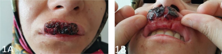
In the differential diagnosis of patients with ulcers on the lips characteristics like the duration of the ulcer, number, size, depth, shape, base, margins, and distribution are considered. Such ulcers arise from many diseases particularly, viral and bacterial infections, malignancies can also be responsible. Classic syphilitic chancres are painless erosions settled on hard papule; these are evident in the genital area in more than 90% of patients. This study describes a case of a 38-year-old female patient presenting with a painful ulcer covering 3 quarters of the upper lip showing settlement on erythematous, edematous, and indurated plaque covered with hemorrhagic crusts. The aim of this study was to consider differences between the classic syphilitic chancre typically found in the genital region from extragenital chancres and to raise awareness of the possibility of primary syphilis when patients present with painful ulcers on the lip.
Figures
Similar articles
-
Syphilitic Chancre of the Lip.Am J Dermatopathol. 2020 Oct;42(10):e143-e146. doi: 10.1097/DAD.0000000000001659. Am J Dermatopathol. 2020. PMID: 32324601
-
Multiple aphthoid syphilitic chancres of the oral cavity.Int J STD AIDS. 2008 Jul;19(7):486-7. doi: 10.1258/ijsa.2007.007262. Int J STD AIDS. 2008. PMID: 18574125
-
Secondary syphilis with persisting hard chancre on the forearm.Acta Derm Venereol. 2013 Mar 27;93(2):236-7. doi: 10.2340/00015555-1355. Acta Derm Venereol. 2013. PMID: 22565505 No abstract available.
-
Syphilitic Chancre of the Lips Transmitted by Kissing: A Case Report and Review of the Literature.Medicine (Baltimore). 2016 Apr;95(14):e3303. doi: 10.1097/MD.0000000000003303. Medicine (Baltimore). 2016. PMID: 27057901 Free PMC article. Review.
-
Primary syphilis.Int J STD AIDS. 2008 Mar;19(3):145-51. doi: 10.1258/ijsa.2007.007258. Int J STD AIDS. 2008. PMID: 18397550 Review.
Cited by
-
A Novel Treponema pallidum Subspecies pallidum Strain Associated With a Painful Oral Lesion Is a Member of a Potentially Emerging Nichols-Related Subgroup.Sex Transm Dis. 2024 Jul 1;51(7):486-492. doi: 10.1097/OLQ.0000000000001971. Epub 2024 Jun 3. Sex Transm Dis. 2024. PMID: 38829929 Free PMC article.
-
[Syphilitic chancre in the mouth: an unusual location. Case report].Rev Med Inst Mex Seguro Soc. 2022 Oct 25;60(6):703-707. Rev Med Inst Mex Seguro Soc. 2022. PMID: 36283073 Free PMC article. Spanish.
References
-
- Dourmishev LA, Dourmishev AL. Syphilis: uncommon presentations in adults. Clin Dermatol. 2005;23:555–564. - PubMed
-
- Domantay-Apostol GP, Handog EB, Gabriel MT. Syphilis: the international challenge of the great imitator. Dermatol Clin. 2008;26:191–202. - PubMed
-
- Serdaroğlu S. Innovations in Serology of Syphilis. Developments in Dermatology-4. Istanbul (TR): Gizben promotion and organization ltd sti; 1999. p. 215.
-
- Chapel TA, Prasad P, Chapel J, Lekas N. Extragenital syphilitic chancres. J Am Acad Dermatol. 1985;13:582–584. - PubMed
-
- Musher DM. Early syphilis. In: Holmes KK, Sparling PF, Mardh PA, Lemon SM, Stamm WE, Piot P, et al., editors. Sexually Transmitted Diseases. 3rd ed. New York (US): McGraw-Hill; 1999. pp. 479–485.
Publication types
MeSH terms
Substances
LinkOut - more resources
Full Text Sources
Other Literature Sources