

Toxicity of antimony, copper, cobalt, manganese, titanium and zinc oxide nanoparticles for the alveolar and intestinal epithelial barrier cells in vitro
- PMID: 27761772
- PMCID: PMC5101306
- DOI: 10.1007/s10616-016-0032-9
Toxicity of antimony, copper, cobalt, manganese, titanium and zinc oxide nanoparticles for the alveolar and intestinal epithelial barrier cells in vitro
Abstract
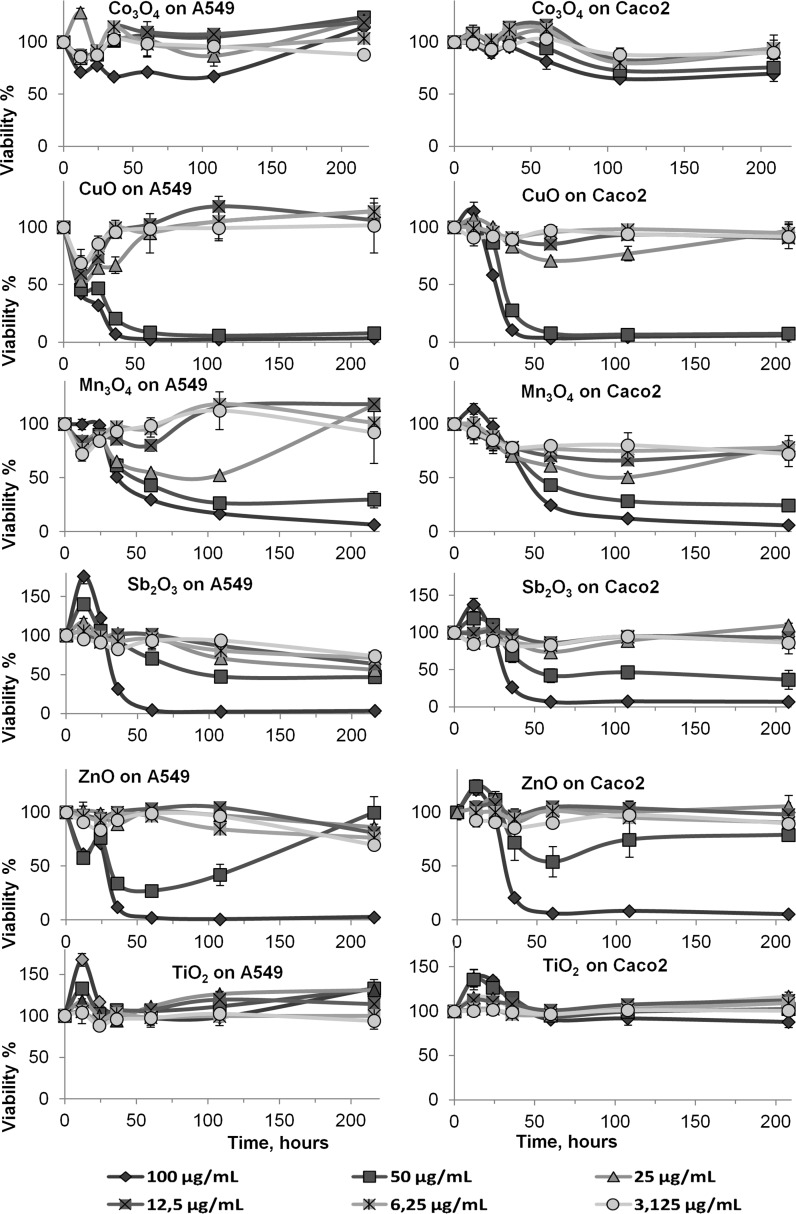
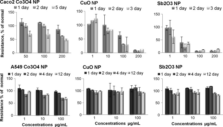
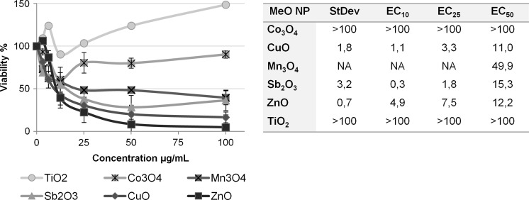
Heavy metals are found naturally on Earth and exposure to them in the living environment is increasing as a consequence of human activity. The toxicity of six different metal oxide nanoparticles (NP) at different points in time was compared using resazurin assay. After incubating Caco2 and A549 cells with 100 μg/mL of Sb2O3, Mn3O4 and TiO2 nanoparticles (NPs) for 24 h no toxic effects were observed while Co3O4 and ZnO NPs had moderate effects and CuO NPs were toxic below 100 μg/mL (24 h EC25 = 11 for A549 and 71 μg/mL for Caco2). The long-term monitoring (up to 9 days) of cells to NPs revealed that the toxic effects of Mn3O4 and Sb2O3 NPs remarkably increased over time. The 9 day EC50 values for Sb2O3 NPs were 22 and 48 μg/mL for A549 and Caco2 cells; and for Mn3O4 NPs were 47 and 29 μg/mL for A549 and Caco2 cells, respectively. In general, the sensitivity of the cell lines in the resazurin assay was comparable. Trans epithelial electrical resistance (TEER) measurements were performed for both cell types exposed to Co3O4, Sb2O3 and CuO NPs. In TEER assay, the Caco2 cells were more susceptible to the toxic effects of these NPs than A549 cells, where the most toxic NPs were the Sb2O3 NPs: the permeability of the Caco2 cell layer exposed to 10 μg/mL Sb2O3 NPs already increased after 24 h of exposure.
Keywords: A549 cells; Caco2 cells; Long term viability; Metal oxide nanoparticles; Non-monotonic dose–response (NMDR); Transepithelial electrical resistance (TEER).
Conflict of interest statement
Complaince with ethical standard Conflict of interest The authors confirm that this article authorship or content has no conflict of interest.
Figures
References
-
- Bartłomiejczyk T, Lankoff A, Kruszewski M, Szumiel I. Silver nanoparticles—allies or adversaries? Ann Agric Environ Med. 2013;20:48–54. - PubMed
-
- Bastian S, Busch W, Kühnel D, Springer A, Meissner T, Holke R, Scholz S, Iwe M, Pompe W, Gelinsky M, Potthoff A, Richter V, Ikonomidou C, Schirmer K. Toxicity of tungsten carbide and cobalt-doped tungsten carbide nanoparticles in mammalian cells in vitro. Environ Health Perspect. 2009;117:530–536. doi: 10.1289/ehp.0800121. - DOI - PMC - PubMed
-
- Bender CP, Hübner N-O, Weltman K-D, Scharf C, Kramer A. Tissue tolerable plasma and polihexanide: are synergistic effects possible to promote healing of chronic wounds? In vivo and in vitro results. In: Machala Z, editor. Plasma for bio-decontamination, medicine and food security. Berlin: Springer; 2011. pp. 324–325.
LinkOut - more resources
Full Text Sources
Other Literature Sources
Miscellaneous
