Review
. 2017 Oct;174(20):3425-3442.
doi: 10.1111/bph.13650. Epub 2016 Nov 17.

The role of perivascular adipose tissue in obesity-induced vascular dysfunction

Affiliations
Review

The role of perivascular adipose tissue in obesity-induced vascular dysfunction

Ning Xia et al. Br J Pharmacol. 2017 Oct.

Abstract

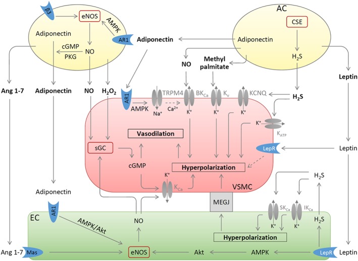
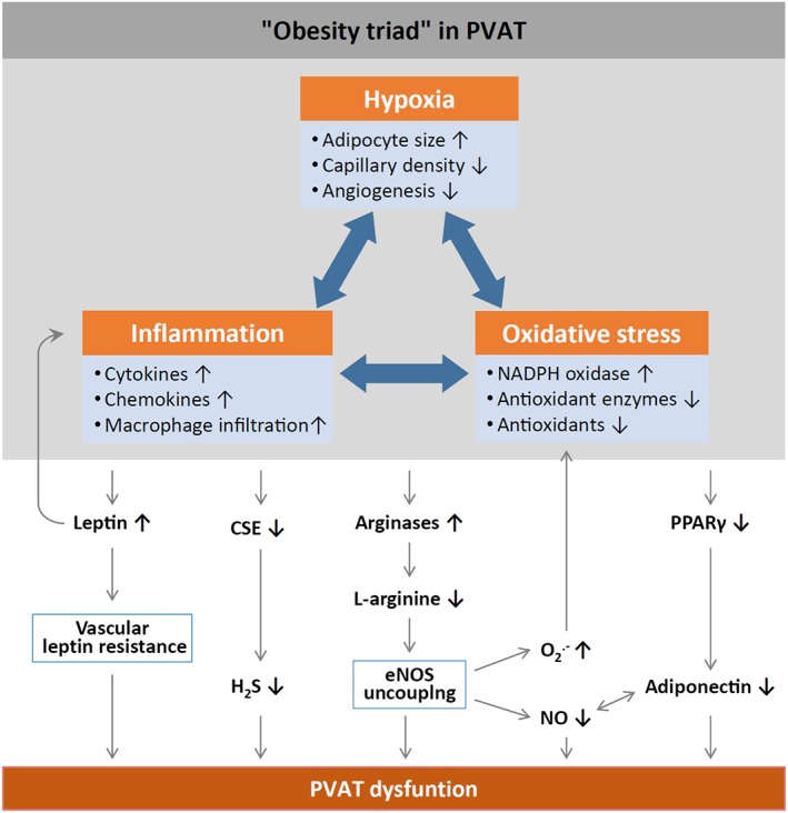
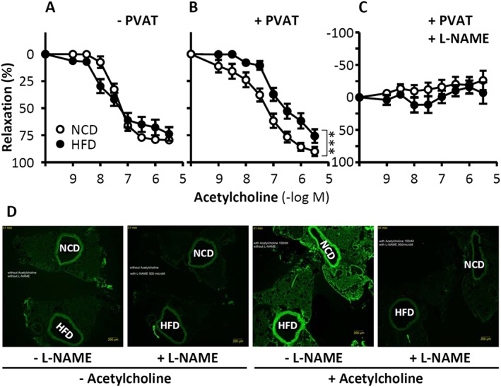
Under physiological conditions, perivascular adipose tissue (PVAT) attenuates agonist-induced vasoconstriction by releasing vasoactive molecules including hydrogen peroxide, angiotensin 1-7, adiponectin, methyl palmitate, hydrogen sulfide, NO and leptin. This anticontractile effect of PVAT is lost under conditions of obesity. The central mechanism underlying this PVAT dysfunction in obesity is likely to be an 'obesity triad' (consisting of PVAT hypoxia, inflammation and oxidative stress) that leads to the impairment of PVAT-derived vasoregulators. The production of hydrogen sulfide, NO and adiponectin by PVAT is reduced in obesity, whereas the vasodilator response to leptin is impaired (vascular leptin resistance). Strikingly, the vasodilator response to acetylcholine is reduced only in PVAT-containing, but not in PVAT-free thoracic aorta isolated from diet-induced obese mice, indicating a unique role for PVAT in obesity-induced vascular dysfunction. Furthermore, PVAT dysfunction has also been observed in small arteries isolated from the gluteal/visceral fat biopsy samples of obese individuals. Therefore, PVAT may represent a new therapeutic target for vascular complications in obesity. A number of approaches are currently being tested under experimental conditions. Potential therapeutic strategies improving PVAT function include body weight reduction, enhancing PVAT hydrogen sulfide release (e.g. rosiglitazone, atorvastatin and cannabinoid CB1 receptor agonists) and NO production (e.g. arginase inhibitors), inhibition of the renin-angiotensin-aldosterone system, inhibition of inflammation with melatonin or cytokine antagonists, activators of AMP-activated kinase (e.g. metformin, resveratrol and diosgenin) and adiponectin releasers or expression enhancers.

Linked articles: This article is part of a themed section on Molecular Mechanisms Regulating Perivascular Adipose Tissue - Potential Pharmacological Targets? To view the other articles in this section visit http://onlinelibrary.wiley.com/doi/10.1111/bph.v174.20/issuetoc.


Figures

Figure 1
Figure 1
PVAT‐derived vasoactive molecules. Methyl palmitate produced by PVAT adipocytes (AC) causes vasodilatation by opening the Kv channels on VSMC. H2S is synthesized in PVAT by CSE and induces VSMC hyperpolarization by stimulating KCNQ‐type Kv or KATP channels. Leptin induces endothelium‐dependent vasodilatation by stimulating leptin receptor (LepR), which leads to activation of eNOS via a pathway involving AMPK and Akt and to H2S production. This H2S functions as an EDHF and activates endothelial small (SKCa) and intermediate (IKCa) conductance calcium‐dependent K+ channels via autocrine mechanisms. The resulting hyperpolarization of endothelial cells can be transmitted to VSMC by electrical coupling through myoendothelial gap junction (MEGJ). Leptin also causes endothelium‐independent vasodilatation by inducing VSMC hyperpolarization through unknown mechanisms. NO and H2O2 released from PVAT can elicit vasodilatation by activating sGC leading to the synthesis of cGMP. Adiponectin released from PVAT AC can be enhanced by stimulation of β3 adrenoceptors (β3) and by the NO‐cGMP‐PKG pathway. Adiponectin exerts multiple vascular effects: it stimulates NO production from PVAT and from endothelial cells and induces VSMC hyperpolarization by activating TRPM4 channels followed by opening BKCa; Ang 1–7 produced by PVAT acts on endothelial Ang 1–7 receptor (Mas; MAS1 receptor) thereby stimulating endothelial NO production. Besides stimulating sGC activity, NO from PVAT and endothelial cells can also induce/potentiate VSMC hyperpolarization through KCa or BKCa. Partly adopted from (Beltowski, 2013; Weston et al., 2013; Withers et al., 2014a).
Figure 2
Figure 2
Mechanisms of PVAT dysfunction in diet‐induced obesity. HFD‐induced adipocyte hypertrophy leads to hypoxia and the production of pro‐inflammatory cytokines and chemokines, activation of NADPH oxidase and down‐regulation of antioxidant enzymes (e.g. superoxide dismutase and peroxiredoxin‐1) and non‐enzymatic antioxidants (e.g. glutathione). Infiltrating immune cells potentiate PVAT inflammation and oxidative stress. Chronic hyperleptinaemia leads to vascular leptin resistance (loss of leptin‐induced vasodilatation) and potentiation of PVAT inflammation. Long‐term obesity decreases PVAT H2S production by down‐regulating CSE expression. The up‐regulation of arginases leads to L‐arginine deficiency and eNOS uncoupling (enhanced superoxide production and reduced NO production by eNOS). PVAT adiponectin expression is reduced in obesity, very likely due to a down‐regulation of PPARγ. Normally, NO stimulates adiponectin secretion and adiponectin increases PVAT NO production. This positive feedback mechanism is impaired in obesity.
Figure 3
Figure 3
Role of PVAT in obesity‐induced vascular dysfunction. C57BL/6J mice were fed a HFD or normal control diet (NCD) for 20 weeks starting at the age of 8 weeks. The vasodilator response to acetylcholine (A–C) was performed in noradrenaline‐precontracted aorta with or without PVAT in the absence or presence of the NO synthase inhibitor L‐NAME. ***P < 0.001, n = 8. To detect PVAT NO production, NCD and HFD aorta samples were mounted back‐to‐back on the same slide to guarantee identical staining conditions for the two samples (D). NO production in PVAT‐containing aorta was determined by 4,5‐diaminofluorescein diacetate (DAF‐2 DA) staining. From Xia et al. (2016) with permission of Wolters Kluwer Health, Inc. Copyright © 2016, Wolters Kluwer Health.

Similar articles

Cited by

References

    1. Agabiti‐Rosei C, De Ciuceis C, Rossini C, Porteri E, Rodella LF, Withers SB et al. (2014). Anticontractile activity of perivascular fat in obese mice and the effect of long‐term treatment with melatonin. J Hypertens 32: 1264–1274. - PubMed
    1. Aghamohammadzadeh R, Greenstein AS, Yadav R, Jeziorska M, Hama S, Soltani F et al. (2013). Effects of bariatric surgery on human small artery function: evidence for reduction in perivascular adipocyte inflammation, and the restoration of normal anticontractile activity despite persistent obesity. J Am Coll Cardiol 62: 128–135. - PMC - PubMed
    1. Aghamohammadzadeh R, Unwin RD, Greenstein AS, Heagerty AM (2015). Effects of obesity on perivascular adipose tissue vasorelaxant function: nitric oxide, inflammation and elevated systemic blood pressure. J Vasc Res 52: 299–305. - PMC - PubMed
    1. Ahmed SR, Johansson BL, Karlsson MG, Souza DS, Dashwood MR, Loesch A (2004). Human saphenous vein and coronary bypass surgery: ultrastructural aspects of conventional and “no‐touch” vein graft preparations. Histol Histopathol 19: 421–433. - PubMed
    1. Alexander SPH, Kelly E, Marrion N, Peters JA, Benson HE, Faccenda E et al. (2015a). The Concise Guide to PHARMACOLOGY 2015/16: Overview. Br J Pharmacol 172: 5729–5743. - PMC - PubMed