

Review
doi: 10.4103/0971-9784.192617.
Left ventricular global systolic function assessment by echocardiography
Affiliations
- PMID: 27762246
- PMCID: PMC5100240
- DOI: 10.4103/0971-9784.192617
Item in Clipboard
Review
Left ventricular global systolic function assessment by echocardiography
Ann Card Anaesth.
2016 Oct.
Abstract
The left ventricle, with its thickened myocardial walls, unlike the right ventricle has no measurable geometric shape. It has a conical apex and its function quantification, needs intensive, 2D, 3D and M mode transesophageal echocardiography, which is described in this review.
Figures
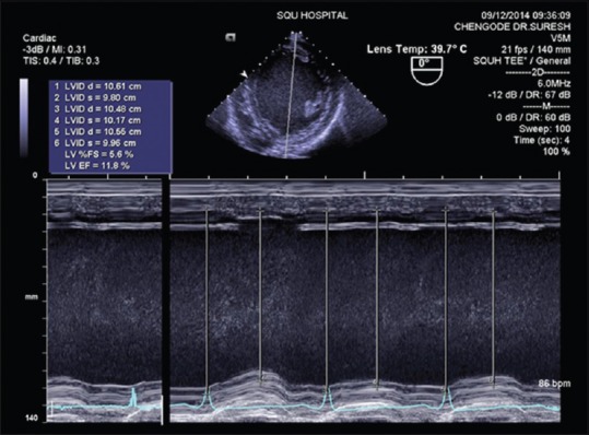
Fractional shortening measurement using M-mode echocardiography and ejection fraction calculated by Teichholz method (severe left ventricle dysfunction)
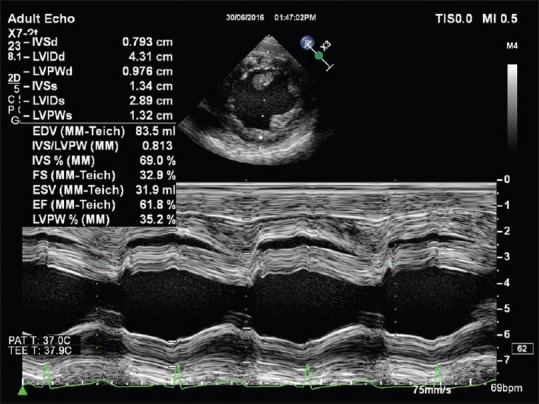
Fractional shortening measurement using M-mode echocardiography and ejection fraction calculated by Teichholz method (good left ventricle function)
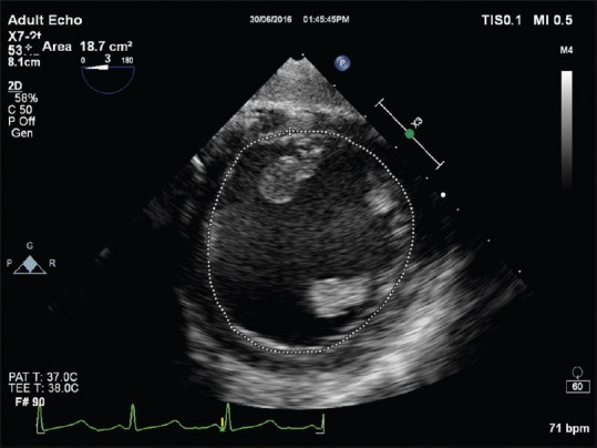
End-diastolic area measurement from transgastric mid-papillary short axis for the calculation of fractional area change
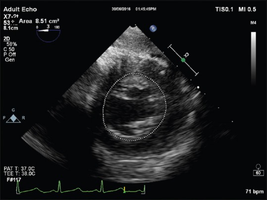
End-systolic area measurement from transgastric mid-papillary short axis for the calculation of fractional area change
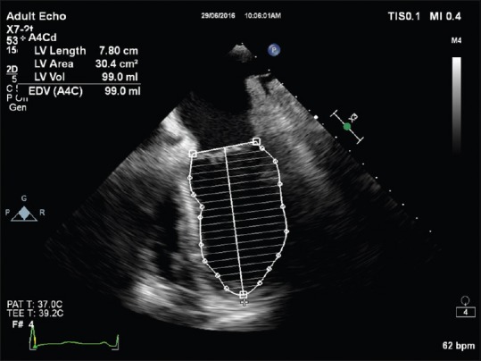
End-diastolic volume measurement using modified Simpson's method
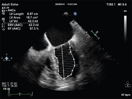
End-systolic volume and ejection fraction calculation using modified Simpson's method
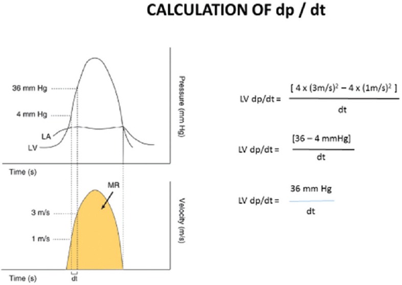
Principle of systolic index of contractility (dP/dt) measurement
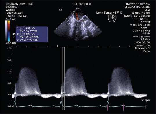
Systolic index of contractility (dP/dt) measurement

Tissue Doppler imaging of lateral mitral annulus. Note E’ velocity of >5.4 cm/s suggesting a good left ventricle function
Similar articles
-
Speckle-tracking strain echocardiography: any place in routine daily practice in 2014?Arch Cardiovasc Dis. 2013 Dec;106(12):629-34. doi: 10.1016/j.acvd.2013.10.001. Epub 2013 Nov 15. Arch Cardiovasc Dis. 2013. PMID: 24246615 No abstract available.
-
Early detection of left ventricular contractility abnormalities by two-dimensional speckle tracking strain in Chagas’ disease.Echocardiography. 2014 May;31(5):623-30. doi: 10.1111/echo.12426. Echocardiography. 2014. PMID: 25232573
-
Left ventricular 4D echocardiogram motion and shape analysis.Ultrasonics. 2002 May;40(1-8):949-54. doi: 10.1016/s0041-624x(02)00244-5. Ultrasonics. 2002. PMID: 12160075
-
Assessment of left ventricular function by three-dimensional echocardiography.Cardiovasc Ultrasound. 2003 Sep 8;1:12. doi: 10.1186/1476-7120-1-12. Cardiovasc Ultrasound. 2003. PMID: 14514356 Free PMC article. Review.
-
Determinants of Discrepancy in the Left Ventricular Systolic Function Evaluation Between Preoperative and Intraoperative Evaluations.Semin Cardiothorac Vasc Anesth. 2020 Dec;24(4):321-327. doi: 10.1177/1089253220936784. Epub 2020 Jul 1. Semin Cardiothorac Vasc Anesth. 2020. PMID: 32605429 Review.
Cited by
-
Doxorubicin Incorporation into Gold Nanoparticles: An In Vivo Study of Its Effects on Cardiac Tissue in Rats.Nanomaterials (Basel). 2024 Oct 14;14(20):1647. doi: 10.3390/nano14201647. Nanomaterials (Basel). 2024. PMID: 39452984 Free PMC article.
-
Cardioprotective effects of exercise training on doxorubicin-induced cardiomyopathy: a systematic review with meta-analysis of preclinical studies.Sci Rep. 2021 Mar 18;11(1):6330. doi: 10.1038/s41598-021-83877-8. Sci Rep. 2021. PMID: 33737561 Free PMC article.
-
Resolution and Speckle Reduction in Cardiac Imaging.IEEE Trans Ultrason Ferroelectr Freq Control. 2021 Apr;68(4):1131-1143. doi: 10.1109/TUFFC.2020.3034518. Epub 2021 Mar 26. IEEE Trans Ultrason Ferroelectr Freq Control. 2021. PMID: 33112742 Free PMC article.
-
Detection of Right and Left Ventricular Dysfunction in Pediatric Patients Using Artificial Intelligence-Enabled ECGs.J Am Heart Assoc. 2024 Nov 5;13(21):e035201. doi: 10.1161/JAHA.124.035201. Epub 2024 Nov 4. J Am Heart Assoc. 2024. PMID: 39494568 Free PMC article.
-
Cardiac Structure and Function in Adults with Down Syndrome.Int J Environ Res Public Health. 2022 Sep 28;19(19):12310. doi: 10.3390/ijerph191912310. Int J Environ Res Public Health. 2022. PMID: 36231610 Free PMC article.
References
-
- Lang RM, Badano LP, Mor-Avi V, Afilalo J, Armstrong A, Ernande L, et al. Recommendations for cardiac chamber quantification by echocardiography in adults: An update from the American Society of Echocardiography and the European Association of Cardiovascular Imaging. J Am Soc Echocardiogr. 2015;28:1–39-e14. - PubMed
-
- Hahn RT, Abraham T, Adams MS, Bruce CJ, Glas KE, Lang RM, et al. Guidelines for performing a comprehensive transesophageal echocardiographic examination: Recommendations from the American Society of Echocardiography and the Society of Cardiovascular Anesthesiologists. J Am Soc Echocardiogr. 2013;26:921–64. - PubMed
-
- Gottdiener JS, Bednarz J, Devereux R, Gardin J, Klein A, Manning WJ, et al. American Society of Echocardiography recommendations for use of echocardiography in clinical trials. J Am Soc Echocardiogr. 2004;17:1086–119. - PubMed
-
- Savage RM, Aronson S, Shernan SK. Comprehensive Textbook of Perioperative Transesophageal Echocardiography. 2nd ed. USA: Lippincott Williams and Wilkins; 2013.
-
- Smith MD, MacPhail B, Harrison MR, Lenhoff SJ, DeMaria AN. Value and limitations of transesophageal echocardiography in determination of left ventricular volumes and ejection fraction. J Am Coll Cardiol. 1992;19:1213–22. - PubMed
Publication types
MeSH terms
LinkOut - more resources
Full Text Sources
Other Literature Sources
Miscellaneous