. 2016 Oct 20:7:13047.
doi: 10.1038/ncomms13047.

Quantitative interaction mapping reveals an extended UBX domain in ASPL that disrupts functional p97 hexamers

Affiliations

Quantitative interaction mapping reveals an extended UBX domain in ASPL that disrupts functional p97 hexamers

Anup Arumughan et al. Nat Commun. .

Abstract

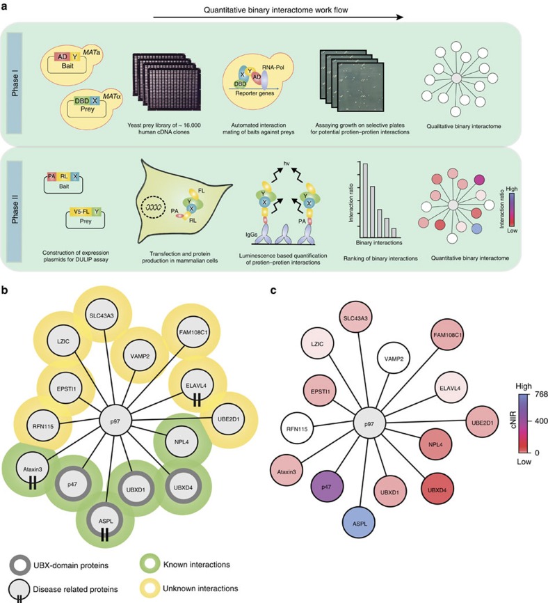
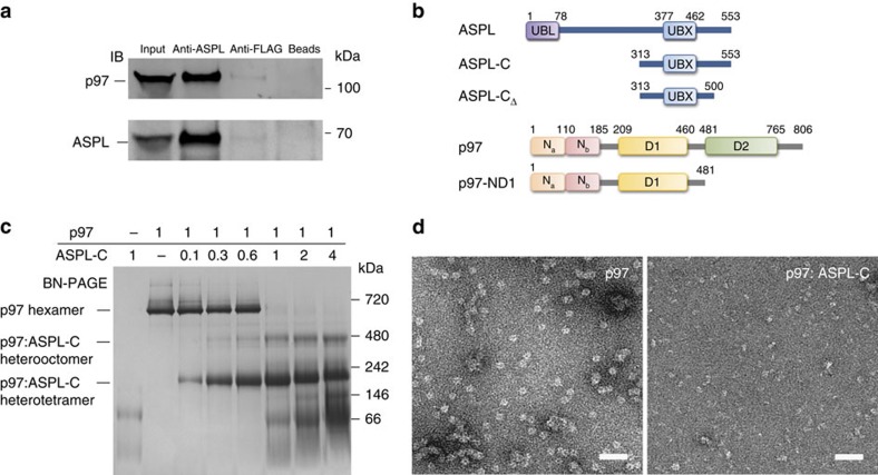
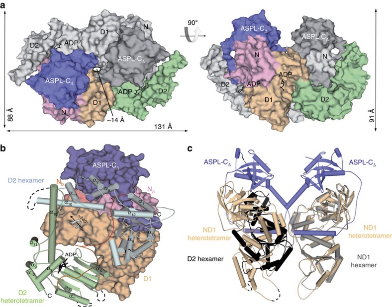
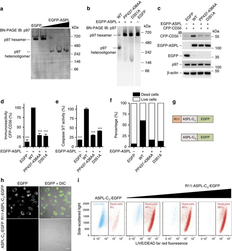
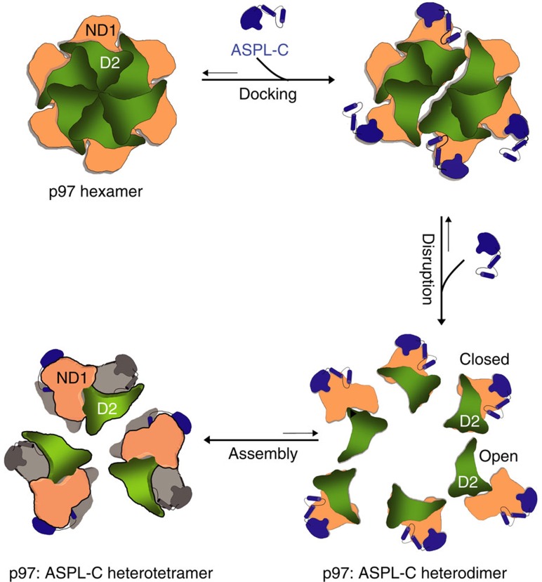
Interaction mapping is a powerful strategy to elucidate the biological function of protein assemblies and their regulators. Here, we report the generation of a quantitative interaction network, directly linking 14 human proteins to the AAA+ ATPase p97, an essential hexameric protein with multiple cellular functions. We show that the high-affinity interacting protein ASPL efficiently promotes p97 hexamer disassembly, resulting in the formation of stable p97:ASPL heterotetramers. High-resolution structural and biochemical studies indicate that an extended UBX domain (eUBX) in ASPL is critical for p97 hexamer disassembly and facilitates the assembly of p97:ASPL heterotetramers. This spontaneous process is accompanied by a reorientation of the D2 ATPase domain in p97 and a loss of its activity. Finally, we demonstrate that overproduction of ASPL disrupts p97 hexamer function in ERAD and that engineered eUBX polypeptides can induce cell death, providing a rationale for developing anti-cancer polypeptide inhibitors that may target p97 activity.


Figures

Figure 1
Figure 1. A quantitative p97 interaction network identifies ASPL as a strong binding partner of p97.
(a) Schematic representation of the workflow for the generation of a quantitative binary p97 PPI interaction network using sequential applications of automated yeast two-hybrid technology (Phase I) and DULIP assays (Phase II). (b) Network graph of protein–protein interactions of p97 identified by automated Y2H assay. (c) Overlay of the DULIP assay data to the p97 Y2H network resulted in the generation of the quantitative binary interaction network for p97. In DULIP assays full-length p97 protein was utilized as bait.
Figure 2
Figure 2. ASPL-C converts p97 hexamers into stable p97:ASPL-C heterooligomers.
(a) Co-immunoprecipitation of a p97:ASPL protein complex from human brain homogenate using an anti-ASPL antibody. Anti-FLAG antibody and beads alone were used as negative controls. (b) Schematic representation of fragments and full-length ASPL and p97. Conserved protein domains are depicted: ubiquitin-like domain (UBL); ubiquitin regulatory-X domain (UBX); N-terminal protein binding domains (Na and Nb); ATPase domains (D1 and D2). (c) Blue-native gel stained with Coomassie Brilliant Blue, demonstrating the remodelling of p97 hexamers by ASPL-C in a concentration-dependent manner. p97 (10 μg) and ASPL-C (0.3, 0.6, 1.8, 3, 6 and 12 μg) were briefly mixed and incubated on ice for 5 min; then protein complexes were analysed by BN-PAGE. A 1:1 molar ratio of p97 monomers and ASPL-C was sufficient to promote the formation of p97:ASPL-C heterooligomers. (d) Negatively stained electron micrographs of purified p97 in the presence and absence of ASPL-C; scale bar 50 nm.
Figure 3
Figure 3. Structure of the ASPL-C:p97-ND1 heterotetramer.
(a) Surface representation of a heterotetrameric p97-ND1:ASPL-C protein complex in two orthogonal orientations. One heterodimeric unit is displayed in shades of grey, the other is shown in multiple colours, blue (ASPL-C), pink (p97-N) and wheat (p97-D1). (b) Domain annotation and secondary structure of ASPL-C. The β-grasp fold (ββαββαβ) of the canonical UBX domain is coloured in light blue, the extensions β0, α3 and α4 in light grey, and the helical lariat in dark blue. (c) Ribbon-type representation of ASPL-C with colours as in b. (d) Structure of a single heterodimeric p97-ND1:ASPL-C unit observed within the heterotetramer. ASPL-C (blue) is shown as cartoon underneath a translucent molecular surface. The Na (salmon colour) and Nb (pink) subdomains and the D1 domain (wheat colour) of p97 are displayed as cartoon structure. The partially disordered p97 D1-D2 linker is highlighted in black. ADP is depicted as stick model with a bound Mg2+ ion (green). (e) Coomassie-stained blue-native gel showing the effects of site-directed mutagenesis of conserved ASPL-C residues on the disassembly of p97 hexamers. ASPL-C (3 μg) or its indicated variants were combined with p97 (10 μg) and incubated on ice for 5 min. Then, samples were analysed by BN-PAGE. (f) ATPase activities of full-length p97 or its indicated variant in the presence or absence of ASPL-C were determined using a high-performance liquid chromatography-based assay. Molar ratios between ASPL-C and p97 are indicated. Wild-type (WT) p97 or the mutated protein p97-A232E were combined with a three-fold molar excess of ASPL-C and briefly incubated on ice. Then, the recombinant proteins were analysed by size-exclusion chromatography and heterotetrameric protein complexes (p97:ASPL-C or p97-A232E:ASPL-C) were isolated. ATPase activities of interacting recombinant proteins were analysed by high-performance liquid chromatography. Data are expressed as mean±s.e. (n=3).
Figure 4
Figure 4. Remodelling of p97 hexamers by ASPL-CΔ induces a rearrangement of the distal ATPase domain D2.
(a) Surface representation of the heterotetrameric p97:ASPL-CΔ protein complex in two orthogonal orientations. One heterodimeric unit is displayed in shades of grey, the other shows ASPL-C in blue, p97-N in pink, p97-D1 in wheat colour and p97-D2 in green. (b) Structural comparison of the orientation of the D2 domain observed in p97 hexamers and p97:ASPL-CΔ heterotetramers. The N and D1 domains in the p97:ASPL-CΔ heterodimeric unit are shown in surface representation in colours as in a, the D1-D2 linker (orange) and the D2 domain (green) are shown in cartoon representation. The relative orientation of the D2 domain in p97 hexamers is shown in light blue with the D1-D2 linker in red. (c) Structural comparison reveals the potential cause for the reorientation of the D2 domain in p97. Superposition of a single protomer taken from the p97 hexamer (N and D1 in grey, D2 in black) onto the p97-ND1:ASPL-C heterotetramer (p97-ND1 in wheat colour, ASPL-C in blue) demonstrates a steric interference between the D2 domain and a p97-ND1:ASPL-C heterodimeric unit.
Figure 5
Figure 5. ASPL-induced disruption of p97 hexamers causes mammalian cell death.
(a) Overproduction of EGFP-ASPL causes concentration-dependent disassembly of endogenous p97 hexamers in HEK293 cells. Protein extracts were analysed by BN-PAGE. p97-containing protein complexes were identified by IB using an anti-p97 antibody. (b) EGFP-ASPL and indicated variants of ASPL were overproduced in HEK293 cells. After 24 h, native protein complexes were resolved by BN-PAGE and immunoblotted to identify p97-containing protein complexes using an anti-p97 antibody. (c) CFP-CD3δ, EGFP-ASPL and indicated variants of ASPL were overproduced in HEK293 cells. After 24 h, total cell lysates were resolved by SDS–PAGE and immunoblotted to detect indicated proteins; antibodies: anti-CD3δ, anti-GFP, anti-p97 and anti-β-actin. (d) The levels of CFP-CD3δ in samples as indicated in c were quantified using an Aida image analyser. Data are expressed as mean±s.e. (n=3). ***P≤0.001 compared with WT-ASPL (Student's t-test). (e) EGFP-ASPL and indicated variants with point mutations were overproduced in HEK293 cells; activation of caspases 3/7 was monitored after 72 h. Data are expressed as mean±s.e.; (n=3). ***P≤0.001 compared with WT-ASPL (Student's t-test). (f) EGFP-ASPL and indicated variants with point mutations were overproduced in HEK293 cells for 72 h; cells were stained with LIVE/DEAD far-red fluorescence stain and live and dead cell populations were quantified using flow cytometry. 10,000 EGFP cells were analysed per each sample. (g) Schematic representation of R11- and EGFP-tagged ASPL-CΔ recombinant proteins. (h) Confocal imaging micrographs of fixed HeLa cells showing the uptake of R11-ASPL-CΔ-EGFP protein into cells. Scale bars, 10 μm. (i) HeLa cells were treated for 24 h with the recombinant proteins R11-ASPL-CΔ-EGFP and ASPL-CΔ-EGFP (control protein), respectively; cells were stained with the LIVE/DEAD far-red fluorescence stain and live and dead cell populations were quantified using flow cytometry.
Figure 6
Figure 6. Structure-based model for the disassembly of p97 hexamers through the interaction with ASPL-C.
ASPL-C-mediated disassembly of p97 hexamers is a multistep process. In the first step, ASPL-C docks onto the p97 surface via its cis-Pro touch-turn motif that is conserved in the eUBX domain. Next, the ASPL-C helical lariat disrupts the association of p97 protomers by targeting the D1 ring and binding to the p97 Na subdomain. This results in the generation of six metastable p97:ASPL-C heterodimer units, which rapidly re-assemble into stable heterotetramers. Concomitantly, the D2 domain in p97 undergoes a ∼140° rotation from a closed to an open conformation, preventing the re-association of protomers into p97 hexamers.

Similar articles

Cited by

References

    1. Vidal M., Cusick M. E. & Barabási A.-L. Interactome networks and human disease. Cell 144, 986–998 (2011). - PMC - PubMed
    1. Barabási A.-L., Gulbahce N. & Loscalzo J. Network medicine: a network-based approach to human disease. Nat. Rev. Genet. 12, 56–68 (2011). - PMC - PubMed
    1. Snider J. et al.. Fundamentals of protein interaction network mapping. Mol. Syst. Biol. 11, 848–848 (2015). - PMC - PubMed
    1. Stelzl U. et al.. A human protein-protein interaction network: a resource for annotating the proteome. Cell 122, 957–968 (2005). - PubMed
    1. Gavin A.-C. et al.. Functional organization of the yeast proteome by systematic analysis of protein complexes. Nature 415, 141–147 (2002). - PubMed

Publication types

MeSH terms