. 2016 Oct 20:6:35857.
doi: 10.1038/srep35857.

Formaldehyde impairs transepithelial sodium transport

Affiliations

Formaldehyde impairs transepithelial sodium transport

Yong Cui et al. Sci Rep. .

Abstract

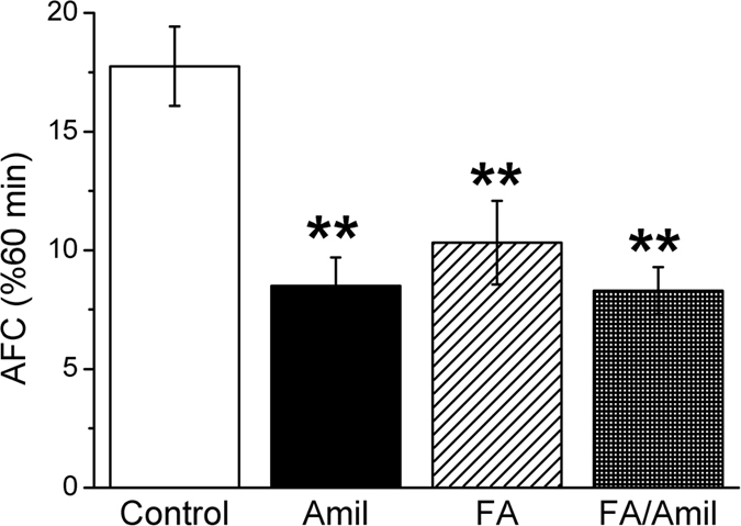
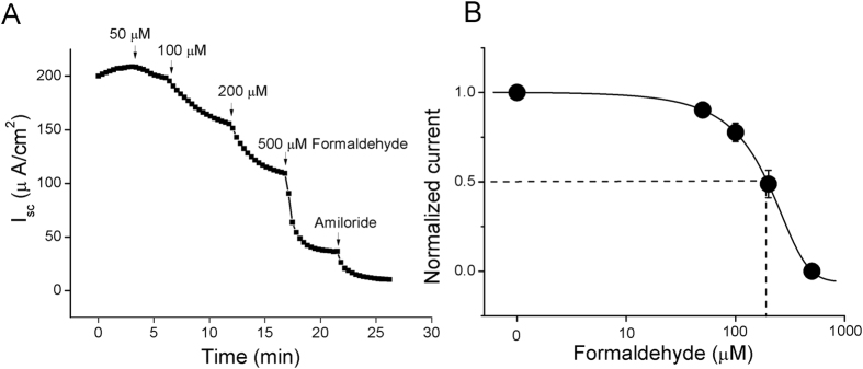
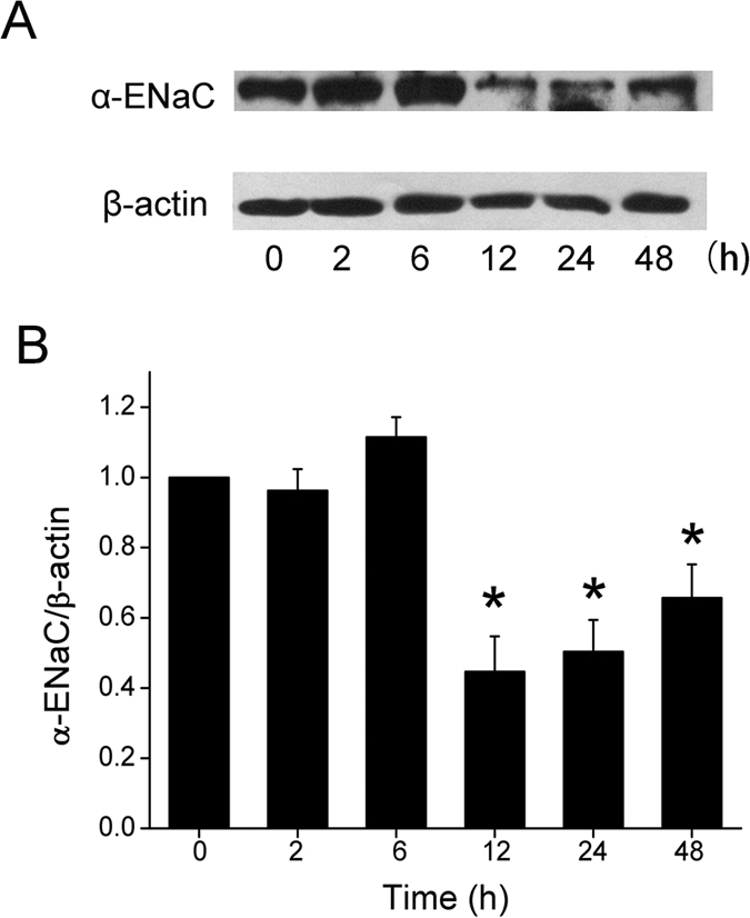
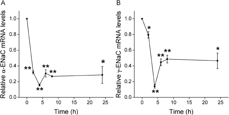
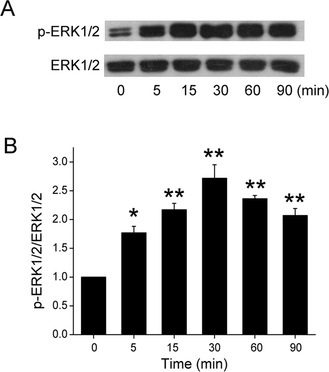
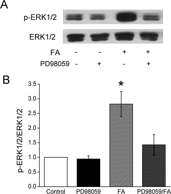
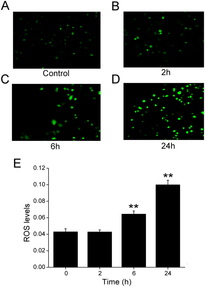
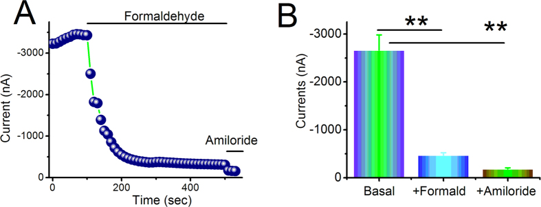
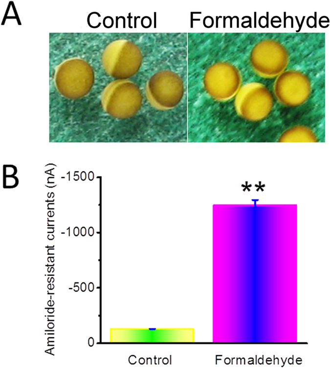
Unsaturated oxidative formaldehyde is a noxious aldehyde in cigarette smoke that causes edematous acute lung injury. However, the mechanistic effects of formaldehyde on lung fluid transport are still poorly understood. We examined how formaldehyde regulates human epithelial sodium channels (ENaC) in H441 and expressed in Xenopus oocytes and exposed mice in vivo. Our results showed that formaldehyde reduced mouse transalveolar fluid clearance in vivo. Formaldehyde caused a dose-dependent inhibition of amiloride-sensitive short-circuit Na+ currents in H441 monolayers and of αβγ-ENaC channel activity in oocytes. α-ENaC protein was reduced, whereas phosphorylation of the extracellular regulated protein kinases 1 and 2 (ERK1/2) increased significantly post exposure. Moreover, both α- and γ-ENaC transcripts were down-regulated. Reactive oxygen species (ROS) was elevated significantly by formaldehyde in addition to markedly augmented membrane permeability of oocytes. These data suggest that formaldehyde contributes to edematous acute lung injury by reducing transalveolar Na+ transport, through decreased ENaC activity and enhanced membrane depolarization, and by elevating ROS production over long-term exposure.


Figures

Figure 1
Figure 1. Downregulation of mouse alveolar fluid clearance in vivo by formaldehyde.
Mouse lungs were instilled with 5% bovine serum albumin dissolved in physiologic saline solution in control group (Control), and in the presence of amiloride (Amil, 1 mM), formaldehyde (FA, 200 μM), and both (FA/Amil) for exposed mice. Reabsorption rate of instillate was computed as the percentage of instilled volume after 60 min (% 60 min). Average AFC values are presented as mean ± SE, One-way ANOVA. **P < 0.01, compared with control group. N = 6.
Figure 2
Figure 2. Formaldehyde reduces short-circuit current level in H441 monolayers in a dose-dependent manner.
(A) Representative short-circuit current (Isc) trace showing applications of a series of concentrations of 50, 100, 200, and 500 μM formaldehyde. Amiloride-sensitive currents are defined as the difference between the total current and the amiloride-resistant current, with the basal amiloride-sensitive current set as 100%. (B) Normalized amiloride-sensitive currents at each concentration were plotted as a dose-response curve, N = 7. The raw data were calculated by fitting with the Boltzmann equation. The IC50 value was 165.6 μM.
Figure 3
Figure 3. Effect of formaldehyde on α-ENaC protein level in H441 cells.
(A) Representative western blot of α-ENaC protein extracted from H441 cells exposed to 200 μM formaldehyde for 0-48 h. Blots were immunostained with antibody to α-ENaC, or to β-actin as a loading control. (B) Graphical representation of data obtained from three sets of western blots and quantified using gray analysis (α-ENaC/β-actin). Data are shown as the mean ± SE, *P < 0.05, compared with control.
Figure 4
Figure 4. Effects of formaldehyde on transcriptional expression of ENaC α- and γ-subunits in H441 cells.
mRNA samples were isolated from H441 cells treated with 200 μM formaldehyde for various periods. Relative levels of mRNA were calculated as α- or γ-ENaC/GAPDH ratios. (A) Real-time PCR results for α-ENaC mRNA. (B) Real-time PCR results for γ-ENaC mRNA. *P < 0.05, **P < 0.01, compared with control. N = 3.
Figure 5
Figure 5. Effects of formaldehyde on ERK1/2 phosphorylation in H441 cells.
(A) Representative western blot of phosphorylated ERK1/2 in protein extracted from H441 cells exposed to 200 μM formaldehyde for 0–90 min. Blots were immunostained with total ERK1/2 as a loading control. (B) Graphical representation of data obtained from three sets of western blots and quantified using gray analysis (p-ERK1/2/ERK1/2). Data are shown as the mean ± SE, *P < 0.05, **P < 0.01, compared with control.
Figure 6
Figure 6. Effects of PD98059 on ERK1/2 phosphorylation in H441 cells.
(A) Representative western blot of phosphorylated ERK1/2 in untreated H441 cells (Control), after pretreatment with PD98059 (PD98059), after treatment with formaldehyde (FA), and after pretreatment with PD98059 for 30 min prior to the addition of formaldehyde (PD98059/FA). Lanes shown in this figure are from the same western blot. (B) Graphical representation of data obtained from three sets of western blots and quantified using gray analysis (p-ERK1/2/ERK1/2). Data are shown as the mean ± SE, *P < 0.05, compared with control.
Figure 7
Figure 7. Effects of formaldehyde on oxidative stress in the H441 cells.
Production of ROS was measured using the fluorogenic substrate 2′,7′-dichlorofluorescein diacetate, which is oxidized to fluorescent 2′,7′-dichlorofluorescein. (A) Air-subjected control cell culture. (BD) Cells exposed to 200 μM formaldehyde for 2 h, 6 h, and 24 h. (E) The ROS levels displays the summary fluorescence intensity measured from the images of H441 cells (N = 25 in each group) treated with different periods of formaldehyde. Background fluorescence level was corrected. Data are shown as the mean ± SE, **P < 0.01, compared with control.
Figure 8
Figure 8. Formaldehyde alters the activity of heterologous human αβγ-ENaC channels expressed in oocytes.
Oocytes were continuously perfused with formaldehyde and amiloride, and whole cell currents were monitored at 10-s intervals. (A) Representative inward current traces in the presence of formaldehyde and amiloride. Currents were digitized at −120 mV. Application of drugs is indicated by solid horizontal lines. (B) Average currents at −120 mV (mean ± SE). **P < 0.01 compared with basal level. N = 10 from 4 different frogs.
Figure 9
Figure 9. Formaldehyde impairs properties of the plasma membrane.
Oocytes expressing human αβγ-ENaC were incubated with formaldehyde for 24 h. (A) The shape of oocytes expressing human αβγ-ENaC observed by microscopy, with and without formaldehyde exposure. (B) Average amiloride-resistant currents at −120 mV (mean ± SE), reflecting oocyte permeability. **P < 0.01 compared with control. N = 10 from 4 different frogs.

Similar articles

Cited by

References

    1. Stedman R. L. The chemical composition of tobacco and tobacco smoke. Chem Rev 68, 153–207 (1968). - PubMed
    1. Shin H. J. et al.. Effect of cigarette filters on the chemical composition and in vitro biological activity of cigarette mainstream smoke. Food Chem Toxicol 47, 192–197 (2009). - PubMed
    1. Nikota J. K. & Stampfli M. R. Cigarette smoke-induced inflammation and respiratory host defense: Insights from animal models. Pulm Pharmacol Ther 25, 257–262 (2012). - PubMed
    1. Moretto N., Volpi G., Pastore F. & Facchinetti F. Acrolein effects in pulmonary cells: relevance to chronic obstructive pulmonary disease. Ann N Y Acad Sci 1259, 39–46 (2012). - PubMed
    1. Sangodkar J., Katz S., Melville H. & Narla G. Lung adenocarcinoma: lessons in translation from bench to bedside. Mt Sinai J Med 77, 597–605 (2010). - PubMed

Publication types

MeSH terms