Review
. 2016 Nov 16;8(45):30649-30664.
doi: 10.1021/acsami.6b09263. Epub 2016 Nov 4.

Nanoyeast and Other Cell Envelope Compositions for Protein Studies and Biosensor Applications

Affiliations
Review

Nanoyeast and Other Cell Envelope Compositions for Protein Studies and Biosensor Applications

Yadveer S Grewal et al. ACS Appl Mater Interfaces. .

Abstract

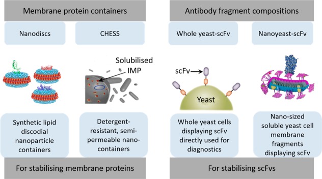
Rapid progress in disease biomarker discovery has increased the need for robust detection technologies. In the past several years, the designs of many immunoaffinity reagents have focused on lowering costs and improving specificity while also promoting stability. Antibody fragments (scFvs) have long been displayed on the surface of yeast and phage libraries for selection; however, the stable production of such fragments presents challenges that hamper their widespread use in diagnostics. Membrane and cell wall proteins similarly suffer from stability problems when solubilized from their native environment. Recently, cell envelope compositions that maintain membrane proteins in native or native-like lipid environment to improve their stability have been developed. This cell envelope composition approach has now been adapted toward stabilizing antibody fragments by retaining their native cell wall environment. A new class of immunoaffinity reagents has been developed that maintains antibody fragment attachment to yeast cell wall. Herein, we review recent strategies that incorporate cell wall fragments with functional scFvs, which are designed for easy production while maintaining specificity and stability when in use with simple detection platforms. These cell wall based antibody fragments are globular in structure, and heterogeneous in size, with fragments ranging from tens to hundreds of nanometers in size. These fragments appear to retain activity once immobilized onto biosensor surfaces for the specific and sensitive detection of pathogen antigens. They can be quickly and economically generated from a yeast display library and stored lyophilized, at room temperature, for up to a year with little effect on stability. This new format of scFvs provides stability, in a simple and low-cost manner toward the use of scFvs in biosensor applications. The production and "panning" of such antibody cell wall composites are also extremely facile, enabling the rapid adoption of stable and inexpensive affinity reagents for emerging infectious threats.

Keywords: affinity reagent; biomaterial; biosensor; cell envelope composition; immunosensor; nanomaterial; nanoyeast.


Conflict of interest statement

The authors declare the following competing financial interest(s): The nanoyeast-scFv work highlighted in this review is under patent as Cell-Free Biofragment Compositions and Related Systems, Devices, and Methods (WO 2014093357 A1).

Figures

Figure 1
Figure 1
Advances in cell envelope compositions for protein studies. Schematic representation of the development of cell envelope compositions for the stabilization of proteins. Nanodiscs: discoidal nanoparticles with a membrane protein assembled with phospholipids and surrounded by membrane scaffold proteins. Reprinted with permission from ref (32). Copyright 2013 Springer Science+Business Media. Cellular high-throughput encapsulation, solubilization, and screening (CHESS): integral membrane proteins (IMP) encapsulated inside semipermeable nanocapsules allowing for proteins to be solubilized in situ while maintaining the genetic information on the protein. Reprinted with permission from ref (23). Copyright 2013 Elsevier. Whole yeast–scFv: scFv displayed on yeast cell surfaces are kept attached to the cell wall during immunoassays, hence maintaining the environment for which they were selected. Reprinted with permission from ref (30). Copyright 2015 Elsevier. Nanoyeast–scFv: scFvs remain attached to nanosized fragments of the yeast cell wall, producing solubilized immunoaffinity reagents. Adapted with permission from ref (28). Copyright 2015 American Chemical Society.
Figure 2
Figure 2
Assembling MPs into nanodiscs. Two different methods (routes 1 and 2) have been developed for the formation of MP into nanodiscs. Route 1 is the traditional method where the target MP (green) is mixed with the MSP. In route 2, the starting membrane is directly solubilized with phospholipids and scaffold protein, which can be purified based on the presence of target MP. Adapted with permission from ref (42). Copyright 2016 Nature Publishing Group.
Figure 3
Figure 3
Schematic representation of the CHESS method. Library of receptor mutants (a) is transformed and expressed in the inner membrane (IM) of E. coli (b). Cells are encapsulated (c) and the cell membrane is permeabilized with detergent (d), leading to solubilization of the receptor. The encapsulation layer serves as a semipermeable barrier, retaining the solubilized receptor and its encoding plasmid within the capsule, where it can bind to functional receptor molecules (e). Capsules containing detergent-stable GPCR mutants are more fluorescent and be sorted from the population using FACS (f). Genetic material is recovered from the sorted capsules (g) and used to either identify desired receptor mutants or serve as a template for further rounds of mutation or selection (h). Figure and caption reprinted with permission from ref (23). Copyright 2013 Elsevier.
Figure 4
Figure 4
Schematic of nanoyeast–scFv particles. (A) Nanoyeast–scFv uses the Boder and Witrrup a-agglutinin display system to express scFv on the cell surface. As with whole yeast scFv, antibody fragments remain covalently attached to the fragments of yeast cell wall, but are filtered to <100 nm in size using a syringe pump size filter. (B) SEM image showing clusters of nanoyeast–scFv specifically immobilized onto a substrate. Scale bar = 100 nm. (C) TEM image showing clusters of nanoyeast–scFv eluted into solution. Nanoyeast–scFv are globular particles each less than 100 nm in size, but aggregating together into larger structures in solution. Scale bar = 200 nm. (D) AFM micrograph showing a nanoyeast–scFv cluster immobilized onto a surface. Scale bar = 500 nm. Arrows indicate the presence of globular nanoyeast–scFv in SEM and TEM images. Adapted with permission from ref (28). Copyright 2015 American Chemical Society.
Figure 5
Figure 5
(A) Traditional antibody production method. The generalized outline shows the first step in inoculating an animal to form an immune response, all the way to the final stage of using the antibody in an assay. This entire process typically takes 2–3 months. (B) Production of nanoyeast–scFv is a simple process once scFvs are displayed on a yeast library. Yeast–scFv can be kept lyophilized a room temperature for up to a year. Once needed they can be simply fragmented using a mortar and pestle, resuspended, and filtered by size using a syringe filter. Nanoyeast–scFv can then be used directly in an immunoassay. This process from the yeast display library to nanoyeast–scFv production takes 2–3 weeks and just minutes from FACS selected yeast–scFv to nanoyeast–scFv. This recombinant production eliminates some of the steps required for generating antibodies.
Figure 6
Figure 6
(A) Example of a Nyquist plot, which plots the real (Z′) and imaginary (Z″) components of impedance measurements. As biomolecules at each step of an immunoassay are immobilized onto the biosensor layer on a conducting or semiconducting electrode, an increase in Ret is observed at each immobilization step. Here, we have an example of five different steps of an immunoassay being immobilized onto an electrode, which corresponds to a Ret increase at each step of i–v. (B) Construction of a nanoyeast–scFv biosensor, layer by layer, which causes the impedance to increase at each layer respectively as shown in the Nyquist plot in panel C. Sensor constructed on gold substrate. Panel B reprinted with permission from ref (26). Copyright 2014 Elsevier. Panel C adapted with permission from ref (25). Copyright 2013 The Royal Society of Chemistry.
Figure 7
Figure 7
Example of SERS spectra. (A) SERS particles have unique spectra, which allow for multiplex detection. Here two sets of SERS reporter labels are shown with their unique spectra and then together, showing multiplex detection. (B) Nanoyeast–scFv affinity reagents were used for duplex detection of two different types of pathogen antigens in a three-channel microfluidic device. False-color images of SERS reporter particles binding to nanoyeast immobilized antigens, and their corresponding average SERS spectra are shown. Channels 1 and 2 each were coated with nanoyeast–scFv that were each specific toward one type of antigen. Channel 3 contained both types of nanoyeast–scFv, allowing for simultaneous duplex detection. Adapted with permission from ref (27). Copyright 2014 American Chemical Society.
Figure 8
Figure 8
Schematic representation of the mechanism of ac-EHD induced surface shear forces for rapid capture and detection of antigen using nanoyeast–scFv as protein capture agents. (A) Optical image of the asymmetric electrode pair containing an inner circular small electrode and a large outer ring electrode with an edge to edge distance of 1000 μm between the electrodes. The diameters of the inner electrode and the width of the outer ring electrode were 250 and 30 μm, respectively. Scale bar = 200 μm. (B) Schematic representation of the mechanism of ac-EHD induced surface shear forces for rapid capture and detection of a pathogenic antigen (not drawn to scale). A confocal microscope visualized detection antibody conjugated with quantum dots. Figure reproduced with permission from ref (29). Copyright 2015 American Chemical Society.
Figure 9
Figure 9
Integrated platform for the rapid deployment of cell envelope diagnostic reagents. (A) Yeast cells displaying scFvs are panned against cell membranes to select yeast–scFv binders onto cell membrane proteins. Transient transfection of alternating cell line CHO or HEK results in expression of the target membrane protein on the surface, with attached intercellular GFP (see inset A′), which increases the target protein density and provides a means to select for cells with high-level expression of cell-surface proteins using FACS. (B) Selected yeast cells are then fragmented into nanoyeast–scFv cell envelope compositions to produce soluble and temperature-stable reagents that can be used in (C) a range of assay platforms for the detection of disease.

Similar articles

Cited by

References

    1. Rask-Andersen M.; Almén M. S.; Schiöth H. B. Trends in the Exploitation of Novel Drug Targets. Nat. Rev. Drug Discovery 2011, 10 (8), 579–590. 10.1038/nrd3478. - DOI - PubMed
    1. Bradbury A.; Plückthun A. Reproducibility: Standardize Antibodies Used in Research. Nature 2015, 518 (7537), 27–29. 10.1038/518027a. - DOI - PubMed
    1. Marx V. Calling the next Generation of Affinity Reagents. Nat. Methods 2013, 10 (9), 829–833. 10.1038/nmeth.2607. - DOI - PubMed
    1. Weisser N. E.; Hall J. C. Applications of Single-Chain Variable Fragment Antibodies in Therapeutics and Diagnostics. Biotechnol. Adv. 2009, 27 (4), 502–520. 10.1016/j.biotechadv.2009.04.004. - DOI - PubMed
    1. Bull S. C.; Doig A. J. Properties of Protein Drug Target Classes. PLoS One 2015, 10 (3), e0117955.10.1371/journal.pone.0117955. - DOI - PMC - PubMed

LinkOut - more resources