Review
. 2016 Oct;4(5):10.1128/microbiolspec.MCHD-0031-2016.
doi: 10.1128/microbiolspec.MCHD-0031-2016.

Myeloid Cell Origins, Differentiation, and Clinical Implications

Affiliations
Review

Myeloid Cell Origins, Differentiation, and Clinical Implications

Kipp Weiskopf et al. Microbiol Spectr. 2016 Oct.

Abstract

The hematopoietic stem cell (HSC) is a multipotent stem cell that resides in the bone marrow and has the ability to form all of the cells of the blood and immune system. Since its first purification in 1988, additional studies have refined the phenotype and functionality of HSCs and characterized all of their downstream progeny. The hematopoietic lineage is divided into two main branches: the myeloid and lymphoid arms. The myeloid arm is characterized by the common myeloid progenitor and all of its resulting cell types. The stages of hematopoiesis have been defined in both mice and humans. During embryological development, the earliest hematopoiesis takes place in yolk sac blood islands and then migrates to the fetal liver and hematopoietic organs. Some adult myeloid populations develop directly from yolk sac progenitors without apparent bone marrow intermediates, such as tissue-resident macrophages. Hematopoiesis also changes over time, with a bias of the dominating HSCs toward myeloid development as animals age. Defects in myelopoiesis contribute to many hematologic disorders, and some of these can be overcome with therapies that target the aberrant stage of development. Furthermore, insights into myeloid development have informed us of mechanisms of programmed cell removal. The CD47/SIRPα axis, a myeloid-specific immune checkpoint, limits macrophage removal of HSCs but can be exploited by hematologic and solid malignancies. Therapeutics targeting CD47 represent a new strategy for treating cancer. Overall, an understanding of hematopoiesis and myeloid cell development has implications for regenerative medicine, hematopoietic cell transplantation, malignancy, and many other diseases.


Figures

Figure 1
Figure 1. General organization of the hematopoietic lineage in mice and humans
The hematopoietic stem cell can give rise to all of the cells of the blood and immune system, with multiple step-wise intermediates arising before developing into fully differentiated cells. The Common Myeloid Progenitor (CMP) and the Common Lymphoid Progenitor (CLP) give rise to the two mains arms of the hematopoietic hierarchy. The CMP can give rise to all myeloid cells. Conventional surface markers for purifying each population are indicated for both mice and humans. Reproduced with permission from Seita and Weissman (2010) Wiley Interdiscip Rev Syst Biol Med (117).
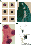
Figure 2
Figure 2. Purification of the first hematopoietic stem cells
A Representative examples of purified HSCs as visualized by microscopy after hematoxylin staining. B Myeloerythroid colonies formed in the spleen formed by the injection of purified HSCs into lethally irradiated mice. C A single lymphoid colony in the thymus formed by the injection of purified HSCs into lethally mice. D Fluorescence-activated cell sorting depicting HSCs as a Thy-1loSca-1+. Reproduced with permission from Spangrude et al. (1988) Science (10).
Figure 3
Figure 3. RNA expression pattern of IL-7Rα throughout the murine hematopoietic lineage
IL-7Rα is a critical surface molecule that helps distinguish the lymphoid arm of the hematopoietic system from HSCs, progenitors, and myeloid cells. Each box represents a different hematopoietic subpopulation. Blue indicates lower expression, pink indicates higher expression. Analysis performed using Gene Expression Commons (46).
Figure 4
Figure 4. GMP frequency is decreased in low risk MDS
Representative example of how hematopoietic progenitor cell populations can be altered in states of disease. A Frequency of GMPs out of total myeloid progenitors in normal, low risk MDS, and non-MDS diseased bone marrow samples. B Frequency of GMPs out of total lineage negative bone marrow mononuclear cells in normal and low risk MDS bone marrow samples. Reproduced with permission from Pang et al. (2013) Proc Nat Acad Sci (77).
Figure 5
Figure 5. CD47-blocking therapies are effective in preclinical models of human cancer
Xenograft studies of mice engrafted with human AML samples that were then treated with anti-CD47 antibodies. A Anti-CD47 antibody treatment decreases leukemia burden, as assessed by the percent of human chimerism in the peripheral blood. B Bone marrow histology showing leukemia infiltration (images 1–2) in control mice, and eradication of disease in mice treated with anti-CD47 antibodies (images 4–5). In some mice with residual tumor burden following treatment with anti-CD47 antibodies, macrophages could be seen in the bone marrow engulfing leukemia cells (images 3–6). Reproduced with permission from Majeti et al. (2009) Cell (92).

References

    1. Ford CE, Hamerton JL, Barnes DW, Loutit JF. Cytological identification of radiation-chimaeras. Nature. 1956 Mar 10;177(4506):452–4. - PubMed
    1. Till JE, Mc CE. A direct measurement of the radiation sensitivity of normal mouse bone marrow cells. Radiat Res. 1961 Feb;14:213–22. - PubMed
    1. Becker AJ, Mc CE, Till JE. Cytological demonstration of the clonal nature of spleen colonies derived from transplanted mouse marrow cells. Nature. 1963 Feb 2;197:452–4. - PubMed
    1. Wu AM, Till JE, Siminovitch L, McCulloch EA. Cytological evidence for a relationship between normal hemotopoietic colony-forming cells and cells of the lymphoid system. J Exp Med. 1968 Mar 1;127(3):455–64. - PMC - PubMed
    1. Chen JY, Miyanishi M, Wang SK, Yamazaki S, Sinha R, Kao KS, Seita J, Sahoo D, Nakauchi H, Weissman IL. Hoxb5 marks long-term haematopoietic stem cells and reveals a homogenous perivascular niche. Nature. 2016 Feb 11;530(7589):223–7. - PMC - PubMed

Publication types