. 2016 Oct 18;17(10):1653.
doi: 10.3390/ijms17101653.

Inducing G2/M Cell Cycle Arrest and Apoptosis through Generation Reactive Oxygen Species (ROS)-Mediated Mitochondria Pathway in HT-29 Cells by Dentatin (DEN) and Dentatin Incorporated in Hydroxypropyl-β-Cyclodextrin (DEN-HPβCD)

Affiliations

Inducing G2/M Cell Cycle Arrest and Apoptosis through Generation Reactive Oxygen Species (ROS)-Mediated Mitochondria Pathway in HT-29 Cells by Dentatin (DEN) and Dentatin Incorporated in Hydroxypropyl-β-Cyclodextrin (DEN-HPβCD)

Al-Abboodi Shakir Ashwaq et al. Int J Mol Sci. .

Abstract

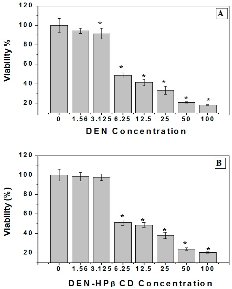
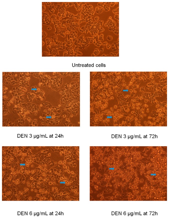
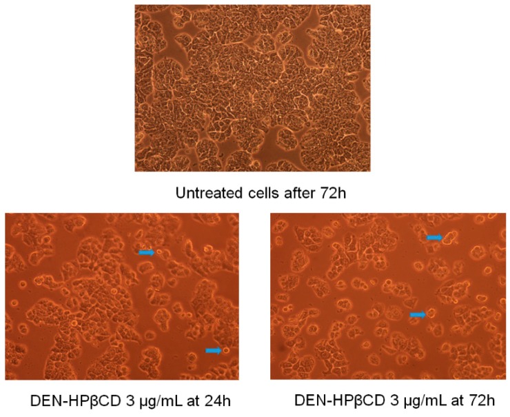
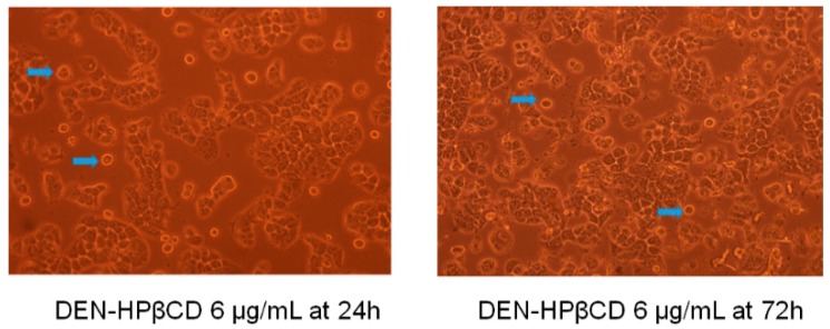
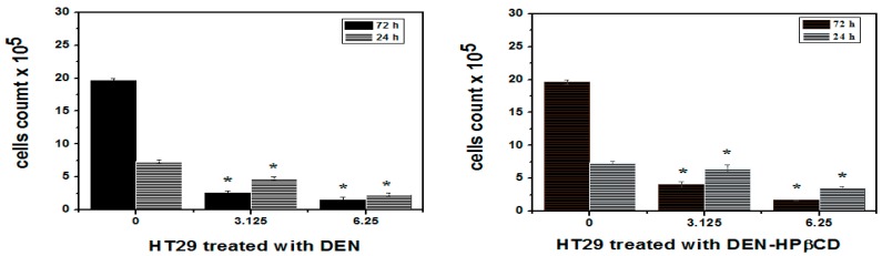
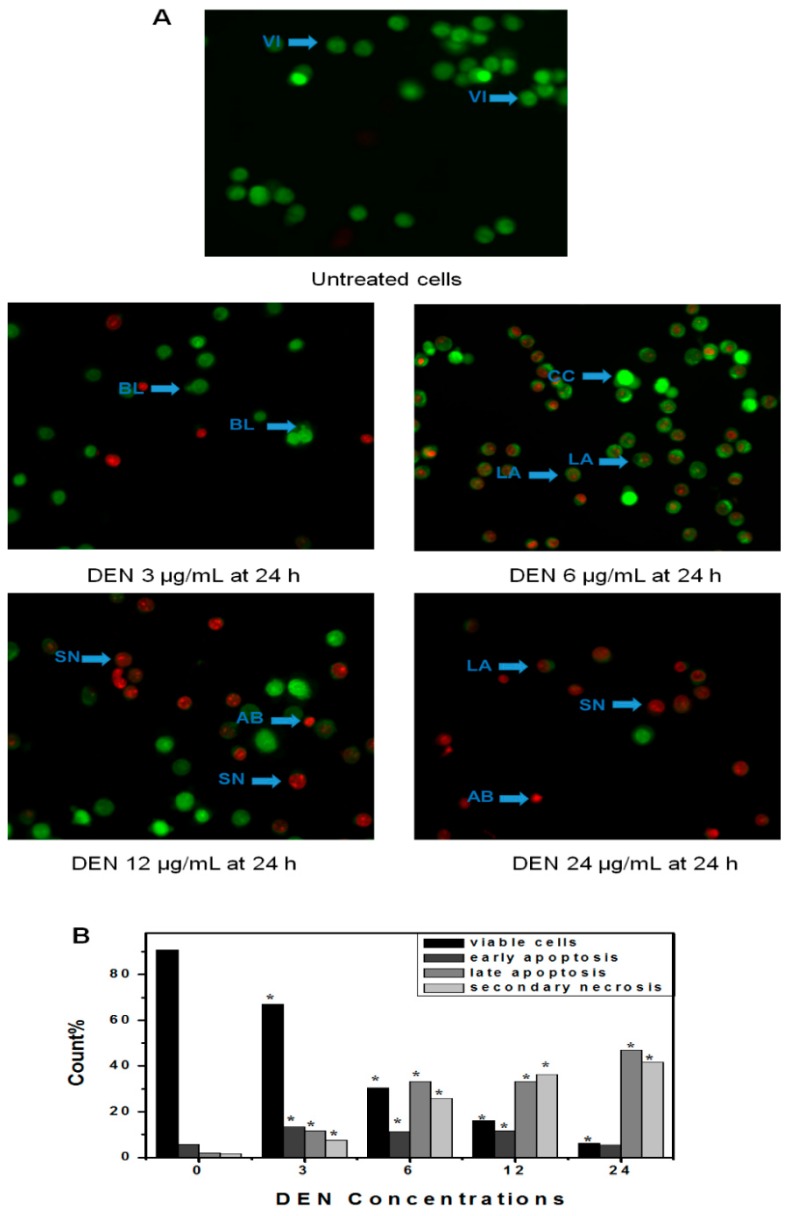
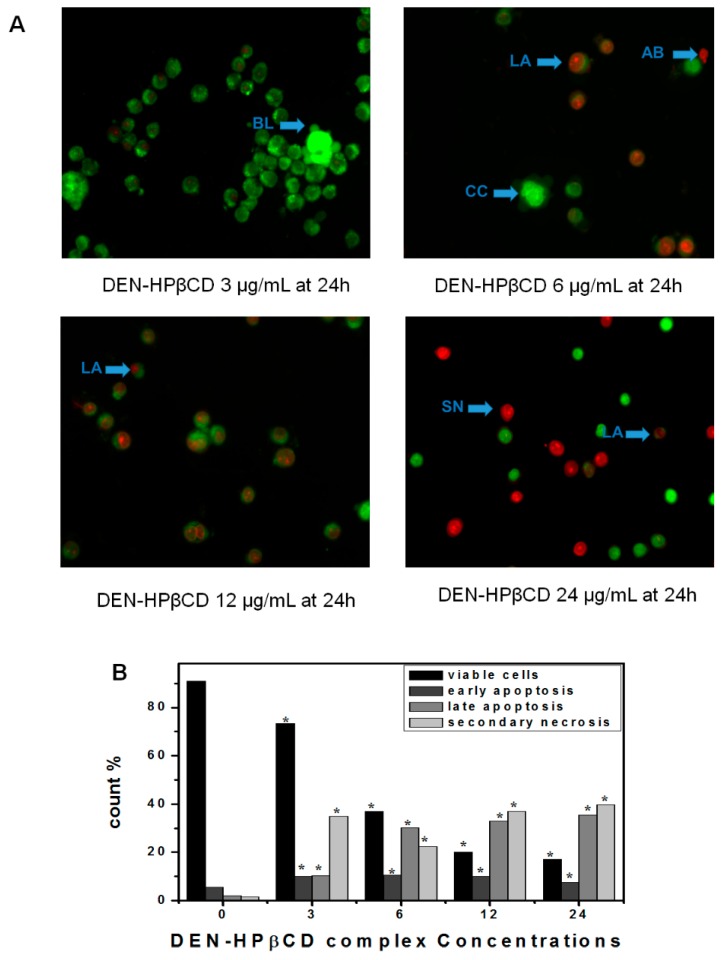
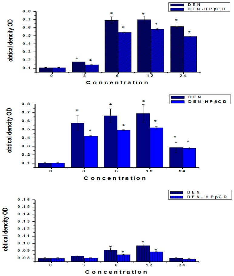
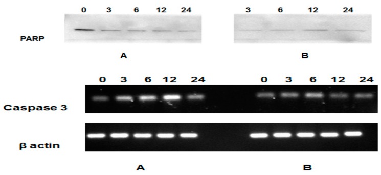
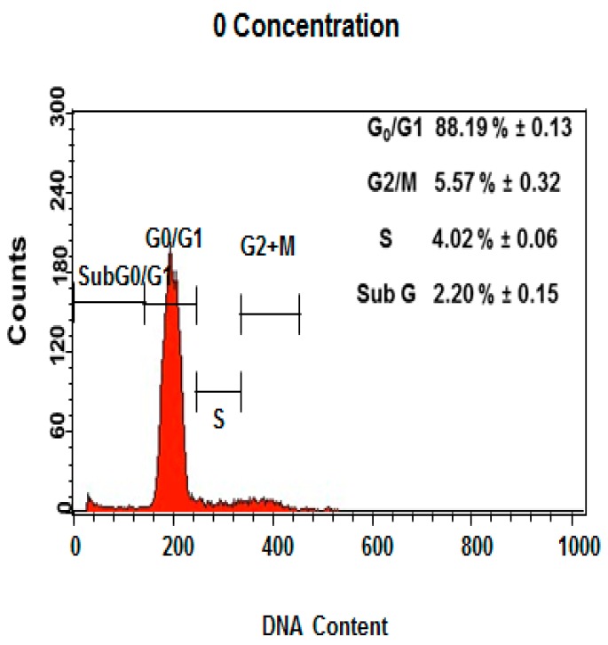
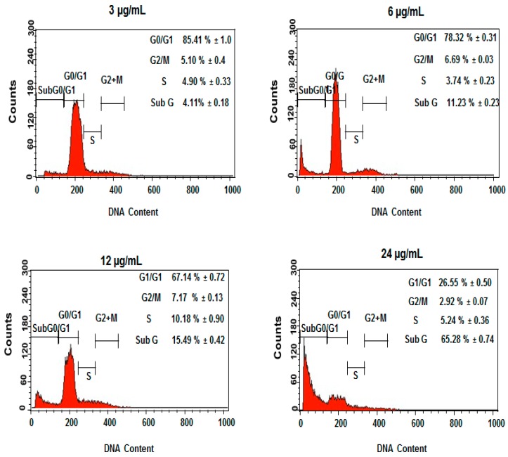
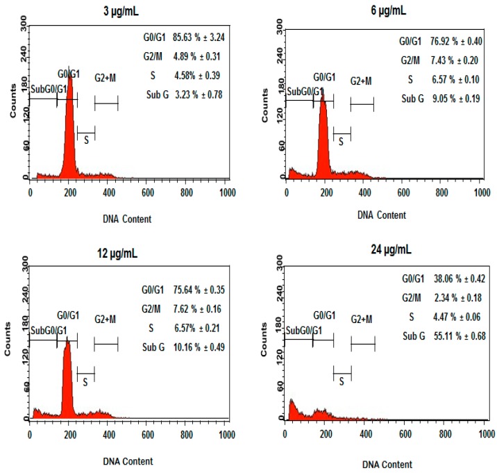
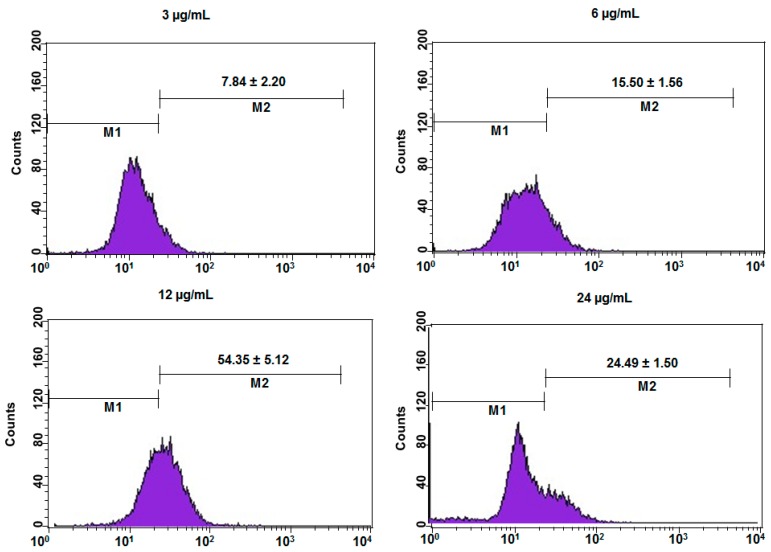
Dentatin (DEN), purified from the roots of Clausena excavata Burm f., has poor aqueous solubility that reduces its therapeutic application. The aim of this study was to assess the effects of DEN-HPβCD (hydroxypropyl-β-cyclodextrin) complex as an anticancer agent in HT29 cancer cell line and compare with a crystal DEN in dimethyl sulfoxide (DMSO). The exposure of the cancer cells to DEN or DEN-HPβCD complex leads to cell growth inhibition as determined by MTT (3-(4,5-dimethylthiazol-2-yl)-2,5-diphenyltetrazolium bromide) assay. To analyze the mechanism, in which DEN or DEN-HPβCD complex causes the death in human colon HT29 cancer cells, was evaluated by the enzyme-linked immunosorbent assay (ELIZA)-based assays for caspase-3, 8, 9, and reactive oxygen species (ROS). The findings showed that an anti-proliferative effect of DEN or DEN-HPβCD complex were via cell cycle arrest at the G2/M phase and eventually induced apoptosis through both mitochondrial and extrinsic pathways. The down-regulation of poly(ADP-ribose) polymerase (PARP) which leaded to apoptosis upon treatment, was investigated by Western-blotting. Hence, complexation between DEN and HPβCD did not diminish or eliminate the effective properties of DEN as anticancer agent. Therefore, it would be possible to resolve the conventional and current issues associated with the development and commercialization of antineoplastic agents in the future.

Keywords: Dentatin; apoptosis; cytostatic; cytotoxicity; reactive oxygen species (ROS).


Conflict of interest statement

The authors declare no conflict of interest.

Figures

Figure 1
Figure 1
(A) Cytotoxicity of DEN (Dentatin) against human colon cancer cells (HT29). The cells were plated in 96-well plates and then exposed to 100, 50, 25, 6.25, 3.125 and 1.25 µg/mL of DEN for 72 h. The viability of treated cells were measured by using an MTT assay. Mean ± standard deviation (SD). (n = 3 well/treatment). * p < 0.05 compared with untreated cells; (B) Cytotoxicity of DEN-HPβCD (hydroxypropyl-β-cyclodextrin) complex against human colon cancer cells (HT29). The cells were plated in 96-well plates and then exposed to 100, 50, 25, 6.25, 3.125 and 1.25 µg/mL of DEN for 72 h. The viability of treated cells were measured by using an MTT assay. Mean ± standard deviation (SD). (n = 3 well/treatment). * p < 0.05 compared with untreated cells.
Figure 2
Figure 2
The morphological changes of HT29 treated cells with (3.125 and 6.25 µg/mL) of DEN dissolved in dimethyl sulfoxide (DMSO) for 24 and 72 h. Note: blue arrows indicate apoptotic cells (200×).
Figure 3
Figure 3
The morphological changes of HT29 treated cells with (3.125 and 6.25 µg/mL) of DEN-HPβCD complex for 24 and 72 h, Note: blue arrows indicate apoptotic cells (200×).
Figure 3
Figure 3
The morphological changes of HT29 treated cells with (3.125 and 6.25 µg/mL) of DEN-HPβCD complex for 24 and 72 h, Note: blue arrows indicate apoptotic cells (200×).
Figure 4
Figure 4
Trypan blue exclusion assay for cell viability of HT29 treated cells after 24 and 72 h of treatment with DEN and /or DEN-HPβCD complex. Mean ± standard deviation (SD). (n = 3 well/treatment). * p < 0.05 compared with untreated cells.
Figure 5
Figure 5
(A) Fluorescent micrographs of double-stained HT29 cells (acridine orange and propidium iodide). Untreated cells appeared normal structure without any features of apoptosis and necrosis. The early apoptosis features were seen after treatment with DEN (3 µg/mL) representing intercalated acridine orange (bright green) with fragmented DNA and Blebbing after 24 h. The hallmarks of late apoptosis which represented by orange colour were noticed after 24 h of treated with (6 µg/mL) which increased with increasing the concentration. Cells with bright red colour and big size representing secondary necrosis were visible after treated with higher concentration at 24 h. VI, viable cells; BL, blebbing of the cell membrane; CC, chromatin condensation; LA, late apoptosis; SN, secondary necrosis; AB, apoptotic bodies. Images are representative of one of the three similar experiment; (B) Count cells on different microphotographs. Note: the magnification is 40×. * p < 0.05 compared with untreated cells.
Figure 6
Figure 6
(A) Fluorescent micrographs of double-stained HT29 cells (acridine orange and propidium iodide). Untreated cells appeared normal structure without any features of apoptosis and necrosis. The early apoptosis features were seen after treatment with DEN-HPβCD complex (3 µg/mL) representing intercalated acridine orange (bright green) with fragmented DNA and Blebbing after 24 h. The hallmarks of late apoptosis which represented by orange colour were noticed after 24 h of treated with (6 µg/mL) which increased with increasing the concentration. Cells with bright red colour and big size representing secondary necrosis were visible after treated with higher concentration at 24 h. VI, viable cells; BL, blebbing of the cell membrane; CC, chromatin condensation; LA, late apoptosis; SN, secondary necrosis; AB, apoptotic bodies. Images are representative of one of the three similar experiment; (B) Count cells on different microphotographs. Note: the magnification is 40×. * p < 0.05 compared with untreated cells.
Figure 7
Figure 7
Treatment of HT29 cells with DEN and/or DEN-HPβCD complex showed in activation of caspase-3, 9 and 8. Cells were treated with 3, 6, 12 and 24 µg/mL for 24 h, and then determination of caspase-3, 9 and 8 activity was done. Mean ± standard deviation (SD). (n = 3 well/treatment). * p < 0.05 compared with untreated cells. RT-PCR analysis for caspase-3 mRNA expression was stimulated in treated and untreated cells. β-actin used as control. Part A represents caspase-3 and β actin mRNA expression in DEN-HT29 treated cells, Part B represents caspase-3 and β actin mRNA expression in DEN-HPβCD-HT29 treated cells. The effect of DEN and DEN-HPβCD complex on apoptosis regulatory proteins in the treated HT29 colon cancer cell line was determined by Western blot assay. Detection of poly(ADP-ribose) polymerase (PARP).
Figure 7
Figure 7
Treatment of HT29 cells with DEN and/or DEN-HPβCD complex showed in activation of caspase-3, 9 and 8. Cells were treated with 3, 6, 12 and 24 µg/mL for 24 h, and then determination of caspase-3, 9 and 8 activity was done. Mean ± standard deviation (SD). (n = 3 well/treatment). * p < 0.05 compared with untreated cells. RT-PCR analysis for caspase-3 mRNA expression was stimulated in treated and untreated cells. β-actin used as control. Part A represents caspase-3 and β actin mRNA expression in DEN-HT29 treated cells, Part B represents caspase-3 and β actin mRNA expression in DEN-HPβCD-HT29 treated cells. The effect of DEN and DEN-HPβCD complex on apoptosis regulatory proteins in the treated HT29 colon cancer cell line was determined by Western blot assay. Detection of poly(ADP-ribose) polymerase (PARP).
Figure 8
Figure 8
The concentrations (3, 6, 12 and 24 µg/mL) of DEN in HT29-treated cells included G2/M cell cycle arrest. Results are represented one of the three independent experiments.
Figure 8
Figure 8
The concentrations (3, 6, 12 and 24 µg/mL) of DEN in HT29-treated cells included G2/M cell cycle arrest. Results are represented one of the three independent experiments.
Figure 9
Figure 9
The concentrations (3, 6, 12 and 24 µg/ml) of DEN-HPβCD complex in HT29-treated cells included G2/M cell cycle arrest. Results are represented one of the three independent experiments.
Figure 10
Figure 10
The concentrations (3, 6, 12 and 24 µg/mL) of DEN in HT29-treated cells induced ROS generation. Results are represented one of the three independent experiments. Note: M1 represented the cells that not producing ROS, M2 represented the cells that actively producing ROS.
Figure 11
Figure 11
The concentrations (3, 6, 12 and 24 µg/mL) of DEN-HPβCD complex in HT29-treated cells induced ROS generation. Results are represented one of the three independent experiments. Note: M1 represented the cells that not producing ROS, M2 represented the cells that actively producing ROS.

Similar articles

Cited by

References

    1. Louis D.N., Ohgaki H., Wiestler O.D., Cavenee W.K., Burger P.C., Jouvet A., Scheithauer B.W., Kleihues P. The 2007 who classification of tumours of the central nervous system. Acta Neuropathol. 2007;114:97–109. doi: 10.1007/s00401-007-0243-4. - DOI - PMC - PubMed
    1. Solanki R., Madat D., Goyal R.K. Anti cancer activity of colon specific osmotic drug delivery of acetone extract of quercus infectoria olivier, fagaceae in 1,2-dimethylhydrazine-induced colon cancer in rats. Mol. Cytogenet. 2014;7:1. doi: 10.1186/1755-8166-7-S1-P14. - DOI - PubMed
    1. Vauzour D., Rodriguez-Mateos A., Corona G., Oruna-Concha M.J., Spencer J.P. Polyphenols and human health: Prevention of disease and mechanisms of action. Nutrients. 2010;2:1106–1131. doi: 10.3390/nu2111106. - DOI - PMC - PubMed
    1. Araújo J.R., Gonçalves P., Martel F. Chemopreventive effect of dietary polyphenols in colorectal cancer cell lines. Nutr. Res. 2011;31:77–87. doi: 10.1016/j.nutres.2011.01.006. - DOI - PubMed
    1. Arbab I.A., Looi C.Y., Abdul A.B., Cheah F.K., Wong W.F., Sukari M.A., Abdullah R., Mohan S., Syam S., Arya A. Dentatin induces apoptosis in prostate cancer cells via Bcl-2, Bcl-xL, survivin downregulation, caspase-9,-3/7 activation, and NF-κB inhibition. Evid. Based Complement. Altern. Med. 2012 doi: 10.1155/2012/856029. - DOI - PMC - PubMed

MeSH terms

LinkOut - more resources