. 2016 Oct 18;8(10):286.
doi: 10.3390/v8100286.

Maporal Hantavirus Causes Mild Pathology in Deer Mice (Peromyscus maniculatus)

Affiliations

Maporal Hantavirus Causes Mild Pathology in Deer Mice (Peromyscus maniculatus)

Amanda McGuire et al. Viruses. .

Abstract

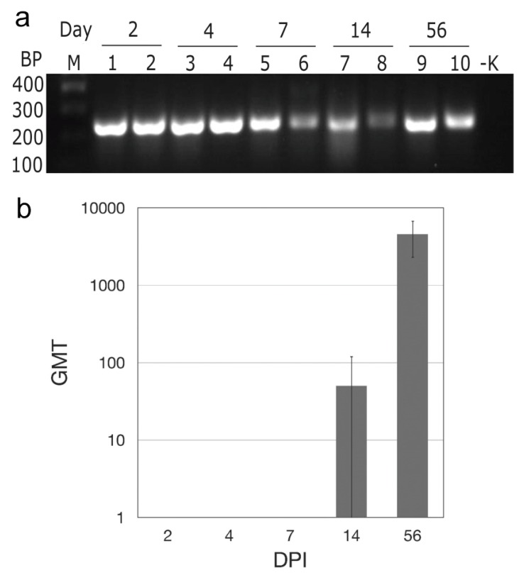
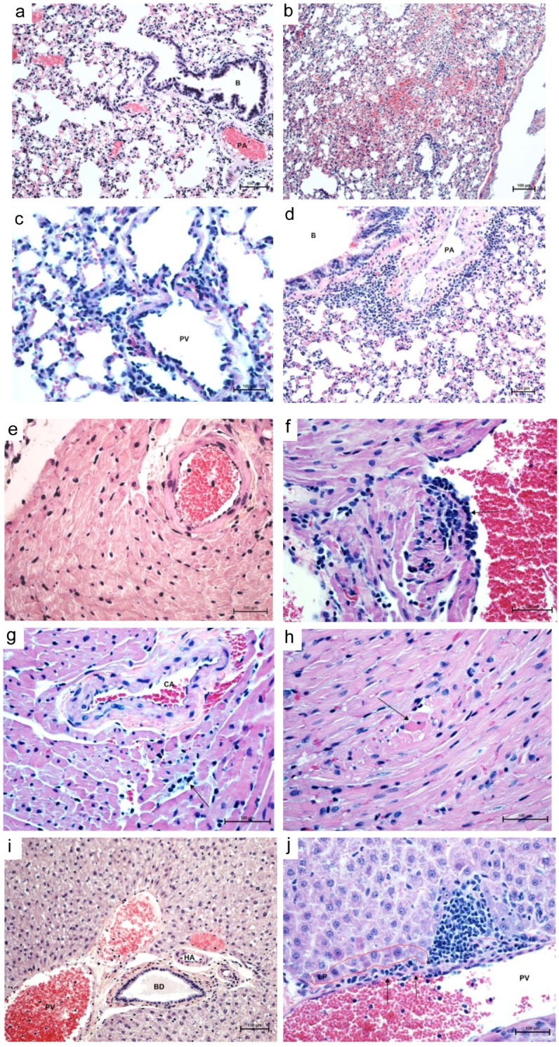
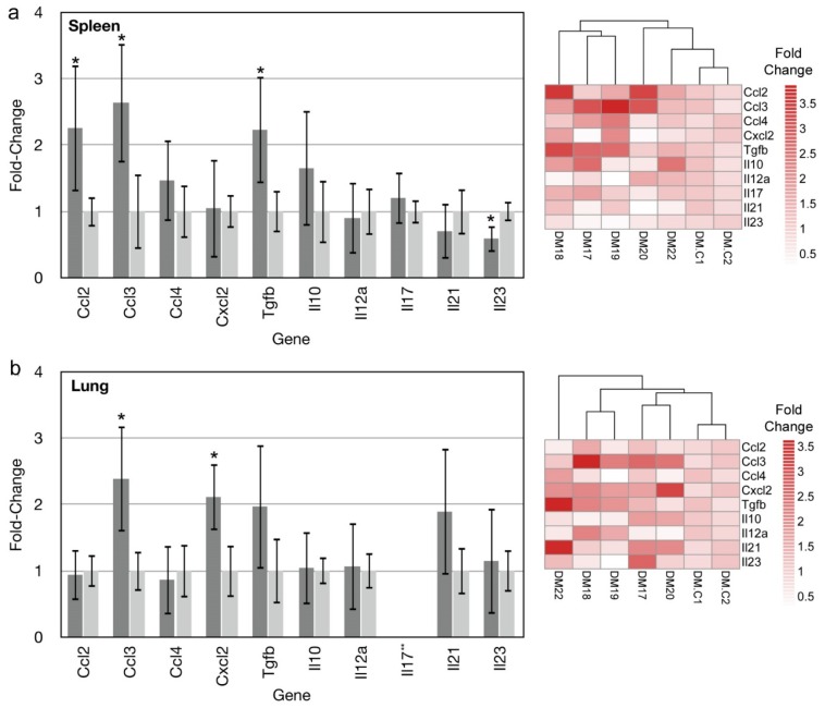
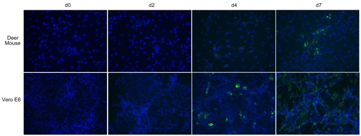
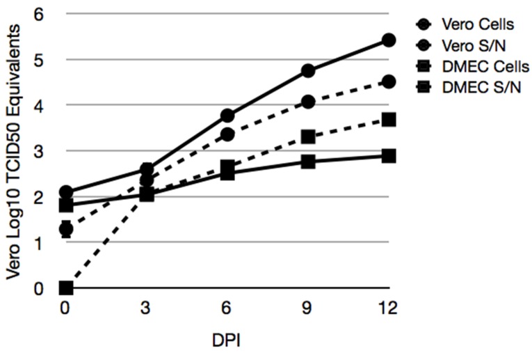
Rodent-borne hantaviruses can cause two human diseases with many pathological similarities: hantavirus cardiopulmonary syndrome (HCPS) in the western hemisphere and hemorrhagic fever with renal syndrome in the eastern hemisphere. Each virus is hosted by specific reservoir species without conspicuous disease. HCPS-causing hantaviruses require animal biosafety level-4 (ABSL-4) containment, which substantially limits experimental research of interactions between the viruses and their reservoir hosts. Maporal virus (MAPV) is a South American hantavirus not known to cause disease in humans, thus it can be manipulated under ABSL-3 conditions. The aim of this study was to develop an ABSL-3 hantavirus infection model using the deer mouse (Peromyscus maniculatus), the natural reservoir host of Sin Nombre virus (SNV), and a virus that is pathogenic in another animal model to examine immune response of a reservoir host species. Deer mice were inoculated with MAPV, and viral RNA was detected in several organs of all deer mice during the 56 day experiment. Infected animals generated both nucleocapsid-specific and neutralizing antibodies. Histopathological lesions were minimal to mild with the peak of the lesions detected at 7-14 days postinfection, mainly in the lungs, heart, and liver. Low to modest levels of cytokine gene expression were detected in spleens and lungs of infected deer mice, and deer mouse primary pulmonary cells generated with endothelial cell growth factors were susceptible to MAPV with viral RNA accumulating in the cellular fraction compared to infected Vero cells. Most features resembled that of SNV infection of deer mice, suggesting this model may be an ABSL-3 surrogate for studying the host response of a New World hantavirus reservoir.

Keywords: Maporal virus; deer mice; hantavirus; reservoir; zoonoses.


Conflict of interest statement

The authors declare no conflict of interest.

Figures

Figure 1
Figure 1
Maporal virus (MAPV) infects deer mice. After subcutaneous inoculation in the hindquarter, MAPV RNA was detected in the lungs two days later and persisted though at least 56 days; M marker, BP base pairs, -K negative control (a). Antibodies to MAPV nucleocapsid appeared as early as 14 days PI and increased in titer by day 56 (b).
Figure 2
Figure 2
Histopathology of cardiopulmonary phase of MAPV infection in deer mice. Control lung (day 14) shows clear airways (bronchiole, B) and air spaces, normal vessels (pulmonary artery, PA) and normal thickness of interstitium (a, 200×). Lungs 14 days PI show alveolar hemorrhage, edema, and mild interstitial pneumonia (b, 100×). Lungs at 14 days PI show prominent perivascular (portal vein, PV, and artery, PA) neutrophilic infiltrates with mild interstitial pneumonia (c, 400×; d, 200×). Control heart shows a normal coronary artery and surrounding myocardium (e, 200×). Left atrium 7 days PI shows endothelial hypertrophy and hyperplasia with lymphocyte infiltration into subjacent myocardium with sarcoplasmic hyalinization and vacuolation of sarcoplasm with loss of cross striations (f, 400×). At 56 days PI, coronary artery (CA) with hypertrophic endothelium and perivascular edema, interstitial lymphocytic myocarditis and cardimyocyte degeneration/necrosis (g, 400×) and necrotic cardiomyocytes appear rounded up with hyperesinophilic sarcoplasm and pyknotic nuclei (h, 400×). Control liver shows intact limiting plate around normal portal vessels, hepatic artery (HA) and bile ductile (BD) (i, 200×). Focal disruption of portal vein (PV) endothelium (arrows) with lymphocytes percolating the limiting plate (LP) into the surrounding parenchyma mixed with a few neutrophils (j, 400×). Stain, hematoxylin-eosin.
Figure 3
Figure 3
Cytokine and chemokine gene expression in spleens and lungs of MAPV-infected deer mice. Fourteen days post infection, spleens and lungs of 5 deer mice were examined for the expression of 10 genes by real-time PCR. Ccl2, Ccl3 and Tgfb were elevated in spleens of infected deer mice (dark gray) compared to uninfected deer mice (light gray), whereas Il23 expression was repressed. Error bars represent 95% confidence intervals and those denoted by * are statistically different from the uninfected control. Heat map indicates fold-change of individual deer mice used in generating the graph of infected deer mice (DM17–DM22) and uninfected controls (DM.C1, DM.C2) (a). In lungs, only Ccl3 and Cxcl2 were elevated. Heat map indicates fold-change of individual deer mice used in generating the graph of infected deer mice (DM17–DM22) and uninfected controls (DM.C1, DM.C2). **Il17 was not detected in either infected or uninfected lung samples (b).
Figure 4
Figure 4
Maporal virus infects deer mouse cells. Deer mouse pulmonary microvascular endothelial cells (PMVEC) or Vero E6 cells were inoculated with 0.1 MOI of MAPV. On days 2, 4 and 7 cells from each were fixed and stained with rabbit antibody specific to nucleocapsid and detected with a mouse anti-rabbit IgG-FITC conjugate (green). Slides were mounted with DAPI to identify nuclei (blue). Viral antigen was detected in some deer mouse PMVEC on day 4 but substantially more cells by day 7, and with punctate characteristics. Virus was detected in few Vero E6 cells on day 2, but on day 4 punctate cells were readily observed. By day 7, many more cells were infected but the pattern was more diffuse and less puntate.
Figure 5
Figure 5
Accumulation of MAPV RNA in cellular fractions of infected deer mouse PMVEC. Vero E6 (circles) and deer mouse PMVEC (squares) were infected with MAPV to examine viral RNA in cells (hatched lines) and supernatants (solid lines) by real-time PCR. Cells were inoculated with 0.1 MOI of MAPV for 1 h, followed by removal of inoculum, 1× wash in PBS and addition of 2% FBS Ham’s F12 medium. The day 0 samples were collected 1 h later. The remaining samples were collected on days 3, 6, 9 and 12 dpi. RNA copies were determined relative to tissue culture infectious dose (TCID50) equivalents.

Similar articles

Cited by

References

    1. Schmaljohn C., Hjelle B. Hantaviruses: A global disease problem. Emerg. Infect. Dis. 1997;3:95–104. doi: 10.3201/eid0302.970202. - DOI - PMC - PubMed
    1. Schountz T., Prescott J. Hantavirus immunology of rodent reservoirs: Current status and future directions. Viruses. 2014;6:1317–1335. doi: 10.3390/v6031317. - DOI - PMC - PubMed
    1. Johnson K.M. Hantaviruses: History and overview. Curr. Top. Microbiol. Immunol. 2001;256:1–14. - PubMed
    1. Jonsson C.B., Figueiredo L.T., Vapalahti O. A global perspective on hantavirus ecology, epidemiology, and disease. Clin. Microbiol. Rev. 2010;23:412–441. doi: 10.1128/CMR.00062-09. - DOI - PMC - PubMed
    1. Clement J., Maes P., Lagrou K., van Ranst M., Lameire N. A unifying hypothesis and a single name for a complex globally emerging infection: Hantavirus disease. Eur. J. Clin. Microbiol. Infect. Dis. 2012;31:1–5. doi: 10.1007/s10096-011-1456-y. - DOI - PMC - PubMed

Publication types

LinkOut - more resources