. 2016 Oct 18;8(10):287.
doi: 10.3390/v8100287.

4EBP-Dependent Signaling Supports West Nile Virus Growth and Protein Expression

Affiliations

4EBP-Dependent Signaling Supports West Nile Virus Growth and Protein Expression

Katherine D Shives et al. Viruses. .

Abstract

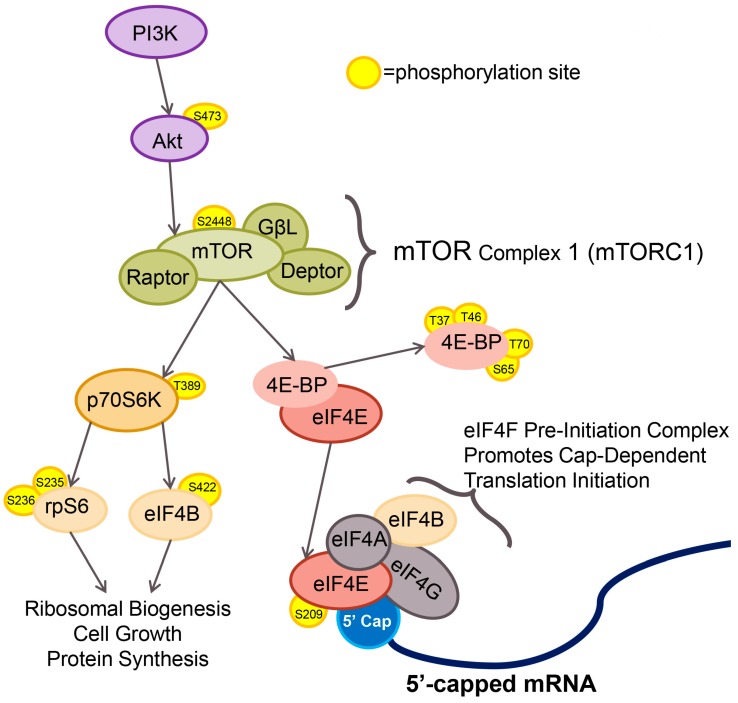
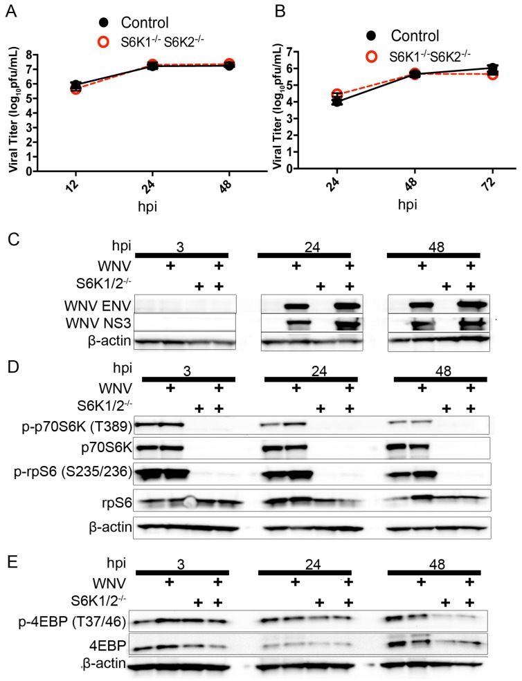
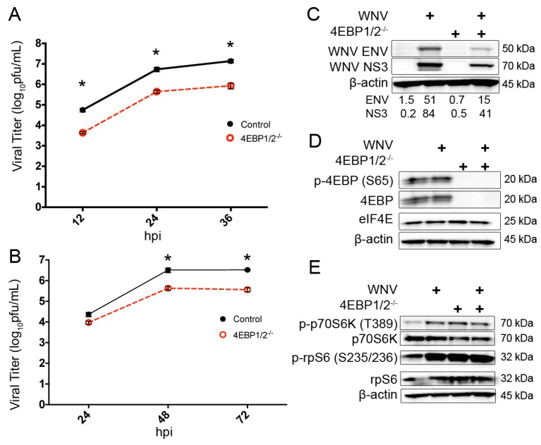
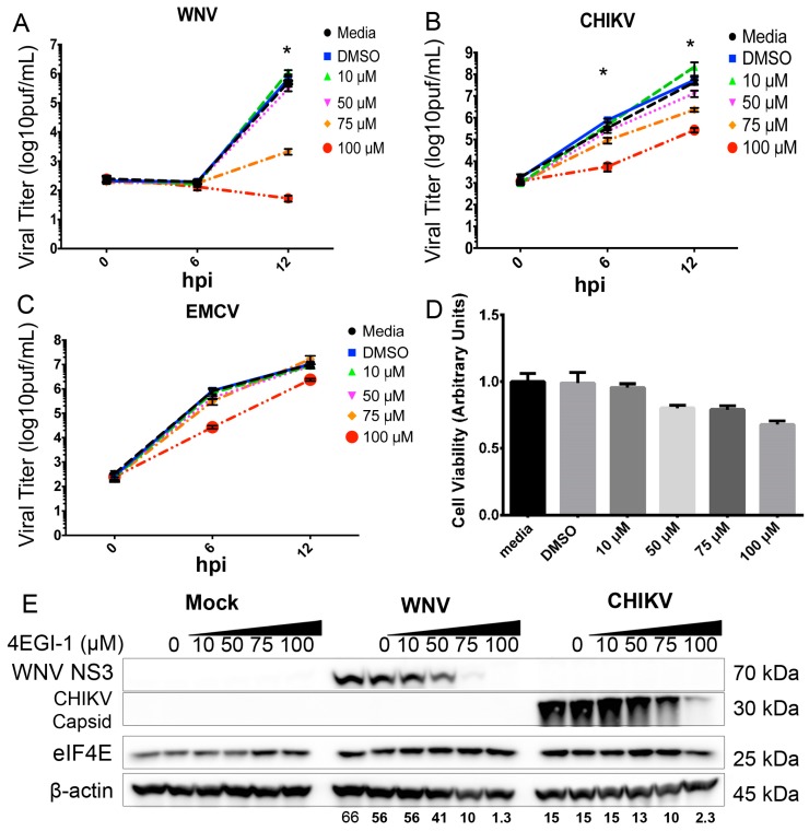
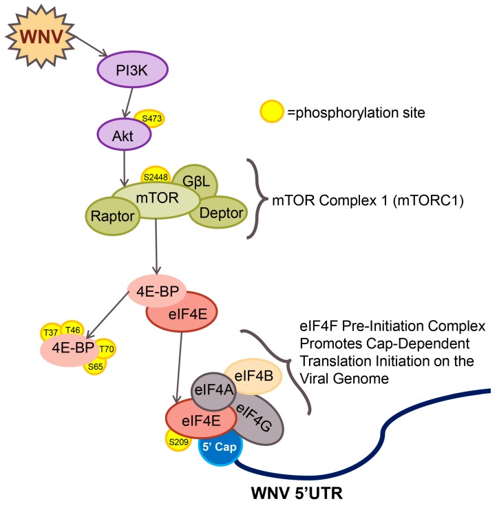
West Nile virus (WNV) is a (+) sense, single-stranded RNA virus in the Flavivirus genus. WNV RNA possesses an m7GpppNm 5' cap with 2'-O-methylation that mimics host mRNAs preventing innate immune detection and allowing the virus to translate its RNA genome through the utilization of cap-dependent translation initiation effectors in a wide variety of host species. Our prior work established the requirement of the host mammalian target of rapamycin complex 1 (mTORC1) for optimal WNV growth and protein expression; yet, the roles of the downstream effectors of mTORC1 in WNV translation are unknown. In this study, we utilize gene deletion mutants in the ribosomal protein kinase called S6 kinase (S6K) and eukaryotic translation initiation factor 4E-binding protein (4EBP) pathways downstream of mTORC1 to define the role of mTOR-dependent translation initiation signals in WNV gene expression and growth. We now show that WNV growth and protein expression are dependent on mTORC1 mediated-regulation of the eukaryotic translation initiation factor 4E-binding protein/eukaryotic translation initiation factor 4E-binding protein (4EBP/eIF4E) interaction and eukaryotic initiation factor 4F (eIF4F) complex formation to support viral growth and viral protein expression. We also show that the canonical signals of mTORC1 activation including ribosomal protein s6 (rpS6) and S6K phosphorylation are not required for WNV growth in these same conditions. Our data suggest that the mTORC1/4EBP/eIF4E signaling axis is activated to support the translation of the WNV genome.

Keywords: RNA; West Nile virus; protein synthesis; translation.


Conflict of interest statement

The authors declare no conflict of interest.

Figures

Figure 1
Figure 1
The mammalian target of rapamycin complex 1 (mTORC1) pathway and translation initiation. Cellular mTORC1 activity is regulated in part by phosphatidylinositol-3-kinase/protein kinase B (PI3K/Akt) signaling. Phosphorylation of mTOR on residue serine 2448 leads to activation of mTOR and phosphorylation of mTORC1 effector proteins, 70 kDa ribosomal protein S6 kinase 1 (p70S6K) and eukaryotic initiation factor 4E binding protein (4EBP). Phosphorylation-induced activity of p70S6K phosphorylates ribosomal protein S6 (rpS6) and eukaryotic initiation factor 4B (eIF4B) resulting in initiation of specific cap-dependent translation events. mTORC1-dependent phosphorylation of 4EBP leads to dissociation of 4EBP from eIF4E. Free eIF4E binds to the 5′ cap of host mRNAs and forms the eIF4F pre-initiation complex.
Figure 2
Figure 2
Raptor expression supports the growth and protein expression of 5′-capped RNA viruses. (A) Multi-step growth curve (MOI = 0.001) of West Nile virus (WNV) in control and Raptor knockout (RapKO) cells. N = 6 replicates per time point, * p < 0.0001; a single-step growth curve (MOI = 3) is shown for (B) Chikungunya virus La Reunion 2006 OPY-1 (CHIKV-LR) and (C) encephalomyocarditis virus (EMCV) in control and RapKO cells as determined by standard plaque assay of the supernatant. The viral titer is presented as log10 pfu/mL at indicated times (hours post-infection (hpi)). N = 6 replicates per time point, * p < 0.005; (D) Cell-associated CHIKV-LR genome copies in RapKO and control murine embryonic fibroblast (MEF) cells at 0 hpi determined by quantitative reverse-transcriptoin PCR (qRT-PCR). Data are presented as log10 CHIKV-LR genomes/µg of RNA; (E) Western blot (WB) analysis of CHIKV-LR protein synthesis in RapKO cells. Cellular lysates were harvested at the indicated time (hpi), normalized for total protein and subjected to WB analysis using antibodies to CHIKV capsid protein and a β-actin loading control. Images are representative of two independent experiments. Densitometry values are provided for CHIKV capsid corrected for β-actin expression; (F) Expression of phospho-eIF4E (serine 209), total eIF4E and β-actin in control and RapKO cells as determined by Western blot analysis. Total protein lysate was collected from cells either mock, WNV or CHIKV-LR infected (MOI = 3), separated by gel electrophoresis and probed for the indicated antibody targets. Images are representative of two independent experiments. Densitometry values are provided for p-eIF4E expression corrected for β-actin expression.
Figure 3
Figure 3
Raptor knockout results in a loss of p70S6K and 4EBP phosphorylation. Cells were either mock- or WNV-inoculated (MOI = 3) in control and iRapKO MEF cells, total cellular protein lysates were collected at the indicated hpi, and probed using antibodies for the indicated target proteins by western blot analysis. (A) Expression of Raptor and eIF4E; (B) Expression of phospho-p70S6K (tyrosine 389) and total p70S6K; (C) Expression of phospho-rpS6 (serine 235/236) and total rpS6. (D) Expression of phospho-eIF4B (serine 422) and total eIF4B; (E) Expression of phospho-4EBP1 (serine 65) and total 4EBP1. All images are representative of two independent experiments. β-actin was used as the loading control for all experiments. Densitometry values are provided for phospho-protein expression and corrected for β-actin expression.
Figure 4
Figure 4
Loss of rpS6 phosphorylation does not impact WNV growth or protein expression. (A) Single-step growth curve of WNV (MOI = 3) in rpS6p−/− and wild-type control MEFs as determined by standard plaque assay of the cell supernatant; (B) expression of the WNV envelope (WNV ENV), phospho-rpS6 (serine 235/236) and total rpS6 as determined by Western blot analysis of mock- and WNV-infected (MOI = 3) rpS6p−/− and wild-type cells. Total cellular protein lysates were collected at the indicated hpi and probed using antibodies against the indicated targets. β-actin was used as the loading control. Images are representative of two independent experiments.
Figure 5
Figure 5
P70S6K activity does not support WNV growth or protein expression. (A) Single-step growth curve of WNV (MOI = 3) in S6K1/2−/− and wild-type control MEFs; (B) Multi-step growth curve (MOI = 0.001) of WNV in S6K1/2−/− and wild-type control MEFs. Supernatants were harvested at the indicated hpi for standard plaque assay. N = 6 replicates per time point; (C) WB analysis of mock- and WNV-infected (MOI = 3) S6K1/2−/− and wild-type cells for WNV protein expression. Cellular lysates were collected at the indicated hpi and probed using antibodies for WNV ENV, WNV NS3, and β-actin loading control. Representative of 2 independent experiments; (D) WB analysis of mock- and WNV-infected (MOI = 3) S6K1/2−/− and wild-type cells for rpS6 and p70S6K expression and phosphorylation. Cellular lysates were collected at the indicated hpi and probed using antibodies against p-p70S6K [T389], total p70S6K, p-rpS6 [S235/236], total rpS6, and β-actin loading control. Representative of two independent experiments; (E) WB analysis of mock- and WNV-infected (MOI = 3) S6K1/2−/− and wild-type cells for 4EBP expression and phosphorylation. Cellular lysates were collected at the indicated hpi and probed using antibodies for p-4EBP [T37/46], total 4EBP, and β-actin loading control. Representative of two independent experiments.
Figure 6
Figure 6
Expression of 4EBP1 and 4EBP2 support WNV growth and protein expression. (A) Single-step growth curve of WNV (MOI = 3) in 4EBP1/2−/− and wild-type control MEFs; (B) Multi-step growth curve of WNV (MOI = 0.001) in 4EBP1/2−/− and wild-type control MEFs. Supernatants were harvested at indicated hpi for standard plaque assay analysis. n = 6, * p < 0.0001; (C) Confirmation of 4EBP1/2−/− (knockout) phenotype. Cellular lysates were harvested at 24 hpi and WB analysis completed using antibodies for total 4EBP, phospho-4EBP (serine 65) and eIF4E. (D) WNV protein synthesis in 4EBP1/2−/− and matched control MEF cells. Cellular lysates were harvested at 24 hpi and subjected to WB analysis using antibodies against WNV NS3 and WNV ENV. (E) S6K pathway activity in 4EBP1/2−/− and matched control MEF cells. Cellular lysates were harvested at 24 hpi for WB analysis using antibodies against total p70S6K, p-p70S6K (threonine 389), total rpS6, and p-rpS6 (serine 235/236). All images representative of two independent experiments. β-actin is shown as a loading control for all western blots. Densitometry values provided for indicated viral proteins corrected for β-actin expression.
Figure 7
Figure 7
eIF4F complex formation supports 5′-capped positive-strand RNA viral growth and protein expression. Single-step growth curves (MOI = 3) for (A) WNV, (B) CHIKV-LR and (C) EMCV in 4EGI-treated Vero cells as determined by standard plaque assay of supernatants. 4EGI-1 was added at the indicated micromolar (µM) concentrations at t = 0 hpi, and supernatants were analyzed by plaque assay at the indicated times. Viral titer data are presented as log10 plaque forming units per mL of supernatant (log10 pfu/mL). N = 9–12 replicates per time point, * p < 0.05; (D) MTT assay for cellular viability of 4EGI-treated Vero cells. MTT cleavage products were solubilized and measured by spectrophotometry at 570 nm. Dimethyl sulfoxide (DMSO) was used as a solvent control, and all groups were normalized to a media control of 1.0 arbitrary units; (E) Western blot analysis of viral protein synthesis in mock, WNV-infected (MOI = 3) and CHIKV-infected (MOI = 3) Vero cells treated with the 4EGI-1 inhibitor. Vehicle control denoted by 4EGI-1 concentration of 0 µM. Images are representative of two independent experiments. Densitometry values provided for indicated viral proteins corrected for β-actin expression.
Figure 8
Figure 8
Working model for WNV activation of mTOR and the effect on viral RNA translation initiation events. WNV infection of host cells leads to activation of mTORC1 resulting in increased phosphorylation of the translation-initiation repressor 4EBP and dissociation from eIF4E. Free eIF4E binds to the 5′ cap on WNV genomic RNA and assembles the eIF4F pre-initiation complex required for cap-dependent translation of viral RNA, resulting in viral protein expression and viral growth.

Similar articles

Cited by

References

    1. Egloff M.-P., Benarroch D., Selisko B., Romette J.-L., Canard B. An RNA cap (nucleoside-2′-O-)-methyltransferase in the flavivirus RNA polymerase NS5: Crystal structure and functional characterization. EMBO J. 2002;21:2757–2768. doi: 10.1093/emboj/21.11.2757. - DOI - PMC - PubMed
    1. Wengler G. The NS 3 nonstructural protein of flaviviruses contains an RNA triphosphatase activity. Virology. 1993;197:265–273. doi: 10.1006/viro.1993.1587. - DOI - PubMed
    1. Bartelma G., Padmanabhan R. Expression, purification, and characterization of the RNA 5′-triphosphatase activity of dengue virus type 2 nonstructural protein 3. Virology. 2002;299:122–132. doi: 10.1006/viro.2002.1504. - DOI - PubMed
    1. Szretter K.J., Daniels B.P., Cho H., Gainey M.D., Yokoyama W.M., Gale M., Jr., Virgin H.W., Klein R.S., Sen G.C., Diamond M.S. 2′-O methylation of the viral mRNA cap by West Nile virus evades IFIT1-dependent and -independent mechanisms of host restriction in vivo. PLoS Pathog. 2012;8:287. doi: 10.1371/journal.ppat.1002698. - DOI - PMC - PubMed
    1. Bowen R.A., Nemeth N.M. Experimental infections with West Nile virus. Curr. Opin. Infect. Dis. 2007;20:293–297. doi: 10.1097/QCO.0b013e32816b5cad. - DOI - PubMed

Substances

LinkOut - more resources