. 2016 Oct 20;7(10):e2425.
doi: 10.1038/cddis.2016.317.

Methamphetamine potentiates HIV-1 gp120-mediated autophagy via Beclin-1 and Atg5/7 as a pro-survival response in astrocytes

Affiliations

Methamphetamine potentiates HIV-1 gp120-mediated autophagy via Beclin-1 and Atg5/7 as a pro-survival response in astrocytes

Lu Cao et al. Cell Death Dis. .

Abstract

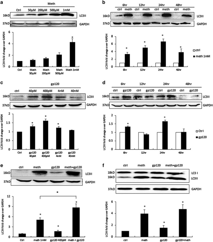
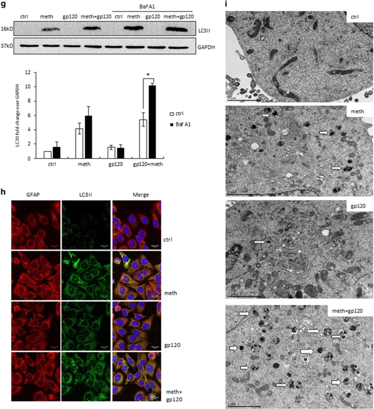
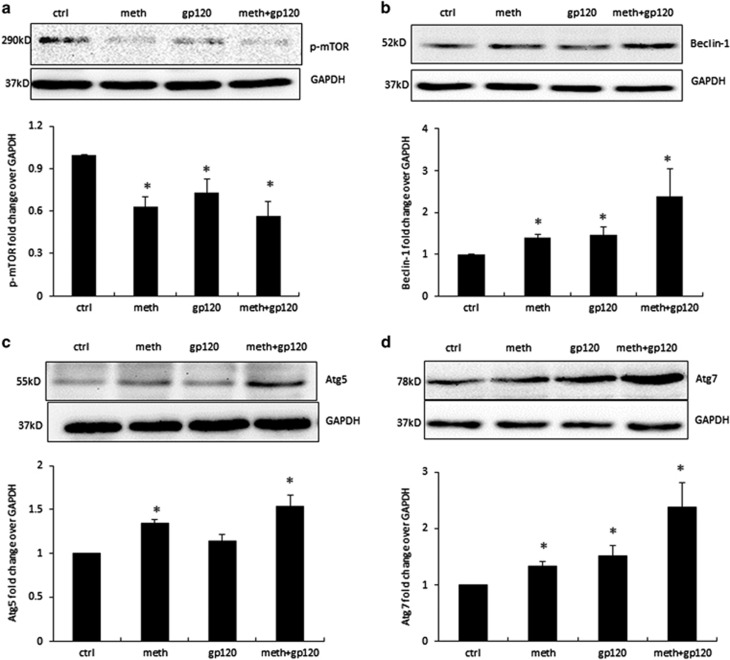
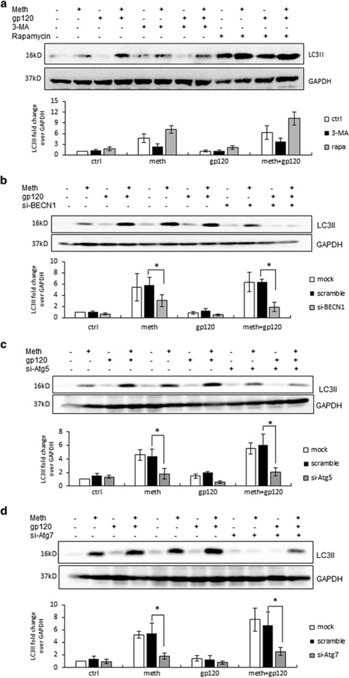
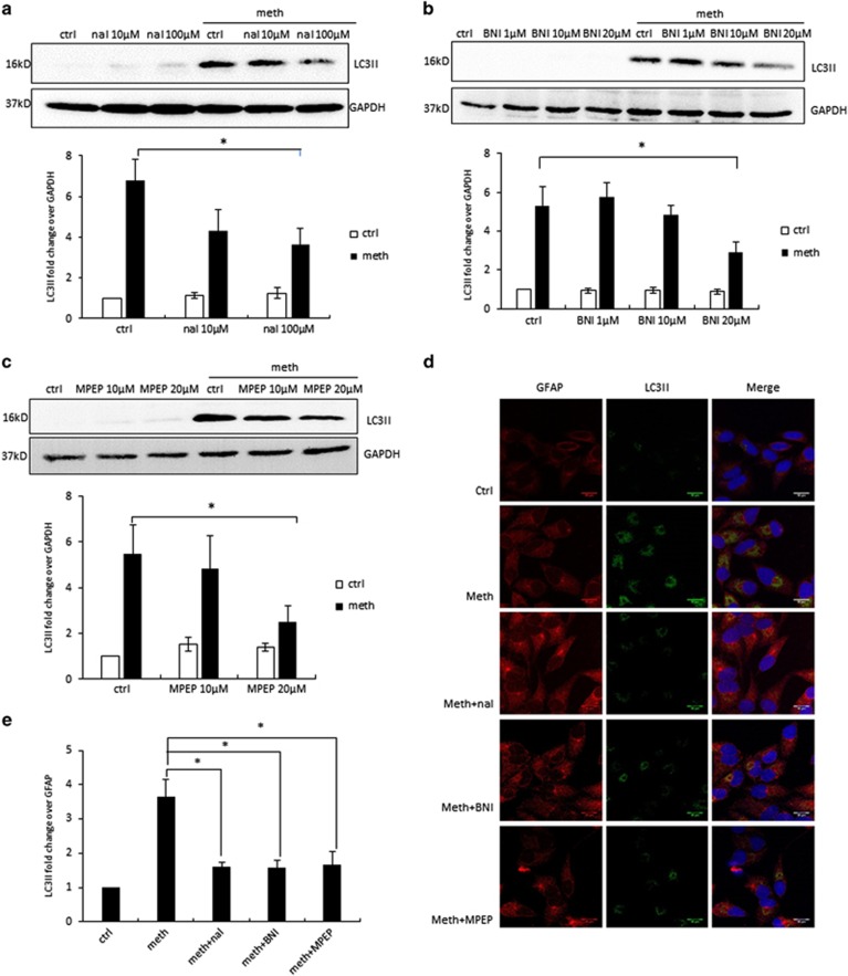
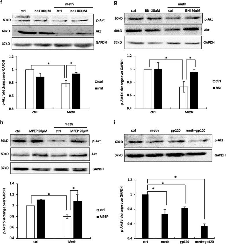
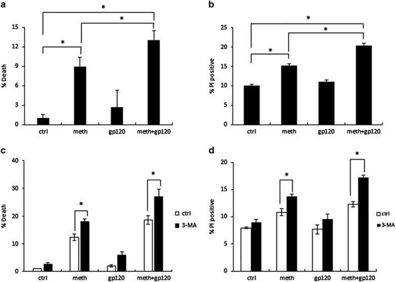
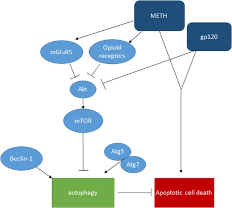
Methamphetamine (METH), a commonly used controlled substance, is known to exacerbate neuropathological dysfunction in HIV-infected individuals. The neuropathological manifestation results from cell death or dysfunction in the central nervous system (CNS) wherein autophagy is expected to have an important role. Autophagy is generally considered protective during deprivation/stress. However, excessive autophagy can be destructive, leading to autophagic cell death. This study was designed to investigate if METH and HIV-1 gp120 interact to induce autophagy in SVGA astrocytes, and whether autophagy is epiphenomenal or it has a role in METH- and gp120-induced cytotoxicity. We found that METH and gp120 IIIb caused an increase in LC3II level in astrocytes in a dose- and time-dependent manner, and the level of LC3II was further increased when the cells were treated with METH and gp120 IIIb in combination. Next, we sought to explore the mechanism by which METH and gp120 induce the autophagic response. We found that METH induces autophagy via opioid and metabotropic glutamate receptor type 5 (mGluR5) receptors. Other than that, signaling proteins Akt, mammalian target of rapamycin (mTOR), Beclin-1, Atg5 and Atg7 were involved in METH and gp120-mediated autophagy. In addition, long-term treatment of METH and gp120 IIIb resulted in cell death, which was exacerbated by inhibition of autophagy. This suggests that autophagy functions as a protective response against apoptosis caused by METH and gp120. This study is novel and clinically relevant because METH abuse among HIV-infected populations is highly prevalent and is known to cause exacerbated neuroAIDS.


Figures

Figure 1
Figure 1
METH and HIV-1 gp120 IIIb induce autophagy in astrocytes. LC3II was analyzed using the western blot and quantified by AlphaEase FC software (Alpha Innotech, San Leandro, CA, USA), which are shown at the bottom of each panel (af). Results are shown as mean±S.E. from three separate experiments. *P<0.05. (a) SVGA cells were exposed to different doses of METH for 24 h, (b) SVGA cells were exposed to 1 mM of METH at varying time periods, (c) SVGA cells were exposed to different doses of HIV-1 gp120 IIIb for 24 h, (d) SVGA cells were exposed to 400 pM gp120 IIIb at varying time periods, (e) SVGA cells were exposed to 1 mM of METH, 400 pM of gp120 IIIb, or both for 24 h, (f) human primary astrocytes were treated with METH and gp120 IIIb as indicated, (g) SVGA cells were exposed to bafilomycin A1 1 h before treatment of METH, gp120 IIIb, or both for 24 h. (h) LC3II punctate dots in METH- and gp120- treated SVGA cells. SVGA cells were treated with 1 mM of METH, 400 pM of gp120 IIIb, or both for 24 h. Cells were fixed with ice-cold methanol: acetone (1 : 1), immunostained with anti-LC3II antibody, and examined by confocal microscopy (scale bar, 20 μm). (i) Electron microscopy images showing the ultrastructure of METH- and gp120-treated SVGA cells. Arrows in the electron micrograph denote presence of autophagosomes (scale bar, 2 μm). Immunostaining and microscopic images are representatives of at least three independent experiments
Figure 1
Figure 1
METH and HIV-1 gp120 IIIb induce autophagy in astrocytes. LC3II was analyzed using the western blot and quantified by AlphaEase FC software (Alpha Innotech, San Leandro, CA, USA), which are shown at the bottom of each panel (af). Results are shown as mean±S.E. from three separate experiments. *P<0.05. (a) SVGA cells were exposed to different doses of METH for 24 h, (b) SVGA cells were exposed to 1 mM of METH at varying time periods, (c) SVGA cells were exposed to different doses of HIV-1 gp120 IIIb for 24 h, (d) SVGA cells were exposed to 400 pM gp120 IIIb at varying time periods, (e) SVGA cells were exposed to 1 mM of METH, 400 pM of gp120 IIIb, or both for 24 h, (f) human primary astrocytes were treated with METH and gp120 IIIb as indicated, (g) SVGA cells were exposed to bafilomycin A1 1 h before treatment of METH, gp120 IIIb, or both for 24 h. (h) LC3II punctate dots in METH- and gp120- treated SVGA cells. SVGA cells were treated with 1 mM of METH, 400 pM of gp120 IIIb, or both for 24 h. Cells were fixed with ice-cold methanol: acetone (1 : 1), immunostained with anti-LC3II antibody, and examined by confocal microscopy (scale bar, 20 μm). (i) Electron microscopy images showing the ultrastructure of METH- and gp120-treated SVGA cells. Arrows in the electron micrograph denote presence of autophagosomes (scale bar, 2 μm). Immunostaining and microscopic images are representatives of at least three independent experiments
Figure 2
Figure 2
METH and gp120 IIIb induce autophagy through mTOR, Beclin-1 and Atg5/7 pathways. SVGA cells were treated with 1 mM METH and 400 pM gp120 IIIb for 24 h and were then subjected to western blotting to measure the signaling proteins p-mTOR (a), Beclin-1 (b), Atg5 (c) and Atg7 (d). The results are shown as mean±S.E. from three independent experiments. Data quantified by AlphaEase FC software are shown at the bottom of each panel. *P<0.05
Figure 3
Figure 3
Involvement of signaling proteins mTOR, PI3K, Beclin-1, Atg5 and Atg7 as confirmed by the use of chemical inhibitors and specific siRNAs. The results are shown as mean±S.E. from three independent experiments. Data from the western blot of LC3II was quantified by AlphaEase FC software and are shown at the bottom of each panel. *P<0.05. (a) SVGA cells were treated with or without 3-MA or rapamycin, and then treated with 1 mM METH and 400pM gp120 IIIb for 24 h. (b) SGVA cells transfected with Beclin-1 siRNA or a scrambled siRNA were treated with 1 mM METH and 400pM gp120 IIIb for 24 h. (c) SGVA cells transfected with Atg5 siRNA or a scrambled siRNA were treated with 1 mM of METH and 400pM of gp120 IIIb for 24 h. LC3II was detected by western blot. (d) SGVA cells transfected with Atg7 siRNA or a scrambled siRNA were treated with 1 mM of METH and 400 pM of gp120 IIIb for 24 h
Figure 4
Figure 4
METH induces autophagy in astrocytes via opioid receptors and mGluR5 receptor. The results are shown as mean±S.E. from three independent experiments. Data from the western blot of LC3II (ac) was quantified by AlphaEase FC software and are shown at the bottom of each panel. *P<0.05. For (fi), cells were subject to western blot analysis with anti-Akt or anti-p-Akt antibody. (a) SVGA cells were treated with different concentrations of naltrexone, and then treated with 1 mM METH for 24 h. (b) SVGA cells were treated with different concentrations of nor-BNI, and then treated with 1 mM METH for 24 h. (c) SVGA cells were treated with different concentrations of MPEP, and then treated with 1 mM METH for 24 h. (d) SVGA cells were treated with or without naltrexone, nor-BNI, and MPEP, and then treated with 1 mM METH for 24 h. LC3II level was examined by confocal microscopy (scale bar, 20 μm). (e) Confocal microscopy data quantified by ImageJ software. (f) SVGA cells were treated with or without 100 μM naltrexone, and then treated with 1 mM METH for 24 h. (g) SVGA cells were treated with or without 20 μM nor-BNI, and then treated with 1 mM METH for 24 h. (h) SVGA cells were treated with or without 20 μM MPEP, and then treated with 1 mM METH for 24 h. (i) SVGA cells were treated with 1 mM METH and 400 pM gp120 IIIb for 24 h
Figure 4
Figure 4
METH induces autophagy in astrocytes via opioid receptors and mGluR5 receptor. The results are shown as mean±S.E. from three independent experiments. Data from the western blot of LC3II (ac) was quantified by AlphaEase FC software and are shown at the bottom of each panel. *P<0.05. For (fi), cells were subject to western blot analysis with anti-Akt or anti-p-Akt antibody. (a) SVGA cells were treated with different concentrations of naltrexone, and then treated with 1 mM METH for 24 h. (b) SVGA cells were treated with different concentrations of nor-BNI, and then treated with 1 mM METH for 24 h. (c) SVGA cells were treated with different concentrations of MPEP, and then treated with 1 mM METH for 24 h. (d) SVGA cells were treated with or without naltrexone, nor-BNI, and MPEP, and then treated with 1 mM METH for 24 h. LC3II level was examined by confocal microscopy (scale bar, 20 μm). (e) Confocal microscopy data quantified by ImageJ software. (f) SVGA cells were treated with or without 100 μM naltrexone, and then treated with 1 mM METH for 24 h. (g) SVGA cells were treated with or without 20 μM nor-BNI, and then treated with 1 mM METH for 24 h. (h) SVGA cells were treated with or without 20 μM MPEP, and then treated with 1 mM METH for 24 h. (i) SVGA cells were treated with 1 mM METH and 400 pM gp120 IIIb for 24 h
Figure 5
Figure 5
METH and gp120 IIIb induce cell death, which is exacerbated when autophagy is inhibited. Results are shown as mean±S.E. from three separate experiments. *P<0.05. (a) SVGA cells were treated with 500 μM METH and 400 pM gp120 IIIb every 24 h for 72 h. The cells were subject to MTT assay. (b) SVGA cells were treated with 500 μM METH and 400pM gp120 IIIb every 24 h for 72 h. The cells were subjected to PI staining. (c) SVGA cells were treated with 3-MA, and then treated with 500 μM METH and 400 pM gp120 IIIb every 24 h for 72 h. The cells were subjected to MTT assay. (d) SVGA cells were treated with 3-MA, and then treated with 500 μM METH and 400 pM gp120 IIIb every 24 h for 72 h. Then cells were subject to PI staining
Figure 6
Figure 6
Schematic diagram showing the signaling pathways leading to METH- and gp120-induced autophagy. METH interacts with mGluR5 and opioid receptors and induce autophagy. METH and gp120 inhibit Akt, which in turn suppresses downstream signal mTOR, and activates Beclin-1- and Atg5/7-dependent autophagy pathway. METH and gp120 coordinate to induce cell death, which is exacerbated when autophagy is inhibited

Similar articles

Cited by

References

    1. Mothobi NZ, Brew BJ. Neurocognitive dysfunction in the highly active antiretroviral therapy era. Curr Opin Infect Dis 2012; 25: 4–9. - PubMed
    1. Rumbaugh JA, Steiner J, Sacktor N, Nath A. Developing neuroprotective strategies for treatment of HIV-associated neurocognitive dysfunction. Futur HIV Ther 2008; 2: 271–280. - PMC - PubMed
    1. Ton H, Xiong H. Astrocyte dysfunctions and HIV-1 neurotoxicity. J AIDS Clin Res 2013; 4: 255. - PMC - PubMed
    1. Thompson KA, McArthur JC, Wesselingh SL. Correlation between neurological progression and astrocyte apoptosis in HIV-associated dementia. Ann Neurol 2001; 49: 745–752. - PubMed
    1. Nath A. Human immunodeficiency virus (HIV) proteins in neuropathogenesis of HIV dementia. J Infect Dis 2002; 186(Suppl 2): S193–S198. - PubMed

MeSH terms