Observational Study
. 2016 Oct 20;11(10):e0164866.
doi: 10.1371/journal.pone.0164866. eCollection 2016.

Asymmetry Analysis of Macular Inner Retinal Layers for Glaucoma Diagnosis: Swept-Source Optical Coherence Tomography Study

Affiliations
Observational Study

Asymmetry Analysis of Macular Inner Retinal Layers for Glaucoma Diagnosis: Swept-Source Optical Coherence Tomography Study

Sang-Yoon Lee et al. PLoS One. .

Abstract

Purpose: To report an asymmetry analysis of macular inner retinal layers using swept-source optical coherence tomography (OCT) and to evaluate the utility for glaucoma diagnosis.

Design: Observational, cross-sectional study.

Participants: Seventy normal healthy subjects and 62 glaucoma patients.

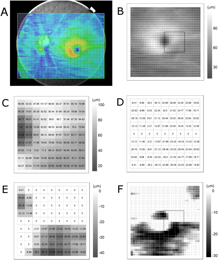
Methods: Three-dimensional scans were acquired from 70 normal subjects and 62 open angle glaucoma patients by swept-source OCT. The thickness of the retinal nerve fiber layer, ganglion cell-inner plexiform layer (GCIPL), ganglion cell complex, and total retina were calculated within a 6.2×6.2 mm macular area divided into a 31×31 grid of 200×200 μm superpixels. For each of the corresponding superpixels, the thickness differences between the subject eyes and contra-lateral eyes and between the upper and lower macula halves of the subject eyes were determined. The negative differences were displayed on a gray-scale asymmetry map. Black superpixels were defined as thickness decreases over the cut-off values.

Results: The negative inter-ocular and inter-hemisphere differences in GCIPL thickness (mean ± standard deviation) were -2.78 ± 0.97 μm and -3.43 ± 0.71 μm in the normal group and -4.26 ± 2.23 μm and -4.88 ± 1.46 μm in the glaucoma group. The overall extent of the four layers' thickness decrease was larger in the glaucoma group than in the normal group (all Ps<0.05). The numbers of black superpixels on all of the asymmetry maps were larger in the glaucoma group than in the normal group (all Ps<0.05). The area under receiver operating characteristic curves of average negative thickness differences in macular inner layers for glaucoma diagnosis ranged from 0.748 to 0.894.

Conclusions: The asymmetry analysis of macular inner retinal layers showed significant differences between the normal and glaucoma groups. The diagnostic performance of the asymmetry analysis was comparable to that of previous methods. These findings suggest that the asymmetry analysis can be a potential ancillary diagnostic tool.


Conflict of interest statement

The authors have declared that no competing interests exist.

Figures

Fig 1
Fig 1. Methods of data processing in the inter-hemisphere asymmetry analysis in present study.
(A) Ganglion cell-inner plexiform layer (GCIPL) thickness map drawn from three-dimensional scans of 12 mm width using swept-source optical coherence tomography. (B) GCIPL thickness map of 6.2 × 6.2 mm macular area divided into a 31 × 31 grid of 200 × 200 μm squares. The black square of 1.8 × 1.8 mm area near the foveal pit was marked as an example for detailed description of the calculation process. (C) GCIPL thickness map in the sample square is shown. (D) Map of inter-hemispheric differences. The thickness difference was calculated for each of the corresponding superpixels across the horizontal reference line drawn through foveal pit center. (E) Only negative values, representing relative decreases compared with corresponding superpixels, were selected for analysis, and positive values were excluded. The negative differences were displayed on a gray-scale asymmetry map. (F) Inter-hemispheric GCIPL asymmetry map of 6.2 × 6.2 mm macular area.
Fig 2
Fig 2. A representative case of glaucoma patient.
A 45-year-old woman was evaluated for possible open angle glaucoma. Best corrected visual acuities were 20/20 in both eyes and intraocular pressures were 15 mmHg in both eyes. (A,B) Retinal nerve fiber layer (RNFL) photography indicates inferotemporal RNFL thinning in the left eye. (C,D,E,F) Optic disc of the left eye shows signs of inferotemporal disc notching. The results of Humphrey visual field testing were normal in the right eye and superior defect in the left eye. The mean deviation was 0.69 dB in the right eye and -3.80 dB in the left eye. The visual field index was 100% and 84% for the right and left eye, respectively. (G) Asymmetry maps from inter-ocular and inter-hemispheric asymmetry analyses showing a cluster of black superpixels corresponding with inferotemporal RNFL defect in left eye. The inter-hemispheric and inter-ocular asymmetry thickness maps of RNFL, ganglion cell-inner plexiform layer (GCIPL), ganglion cell complex and total retina were arrayed in a 4 × 4 matrix form. The maps of the first and fourth columns were constructed from inter-hemispheric asymmetry analyses of the right and left eye, respectively. The maps of the second and third columns were obtained from inter-ocular asymmetry analyses of the right and left eye, respectively. The rows of the maps are arrayed in the order RNFL, GCIPL, ganglion cell complex, total retina.
Fig 3
Fig 3. A representative case of unilateral glaucoma patient with false positive findings on conventional deviation map.
A 58-year-old man was evaluated for open angle glaucoma. Best corrected visual acuities were 20/20 in both eyes and intraocular pressures were 18 mmHg in the right eye and 16 mmHg in the left eye. The refractive errors were -5.50 diopters in both eyes. (A,B) Retinal nerve fiber layer (RNFL) photography presents inferotemporal RNFL thinning in right eye. (C) Topographic profile of peripapillary RNFL thickness measured by Cirrus OCT presented temporal deviation of peaks of RNFL thicknesses. (D) The results of Humphrey visual-field testing were superior defect in right eye and normal in left eye. (E) The peripapillary RNFL thickness deviation map of Cirrus optical coherence tomography (OCT) showed abnormal color-coded area consistent with superotemporal and inferotemporal RNFL thinning in both eyes. (F) The ganglion cell analysis deviation map of Cirrus OCT also showed abnormal color-coded area in the inferior macular area of the right eye and in the superior and inferior macular area of the left eye. The abnormal color-coded areas of the left eye were considered as false-positive because of normal findings on RNFL photography and visual-field testing. (G) Inter-hemispheric and inter-ocular asymmetry thickness maps of RNFL, ganglion cell-inner plexiform layer, ganglion cell complex and total retina were arrayed in 4 × 4 matrix form in same order as Fig 2G. The asymmetry maps from the inter-ocular and inter-hemispheric asymmetry analyses showed a cluster of black superpixels corresponding to an inferotemporal RNFL defect in the right eye. However, in the left eye, the number of black superpixels was small and the map showed only a scattered distribution of black superpixels. The asymmetry maps suggest that this case has significant structural changes only in the right eye, which corresponded well with the results for RNFL photography and visual-field testing.
Fig 4
Fig 4. Receiver operating characteristic curves of six parameters for glaucoma diagnosis.
The parameters are the inter-hemispheric negative difference of retinal nerve fiber layer (RNFL) and ganglion cell-inner plexiform layer (GCIPL) thicknesses, inter-ocular negative difference of RNFL and GCIPL thicknesses, and average RNFL and GCIPL thicknesses. The average RNFL and GCIPL thicknesses were measured using Cirrus optical coherence tomography.

References

    1. Essock EA, Sinai MJ, Fechtner RD. Interocular symmetry in nerve fiber layer thickness of normal eyes as determined by polarimetry. J Glaucoma. 1999;8:90–8. - PubMed
    1. Park JJ, Oh DR, Hong SP, Lee KW. Asymmetry analysis of the retinal nerve fiber layer thickness in normal eyes using optical coherence tomography. Korean J Ophthalmol. 2005;19:281–7. 10.3341/kjo.2005.19.4.281 - DOI - PubMed
    1. Budenz DL. Symmetry between the right and left eyes of the normal retinal nerve fiber layer measured with optical coherence tomography (an AOS thesis). Trans Am Ophthalmol Soc. 2008;106:252–75. - PMC - PubMed
    1. Mwanza JC, Durbin MK, Budenz DL. Interocular symmetry in peripapillary retinal nerve fiber layer thickness measured with the cirrus HD-OCT in healthy eyes. Am J Ophthalmol. 2011;151:514–21.e1. 10.1016/j.ajo.2010.09.015 - DOI - PMC - PubMed
    1. Dalgliesh JD, Tariq YM, Burlutsky G, Mitchell P. Symmetry of retinal parameters measured by spectral-domain OCT in normal young adults. J Glaucoma. 2015;24:20–4. 10.1097/IJG.0b013e318287ac2f - DOI - PubMed

Publication types