. 2016 Oct 20;11(10):e0164987.
doi: 10.1371/journal.pone.0164987. eCollection 2016.

Characterization of In-Body to On-Body Wireless Radio Frequency Link for Upper Limb Prostheses

Affiliations

Characterization of In-Body to On-Body Wireless Radio Frequency Link for Upper Limb Prostheses

Antonietta Stango et al. PLoS One. .

Abstract

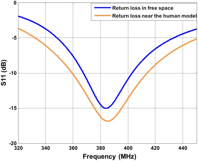
Wireless implanted devices can be used to interface patients with disabilities with the aim of restoring impaired motor functions. Implanted devices that record and transmit electromyographic (EMG) signals have been applied for the control of active prostheses. This simulation study investigates the propagation losses and the absorption rate of a wireless radio frequency link for in-to-on body communication in the medical implant communication service (MICS) frequency band to control myoelectric upper limb prostheses. The implanted antenna is selected and a suitable external antenna is designed. The characterization of both antennas is done by numerical simulations. A heterogeneous 3D body model and a 3D electromagnetic solver have been used to model the path loss and to characterize the specific absorption rate (SAR). The path loss parameters were extracted and the SAR was characterized, verifying the compliance with the guideline limits. The path loss model has been also used for a preliminary link budget analysis to determine the feasibility of such system compliant with the IEEE 802.15.6 standard. The resulting link margin of 11 dB confirms the feasibility of the system proposed.


Conflict of interest statement

The authors have declared that no competing interests exist.

Figures

Fig 1
Fig 1. System overview.
Fig 2
Fig 2. MICS implanted antenna and antenna positioning.
a) front view of the implanted antenna; b) human model with the implanted antennas in the red squares and the external dipole; c) cross section of the arm with the implanted antennas.
Fig 3
Fig 3. Return Loss of the implanted antenna.
S11 is about -10 dB at 403.5 MHz.
Fig 4
Fig 4. 3D Gain polar and radiation pattern plots of the implanted antenna.
a) 3 D gain polar plot, maximum gain equal to -55.37 dBi; b) 3D normalized radiation pattern polar plot; c) values of Eφ (dB) and Eθ (dB) in three different planes. The coordinate system is shown in figure. Maximum value -41.04 dB.
Fig 5
Fig 5. Return Loss of the dipole antenna.
S11 is ~-12 dB at 403.5 MHz near the human body.
Fig 6
Fig 6. 3D Gain polar and radiation pattern plots of the dipole antenna.
a) 3D gain polar plot, maximum gain equal to -3.20 dBi; b) 3D normalized radiation pattern plot; c) Values of Eφ (dB) and Eθ (dB) in three different planes. The coordinate system is shown in figure. Maximum value 11.35 dB.
Fig 7
Fig 7. Helical antenna.
a) helical antenna dimensions. b) position of the helical antenna near the human body.
Fig 8
Fig 8. Return loss of the helical antenna.
S11 is ~-11 dB at 403.5 MHz near the human body (orange curve).
Fig 9
Fig 9. 3D Gain polar and radiation pattern plots of the helical antenna.
a) 3D gain polar plot, maximum gain equal to -7.06 dBi; b) 3D normalized radiation pattern polar plot; c) Values of Eφ (dB) and Eθ (dB) in three different planes. The coordinate system is shown in figure. Maximum value 7.34 dB.
Fig 10
Fig 10. Path loss.
Path loss values as function of the distance between the implanted antenna and the external antenna and representation of the fitted model.
Fig 11
Fig 11. psaSAR values with the half wave dipole.
a) psaSAR related to the external half-wave dipole positioned in front of the arm (scenario 1); b) psaSAR related to implant 1 (scenario 1); c) psaSAR related to implant 2 (scenario1); d) psaSAR related to the external half-wave dipole positioned on the back of the arm (scenario2); e) psaSAR related to implant 1(scenario2); f) psaSAR related to implant 2 (scenario2).
Fig 12
Fig 12. psaSAR values with the helical dipole.
a) psaSAR related to the external helical dipole; b) psaSAR related to implant 1.

References

    1. Movassaghi S, Member S, Abolhasan M, Member S. Wireless Body Area Networks : A Survey. IEEE Commun Surv Tutorials. 2014;16(3):1658–86.
    1. Bazaka K, Jacob M V. Implantable Devices: Issues and Challenges. Electronics. 2012;2(1):1–34.
    1. Hall PS and Hao Y. Antennas and Propagation for Body-Centric Wireless Communications. Boston, MA: Artech House; 2006.
    1. Farrell TR, Weir RF. A comparison of the effects of electrode implantation and targeting on pattern classification accuracy for prosthesis control. IEEE Trans Biomed Eng. 2008. September;55(9):2198–211 10.1109/TBME.2008.923917 - DOI - PMC - PubMed
    1. Weir RF, Troyk PR, DeMichele GA, Kerns DA, Schorsch JF, Maas H. Implantable myoelectric sensors (IMESs) for intramuscular electromyogram recording. IEEE Trans Biomed Eng. 2009;56(1):159–71. 10.1109/TBME.2008.2005942 - DOI - PMC - PubMed