. 2016 Dec;15(23):3251-3267.
doi: 10.1080/15384101.2016.1242534. Epub 2016 Oct 20.

Leucine Zipper Down-regulated in Cancer-1 (LDOC1) interacts with Guanine nucleotide binding protein-like 3-like (GNL3L) to modulate Nuclear Factor-kappa B (NF-κB) signaling during cell proliferation

Affiliations

Leucine Zipper Down-regulated in Cancer-1 (LDOC1) interacts with Guanine nucleotide binding protein-like 3-like (GNL3L) to modulate Nuclear Factor-kappa B (NF-κB) signaling during cell proliferation

Indu Jose Thoompumkal et al. Cell Cycle. 2016 Dec.

Abstract

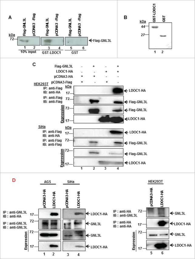
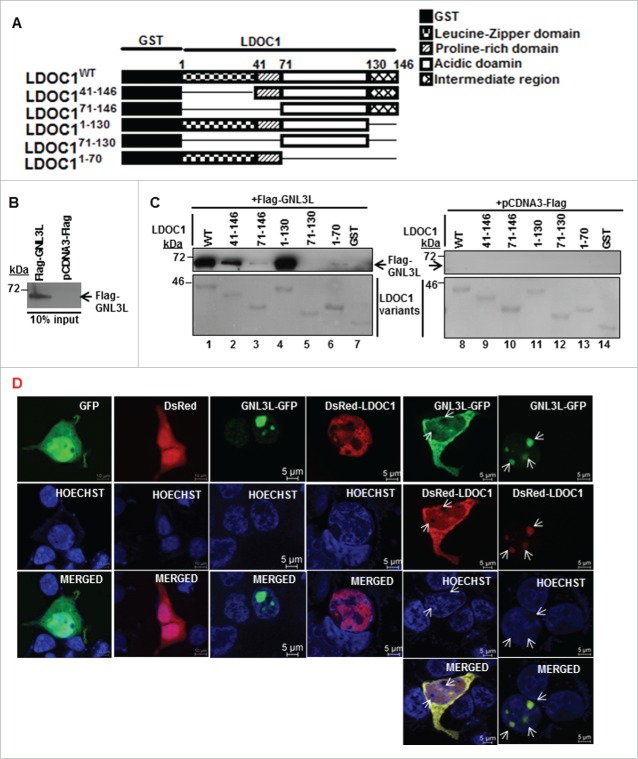
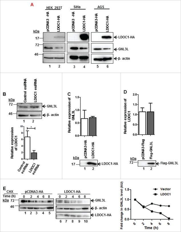
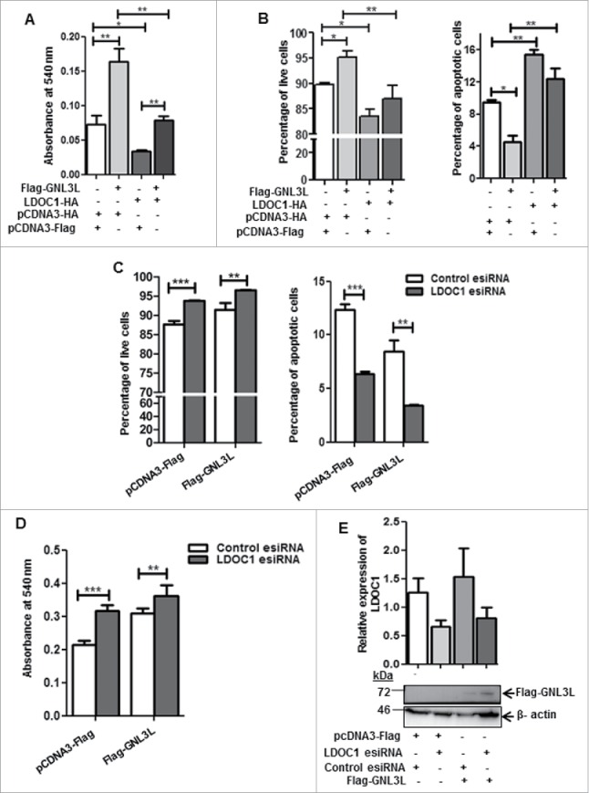
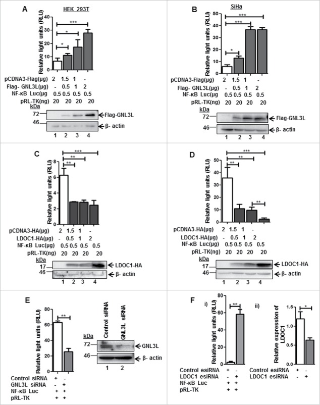
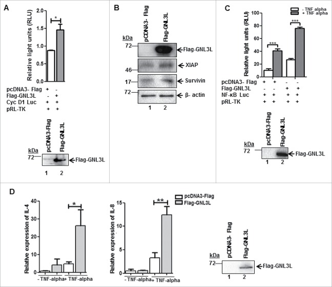
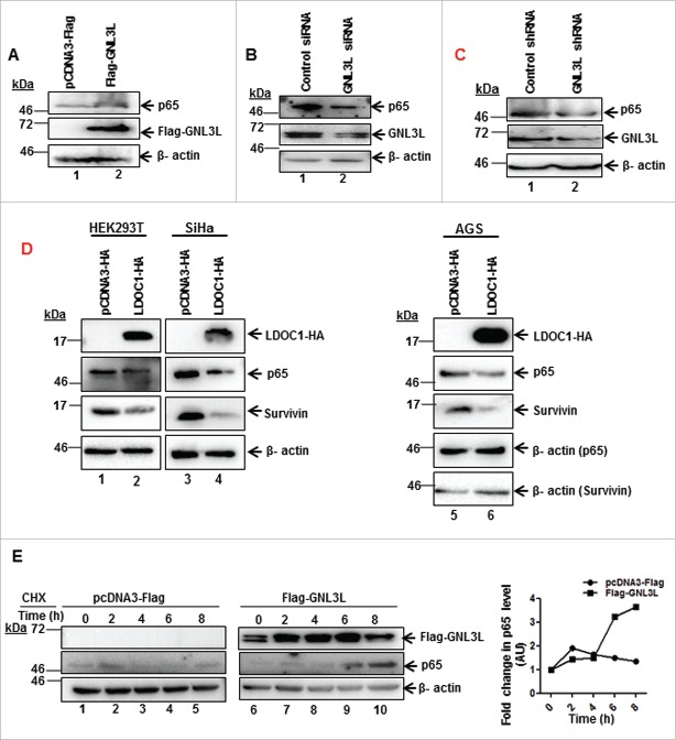
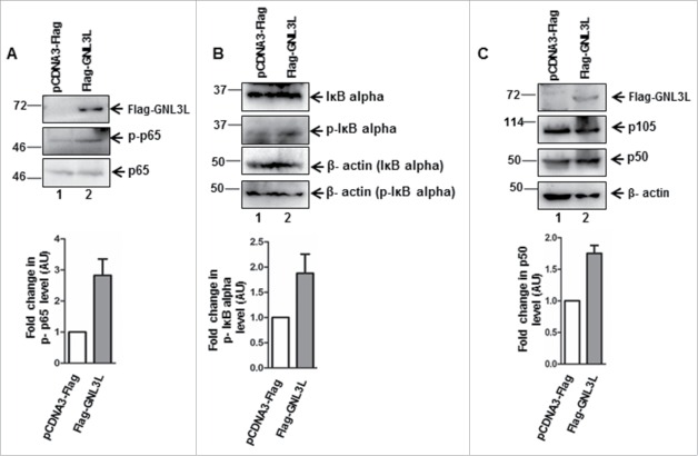
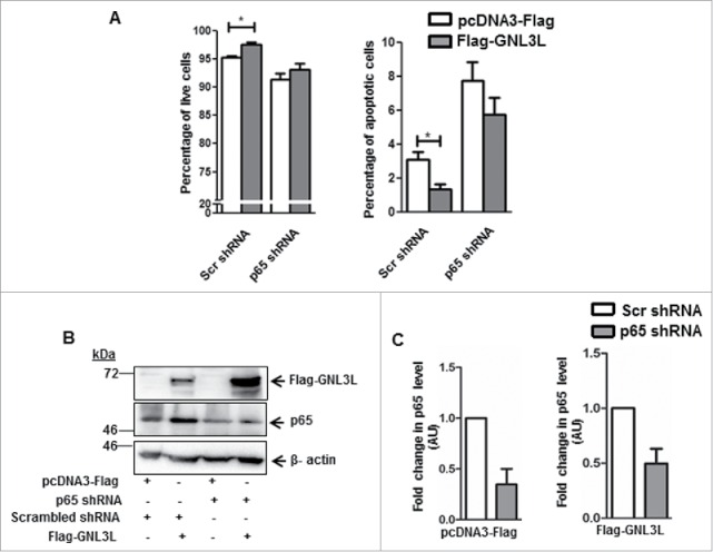
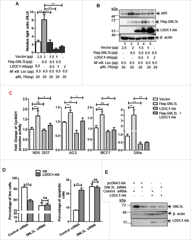
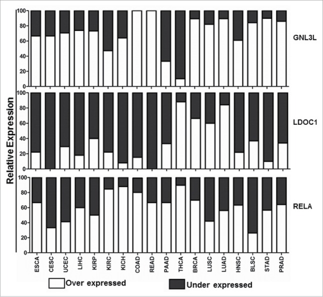
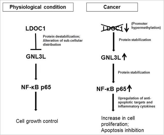
Guanine nucleotide binding protein-like 3-like (GNL3L) is an evolutionarily conserved putative nucleolar GTPase belonging to the HSR1-MMR1 family. In the present study, using protein-protein interaction assays, we show that Leucine Zipper Down-regulated in Cancer-1 (LDOC1) is a novel interacting partner of GNL3L. Furthermore, our results reveal that ectopic expression of LDOC1 destabilizes endogenous GNL3L levels and down modulates GNL3L-induced cell proliferation, in contrast, the knockdown of LDOC1 potentiates cell proliferation upon GNL3L expression. Interestingly, GNL3L upregulates NF-κB dependent transcriptional activity by modulating the expression of NF-κB subunit p65, which is reversed upon co-expression of LDOC1 with GNL3L. GNL3L also potentiates TNF-α mediated NF-κB activity. In addition, anti-apoptotic function of GNL3L is impaired upon p65 knockdown, suggesting its critical role in GNL3L mediated cell proliferation/survival. An inverse correlation of GNL3L and LDOC1 expression profiles in various tumor tissues from BioXpress database indicate their critical role in cancer. Collectively, our data provides evidence that GNL3L-LDOC1 interplay regulates cell proliferation through the modulation of NF-κB pathway during tumorigenesis.

Keywords: Apoptosis; GNL3L; LDOC1; NF-κB transcription factor; cell signaling; proliferation; protein-protein interaction.


Figures

Figure 1.
Figure 1.
GNL3L interacts with LDOC1. HEK293T cells were transfected with Flag-GNL3L or the control vector using PEI. After 48 hrs of transfection, cell lysates were prepared and GST pull-down assay (A) was performed with GST-LDOC1 followed by western blot analysis using anti-Flag antibody. GST was used as negative control. The expression of Flag-GNL3L was confirmed by western blot analysis using anti-Flag antibody. (B) The expression of GST-LDOC1 and GST was confirmed using Coomassie blue staining. (C) HEK293T and SiHa cell lysates expressing Flag-GNL3L and LDOC1-HA were subjected to co-immunoprecipitation using anti-Flag antibody. Complexes were eluted and separated on SDS-12%PAGE followed by western blot analysis with anti-HA antibody. The efficiency of immunoprecipitation was confirmed by western blot analysis using anti-Flag antibody. (D) HEK293T cell lysates expressing LDOC1-HA were subjected to co-immunoprecipitation using anti-HA antibody whereas SiHa or AGS cells expressing LDOC1-HA were co-immunoprecipitated with anti-GNL3L antibody. Complexes were eluted and separated on SDS-12%PAGE followed by western blot analysis with anti-GNL3L or anti-HA antibodies.
Figure 2.
Figure 2.
Identification of GNL3L interaction domain in LDOC1. (A) Schematic representation of wild type and deletion mutants of LDOC1. (B) HEK293T cells were transfected with plasmid encoding Flag-GNL3L or control vector and the expression of GNL3L was confirmed by western blot analysis using anti-Flag antibody. (C) HEK293T cells lysates containing Flag-GNL3L were used in a GST pull-down assay with wild type or indicated variants of GST-LDOC1. GST served as the negative control. Specific interaction between Flag-GNL3L and LDOC1proteins was confirmed by western blot analysis using anti-Flag antibody. Ponceau-S staining indicated the integrity and purity of LDOC1 variants. (D) SiHa cells were transfected with GNL3L-GFP, DsRed-LDOC1 or indicated control vectors and their sub-cellular localization was analyzed using confocal microscopy. The arrow marks represent nucleoli.
Figure 3.
Figure 3.
LDOC1 downregulates GNL3L. (A) HEK293T or SiHa or AGS cells were transfected with LDOC1-HA or the corresponding control vector. After 48 hrs of transfection, the expression levels of ectopically expressed LDOC1 and endogenous GNL3L were determined by western blot analysis using anti-HA and anti-GNL3L antibodies, respectively. (B) HEK293T cells were transfected with 80 pM of LDOC1 esiRNA or the universal negative control esiRNA and endogenous GNL3L expression was assessed by western blot analysis using anti-GNL3L antibody. Knockdown of LDOC1 was confirmed by RT-qPCR analysis using specific primers. Total RNA isolated from HEK293T cells transfected with LDOC1-HA (C) or Flag-GNL3L (D) was reverse-transcribed and RT-qPCR was carried out using specific primers to analyze the endogenous mRNA expression of GNL3L and LDOC1, respectively. (E) HEK293T cells transfected with LDOC1-HA or the control vector were treated with cycloheximide (CHX) for indicated time periods. The expression of endogenous GNL3L was determined by western blot using anti-GNL3L antibody. Beta-actin levels served as the internal control. Densitometry analysis of a representative western blot depicting the endogenous GNL3L levels at various time points.
Figure 4.
Figure 4.
LDOC1 inhibits GNL3L function. (A) MTT and (B) Annexin-V binding assays were carried out after 48 hrs of transfection of HEK293T cells with Flag-GNL3L, LDOC1-HA or corresponding control vectors. HEK293T cells were transfected with Flag-GNL3L or control vector along with LDOC1 esiRNA or control esiRNA. After 48 hrs of transfection, (C) Annexin-V binding as well as (D) MTT assays were performed. The knockdown of LDOC1 was confirmed by RT-qPCR analysis using specific primers. (E) The expression of Flag-GNL3L was analyzed by western blot analysis using anti-Flag antibody.
Figure 5.
Figure 5.
LDOC1 and GNL3L exhibit opposing effects on NF-κB dependent transcriptional activity. GNL3L upregulates NF-κB dependent transcriptional activity. (A) HEK293T and (B) SiHa cells were co-transfected with Flag-GNL3L or control vector along with pGL4.32[luc2P/NF-κB-RE/Hygro] and pRL-TK-Renilla luciferase (internal control) plasmids. After 48 hrs of transfection, luciferase assay was performed to check the NF-κB dependent transcriptional activity. LDOC1 downregulates NF-κB dependent transcriptional activity. (C) HEK293T and (D) SiHa cells were co-transfected with LDOC1-HA or control vector along with pGL4.32[luc2P/NF-κb-RE/Hygro] and pRL-TK-Renilla luciferase (internal control) plasmids. After 48 hrs of transfection, luciferase assay was performed to check basal level of NF-κB dependent transcriptional activity. The expression of Flag-GNL3L and LDOC1-HA was confirmed by western blot analysis using anti-Flag and anti-HA antibodies, respectively. Beta-actin was used as internal control. (E) HEK293T cells were transfected with appropriate amount of GNL3L siRNA or control siRNA along with pGL4.32[luc2P/NF-κB-RE/Hygro] and pRL-TK-Renilla luciferase plasmids. Luciferase assay was performed after 48 hrs of transfection to analyze NF-κB dependent transcriptional activity. (F-i) HEK293T cells were transfected with LDOC1 esiRNA or control esiRNA along with pGL4.32[luc2P/NF-κB-RE/Hygro] and pRL-TK-Renilla luciferase plasmids. Luciferase assay was performed after 48 hrs of transfection to analyze NF-κB dependent transcriptional activity. (F-ii) Knockdown of LDOC1 was analyzed by RT-qPCR using specific primers.
Figure 6.
Figure 6.
GNL3L alters the expression of NF-κB target genes. (A) HEK293T cells were co-transfected with Flag-GNL3L or control vector along with cyclin D1-promoter luciferase construct and pRL-TK-Renilla luciferase plasmids. After 48 hrs of transfection, luciferase assay was performed to check the cyclin D1 promoter activity. (B) GNL3L upregulates anti-apoptotic NF-κB target proteins. SiHa cells were transfected with Flag-GNL3L or control vector and the expression levels of endogenous XIAP and Survivin were analyzed using specific antibodies. Beta-actin was used as the loading control. (C) HEK293T cells were co-transfected with Flag-GNL3L or control vector along with pGL4.32[luc2P/NF-κB-RE/Hygro] and pRL-TK-Renilla luciferase plasmids. After 42 hrs of transfection, TNF-α was added (20 ng/mL final concentration) and luciferase assay was performed at the end of 48 hrs to check the NF-κB dependent transcriptional activity. (D) The mRNA expression levels of IL-4 and IL-8 were analyzed post TNF-α treatment by RT-qPCR in HEK293T cells transfected with Flag-GNL3L or control vector.
Figure 7.
Figure 7.
GNL3L modulates the expression and stability of NF-κB subunit p65. GNL3L upregulates p65 protein levels. (A) HEK293T cells were transfected with Flag-GNL3L or control vector and the expression levels of endogenous p65 and ectopically expressed Flag-GNL3L were confirmed by western blot analysis using anti-p65 and anti-Flag antibodies, respectively. GNL3L specific siRNA (B) or shRNA (C) or control siRNA or shRNA were transfected into HEK293T cells and the expression levels of endogenous p65 and GNL3L were confirmed by western blot analysis using anti-p65 and anti-GNL3L antibodies, respectively. (D) LDOC1 downregulates p65 protein levels. HEK293T or SiHa or AGS cells were transfected with LDOC1-HA or control vector and the expression levels of endogenous p65, survivin and ectopically expressed LDOC1-HA were confirmed by western blot analysis using anti-p65, anti-survivin, and anti-HA antibodies, respectively. (E) HEK293T cells transfected with Flag-GNL3L or the control vector were treated with cycloheximide (CHX) for different time periods. The expression of Flag-GNL3L and endogenous p65 was determined by western blot using anti-Flag and anti-p65 antibodies, respectively. Densitometry analysis of a representative western blot shows the endogenous p65 levels at different time points.
Figure 8.
Figure 8.
GNL3L modulates p65 phosphorylation levels. HEK293T cells were transfected with Flag-GNL3L or control vector and the expression levels of Flag-GNL3L, endogenous p65, and phospho-p65 (A); IκB-α (B) and phospho-IκB-α and p105 and p50 (C) were analyzed by western blot using appropriate antibodies. Quantitative measurements of respective western blots by densitometry analysis are provided below.
Figure 9.
Figure 9.
Anti-apoptotic function of GNL3L is p65-dependent. (A) Annexin-V binding assay was performed in HEK293T cells co-transfected with Flag-GNL3L or vector along with an equal concentration of p65 specific or scrambled shRNA constructs. Scr: scrambled. (B) Expression of Flag-GNL3L as well as the extent of p65 knockdown was analyzed by western blot analysis using anti-Flag and anti-p65 antibodies, respectively. Beta-actin was used as the loading control in all these experiments. (C) Densitometry analysis of the western blot was performed to quantitate the extent of p65 knockdown.
Figure 10.
Figure 10.
LDOC1 downregulates GNL3L-mediated NF-κB dependent transcriptional activity by modulating p65 expression. (A) HEK293T cells were co-transfected with Flag-GNL3L and increasing amounts of LDOC1-HA along with pGL4.32[luc2P/NF-κB-RE/Hygro] and pRL-TK-Renilla luciferase (internal control) plasmids. The NF-κB dependent transcriptional activity was analyzed by luciferase assay after 48 hrs of transfection. (B) The expression levels of Flag-GNL3L, LDOC1-HA and endogenous p65 were assessed by western blot analysis using specific antibodies. LDOC1 mediated inhibition of apoptosis is GNL3L-dependent. (C) HEK293T or AGS or MCF-7 or SiHa cells were co-transfected with Flag-GNL3L or LDOC1-HA in combination with cyclin D1-promoter luciferase and pRL-TK-Renilla luciferase plasmids. Luciferase assay was performed at the end of 48 hours of transfection to assess the cyclin D1 promoter dependent transcriptional activity. (D) Annexin-V binding assay was performed in HEK293T cells co-transfected with LDOC1-HA or vector along with GNL3L siRNA or control siRNA. (E) The expression of LDOC1-HA and the level of GNL3L knockdown was analyzed by western blot analysis using anti-HA and anti-GNL3L antibodies, respectively.
Figure 11.
Figure 11.
Expression profile of GNL3L, LDOC1 and RELA in different cancers. Gene expression analysis of GNL3L, LDOC1 and RELA were carried out from data set of 18 cancers and respective normal tissue expression profiles available in BioXpress database (https://hive.biochemistry.gwu.edu/tools/bioxpress/).
Figure 12.
Figure 12.
Functional significance of LDOC1-GNL3L interaction during cell proliferation. Under normal physiological conditions, LDOC1 exerts an inhibitory control over GNL3L. LDOC1 alters GNL3L protein stability and subcellular localization to modulate NF-κB signaling to balance the cell growth and apoptosis. In cancerous conditions, GNL3L is relieved from the inhibitory action of LDOC1 possibly due to its promoter hyper methylation. This results in up-regulation of NF-κB dependent transcriptional activity by GNL3L through p65 protein stabilization. The upregulation of NF-κB specific targets may resulted in increased cell proliferation and inhibition of apoptosis.

Similar articles

Cited by

References

    1. Subba Rao MRK, Kumari G, Balasundaram D, Sankaranarayanan R, Mahalingam SA. Novel lysine-rich domain and gtp binding motifs regulate the nucleolar retention of human guanine nucleotide binding protein, GNL3L. J Mol Biol 2006; 364:637-54; PMID:17034816; http://dx.doi.org/10.1016/j.jmb.2006.09.007 - DOI - PubMed
    1. Meng L, Zhu Q, Tsai R. Nucleolar trafficking of nucleostemin family proteins: common versus protein-specific mechanisms. Mol Cell Biol 2007; 27:8670-82; PMID:17923687; http://dx.doi.org/10.1128/MCB.00635-07 - DOI - PMC - PubMed
    1. Thoompumkal IJ, Subba Rao MRK, Kumaraswamy A, Krishnan R, Mahalingam S. GNL3L is a nucleo-cytoplasmic shuttling protein-Role in cell cycle regulation. PLoS One 2015; 10(8):e0135845. - PMC - PubMed
    1. Meng L, Hsu JK, Tsai RYL. GNL3L depletion destabilizes MDM2 and induces p53-dependent G2/M arrest. Oncogene 2010; 30:1716-26; PMID:21132010; http://dx.doi.org/10.1038/onc.2010.550 - DOI - PMC - PubMed
    1. Yasumoto H, Meng L, Lin T, Zhu Q, Tsai R. GNL3L inhibits activity of estrogen-related receptor γ by competing for co-activator binding. J Cell Sci 2007; 120:2532-43; PMID:17623774; http://dx.doi.org/10.1242/jcs.009878 - DOI - PMC - PubMed

MeSH terms

LinkOut - more resources