. 2016 Nov 29;7(48):79372-79387.
doi: 10.18632/oncotarget.12676.

A combined gene expression and functional study reveals the crosstalk between N-Myc and differentiation-inducing microRNAs in neuroblastoma cells

Affiliations

A combined gene expression and functional study reveals the crosstalk between N-Myc and differentiation-inducing microRNAs in neuroblastoma cells

Zhenze Zhao et al. Oncotarget. .

Abstract

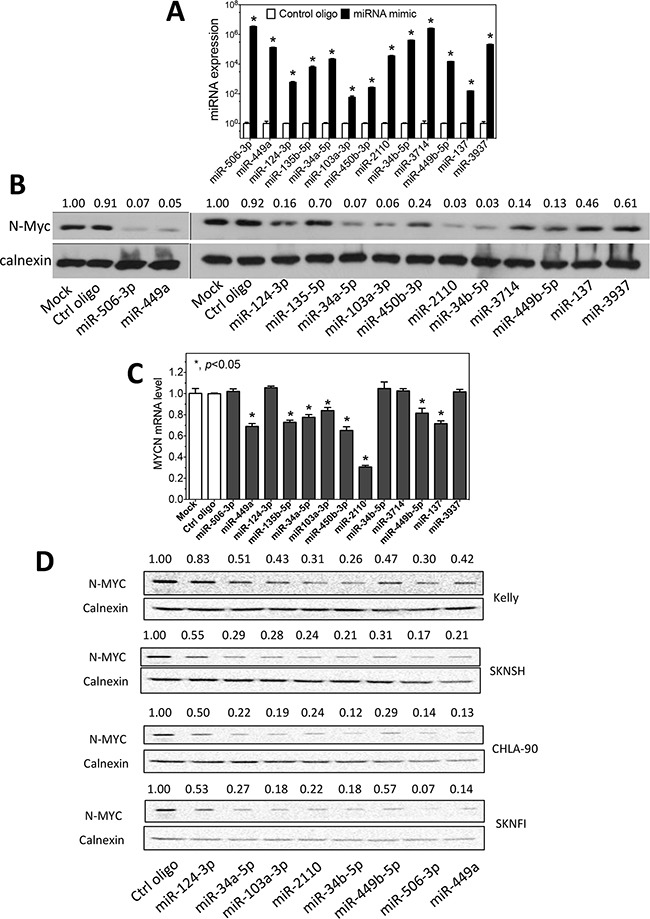
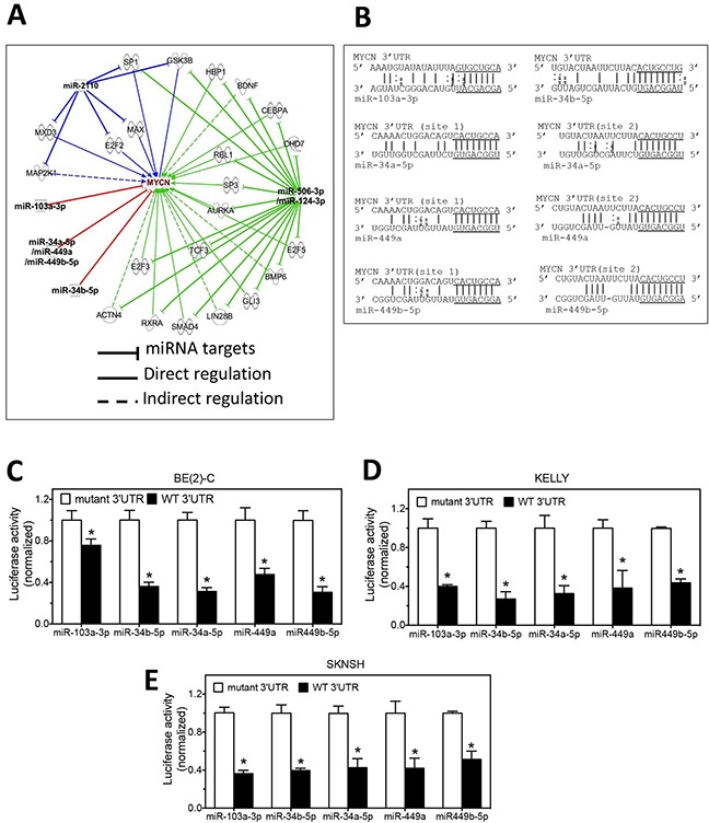
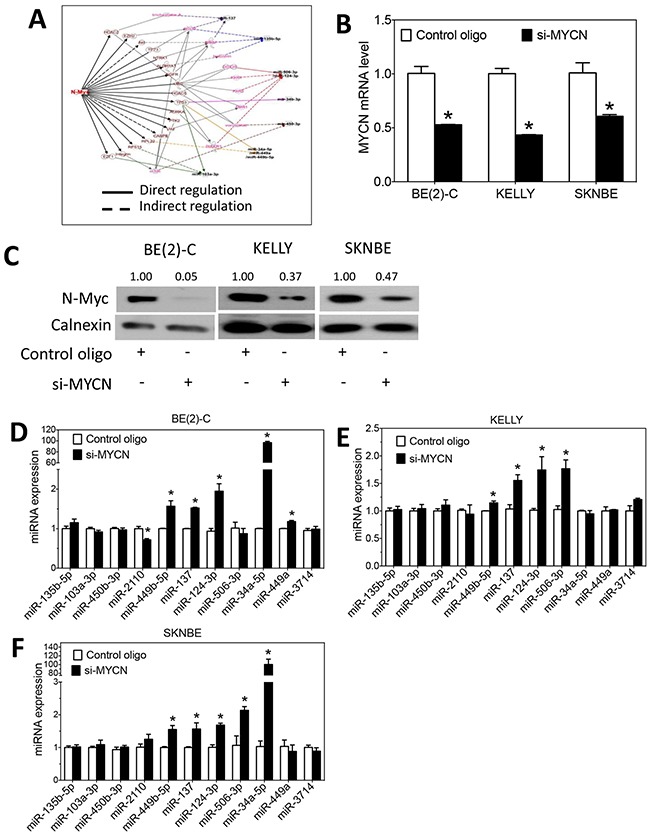
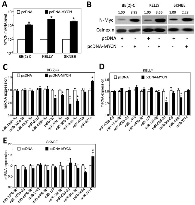
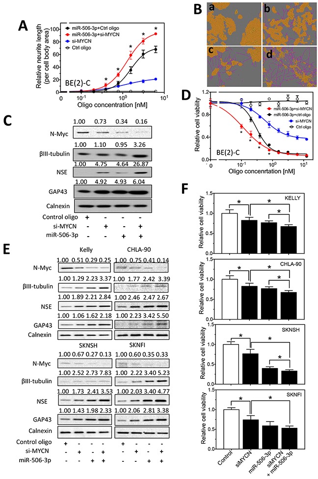
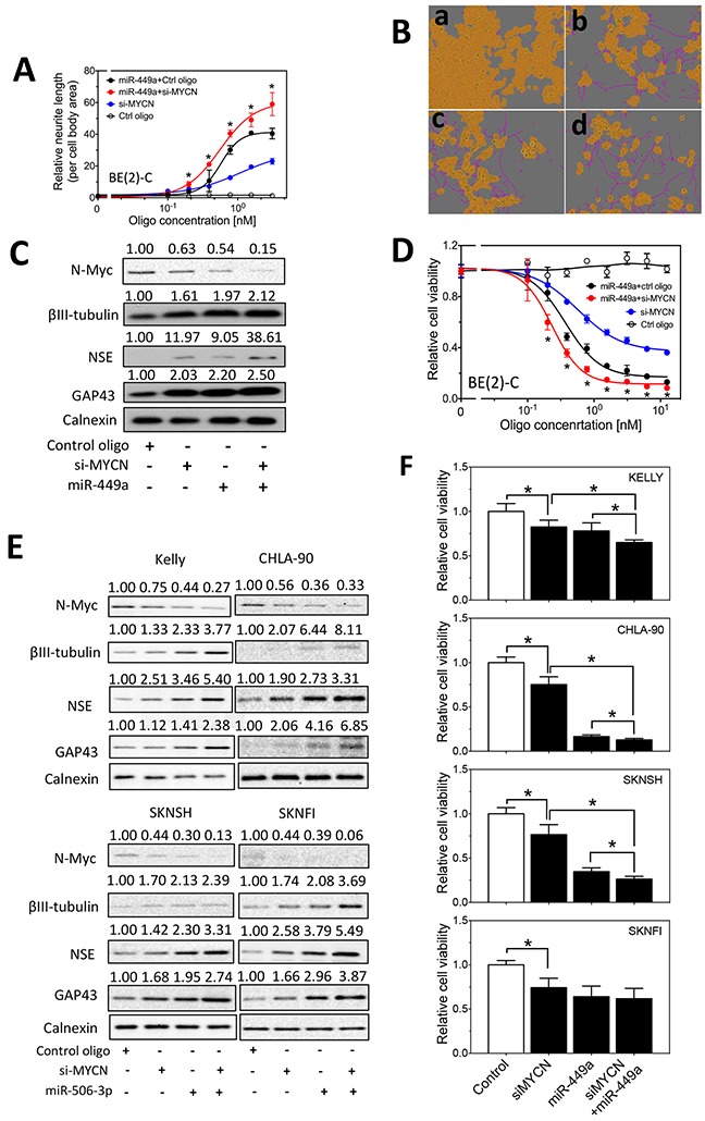
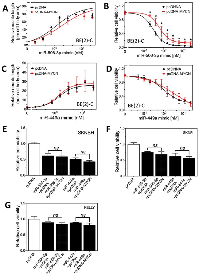
MYCN amplification is the most common genetic alteration in neuroblastoma and plays a critical role in neuroblastoma tumorigenesis. MYCN regulates neuroblastoma cell differentiation, which is one of the mechanisms underlying its oncogenic function. We recently identified a group of differentiation-inducing microRNAs. Given the demonstrated inter-regulation between MYCN and microRNAs, we speculated that MYCN and the differentiation-inducing microRNAs might form an interaction network to control the differentiation of neuroblastoma cells. In this study, we found that eight of the thirteen differentiation-inducing microRNAs, miR-506-3p, miR-124-3p, miR-449a, miR-34a-5p, miR-449b-5p, miR-103a-3p, miR-2110 and miR-34b-5p, inhibit N-Myc expression by either directly targeting the MYCN 3'UTR or through indirect regulations. Further investigation showed that both MYCN-dependent and MYCN-independent pathways play roles in mediating the differentiation-inducing function of miR-506-3p and miR-449a, two microRNAs that dramatically down-regulate MYCN expression. On the other hand, we found that N-Myc inhibits the expression of multiple differentiation-inducing microRNAs, suggesting that these miRNAs play a role in mediating the function of MYCN. In examining the published dataset collected from clinical neuroblastoma specimens, we found that expressions of two miRNAs, miR-137 and miR-2110, were significantly anti-correlated with MYCN mRNA levels, suggesting their interactions with MYCN play a clinically-relevant role in maintaining the MYCN and miRNA expression levels in neuroblastoma. Our findings altogether suggest that MYCN and differentiation-inducing miRNAs form an interaction network that play an important role in neuroblastoma tumorigenesis through regulating cell differentiation.

Keywords: MYCN; differentiation; microRNA; neuroblastoma.


Conflict of interest statement

COMPETING INTERESTS

The authors declare that they have no competing interests.

Figures

Figure 1
Figure 1. Regulation of N-myc expression by differentiation-inducing miRNAs
A. Overexpression of miRNAs by the corresponding miRNA mimics in BE(2)-C cells. Cells were transfected with 25 nM mimics or control oligos. After two days, RNA was collected and miRNA levels were measured by qRT-PCR. Shown are the relative expression levels of miRNAs in cells transfected with the corresponding miRNA mimics normalized to those in cells transfected with control oligos. B. Effect of miRNA overexpression on N-Myc protein expression in BE(2)-C. N-Myc protein levels were measured by Western blots with calnexin as a loading control. Shown above the blots are the relative intensities of the corresponding bands. C. Effect of the miRNA overexpression on MYCN mRNA expression. MYCN expression was quantified using qRT-PCR and normalized with GAPDH mRNA as a loading control. Shown are fold changes of MYCN mRNA levels induced by miRNA mimics relative to mock transfection. *, p<0.05. D. Effect of miRNA overexpression on N-Myc protein expression in KELLY, SKNSH, CHLA-90 and SKNFI cells. N-Myc protein levels were measured as above.
Figure 2
Figure 2. miRNAs regulate MYCN expression through direct and indirect pathways
A. IPA analysis identifies the potential mechanisms by which the differentiation-inducing miRNAs regulate MYCN expression. B. The predicted interactions of miRNAs with the targets sites in the 3′UTR of MYCN mRNA. The seed sequences are underlined. C-E. Validation of the target sites of the miRNAs in the 3′UTRs of MYCN mRNA by luciferase reporter assay in BE(2)-C, KELLY and SKNSH cells. Cells were co-transfected with the luciferase reporter vector expressing either the wildtype 3′UTR of MYCN (WT 3′UTR) or mutated MYCN 3′UTR (mutant 3′UTR) and miRNA mimics. After 72 h of transfection, cells were lysed and luciferase activity was measured. Shown are normalized luciferase activities with the luciferase activity associated with the WT 3′UTR normalized to those associated with the corresponding mutant 3′UTRs. *, p<0.05 compared to the mutant 3′UTR.
Figure 3
Figure 3. N-Myc regulates the expression of differentiation-inducing miRNAs
A. IPA analysis identifies the potential mechanisms by whichN-Myc regulates the expression of the ten differentiation-inducing miRNAs. B-C. MYCN mRNA levels and N-Myc protein expression as a function of MYCN depletion. Cells were transfected with MYCN siRNA (si-MYCN) or control oligos for three days. (B) RNA was isolated for measuring MYCN mRNA levels as above, and (C) cell lysates were harvested for measuring N-Myc protein levels by Western blots as above. *, p<0.05 compared to control. D-F. Effect of MYCN knockdown on the expression of miRNAs. Cells were transfected with 25 nM of MYCN siRNA or control oligos. After 72 hours, RNA was collected, and miRNA levels were measured by qRT-PCR as above.
Figure 4
Figure 4. Effect of MYCN overexpression on the expression of differentiation-inducing miRNAs
A-B. MYCN mRNA and N-Myc protein expression as a function of MYCN overexpression using the pcDNA-MYCN expression vector. Cells transfected with empty vector (pcDNA) were used as a negative control. Cells were transfected with the above vectors for three days, and (A) RNA was isolated for measuring MYCN mRNA levels as above, and (B) cell lysates were harvested for measuring N-Myc protein levels by Western blots as above. C-E. Effect of MYCN overexpression on the expression of miRNAs. Cells were transfected as above for 3 days, and miRNA levels were measured as above. (*, p<0.05, compared to control)
Figure 5
Figure 5. Effect of MYCN knockdown and miR-506-3p overexpression on cell differentiation and cell survival in neuroblastoma cells
A-B. Effect of MYCN knockdown and miR-506-3p overexpression on neurite outgrowth in BE(2)-C cells. Cells were transfected with different concentrations of the indicated oligos. After 4 days, relative neurite outgrowth was measured. (A) Quantification of neurites showing the dose-dependent effect of MYCN siRNA (si-MYCN) and miR-506-3p mimic on neurite outgrowth. (B) Representative images, with the cell body area (yellow) and neurites (pink) defined, showing the effect of si-MYCN and miR-506-3p mimic on neurite outgrowth. Shown are cell images for (a) control oligo (6 nM), (b) si-MYCN (3 nM), (c) miR-506-3p mimic (3 nM) and (d) si-MYCN (3 nM)+miR-506-3p mimic (3 nM). C. Effect of MYCN knockdown and miR-506-3p overexpression on the expression of N-Myc and differentiation markers in BE(2)-C cells. Cells were transfected with the indicated oligos. After 4 days, cell lysates were collected, and protein levels of N-Myc, as well as protein levels of the differentiation markers, βIII–tubulin, NSE and GAP43 were measured by Western blots with calnexin levels as a loading control. D. Effect of MYCN knockdown and miR-506-3p overexpression on cell viability of BE(2)-C cells. Cells were transfected with different concentrations of the indicated oligos. After 4 days, cell viability were measured as described in the Material and Methods. *, p<0.05 compared cells co-transfected with si-MYCN and miR-506-3p mimic to cells co-transfected with control oligo and miR-506-3p mimic. E. Effect of MYCN knockdown and miR-506-3p overexpression on expression of N-Myc and differentiation markers in KELLY, CHLA-90, SKNSH and SKNFI cells. F. Effect of MYCN knockdown and miR-506-3p overexpression on cell viability in KELLY, CHLA-90, SKNSH and SKNFI cells. *, p<0.05.
Figure 6
Figure 6. Effect of MYCN knockdown and miR-449a overexpression on cell differentiation and cell survival in neuroblastoma cells
A-D. Effect of MYCN knockdown and miR-449a overexpression on neurite outgrowth (A-B), on expression of N-Myc and differentiation markers (C), and on cell viability (D) in BE(2)-C cells as measured as above. Shown in (B) are cell images for (a) control oligo (6 nM), (b) si-MYCN (3 nM), (c) miR-449a mimic (3 nM) and (d) si-MYCN (3 nM)+miR-449a mimic (3 nM). *, p<0.05 compared cells co-transfected with si-MYCN and miR-506-3p mimic to cells co-transfected with control oligo and miR-506-3p mimic. E. Effect of MYCN knockdown and miR-449a overexpression on expression of N-Myc and differentiation markers in KELLY, CHLA-90, SKNSH and SKNFI cells. F. Effect of MYCN knockdown and miR-449a overexpression on cell viability in KELLY, CHLA-90, SKNSH and SKNFI cells. *, p<0.05.
Figure 7
Figure 7. Effect of MYCN overexpression on the differentiation-inducing effect of miR-506-3p and miR-449a in neuroblastoma cells
A-B. Effect of MYCN overexpression on miR-506-3p mimic-induced cell differentiation and cell survival reduction in BE(2)-C cells. Cells expressing pcDNA-MYCN or pcDNA were transfected with different concentrations of miR-506-3p mimic. After 4 days, neurite outgrowth (A) and cell viability (B) were measured as above. C-D. Effect of MYCN overexpression on miR-449a mimic-induced cell differentiation and cell survival reduction in BE(2)-C cells. Cells were transfected with different concentrations of miR-449a mimic, and neurite outgrowth (C) and cell viability (D) were measured as above. E-G. Effect of MYCN overexpression on miR-506-3p mimic- and miR-449a-induced cell survival reduction in SKNSH, SKNFI and Kelly cells. *, p<0.05 compared to the control pcDNA.

References

    1. Brodeur GM, Seeger RC, Schwab M, Varmus HE, Bishop JM. Amplification of N-myc in untreated human neuroblastomas correlates with advanced disease stage. Science. 1984;224:1121–1124. - PubMed
    1. Brodeur GM. Neuroblastoma: biological insights into a clinical enigma. Nature reviews Cancer. 2003;3:203–216. - PubMed
    1. Maris JM. Recent advances in neuroblastoma. N Engl J Med. 2010;362:2202–2211. - PMC - PubMed
    1. Seeger RC, Brodeur GM, Sather H, Dalton A, Siegel SE, Wong KY, Hammond D. Association of multiple copies of the N-myc oncogene with rapid progression of neuroblastomas. N Engl J Med. 1985;313:1111–1116. - PubMed
    1. Weiss WA, Aldape K, Mohapatra G, Feuerstein BG, Bishop JM. Targeted expression of MYCN causes neuroblastoma in transgenic mice. Embo J. 1997;16:2985–2995. - PMC - PubMed