Review
. 2016 Oct 21;20(1):319.
doi: 10.1186/s13054-016-1485-0.

Regulation of blood flow and volume exchange across the microcirculation

Affiliations
Review

Regulation of blood flow and volume exchange across the microcirculation

Matthias Jacob et al. Crit Care. .

Abstract

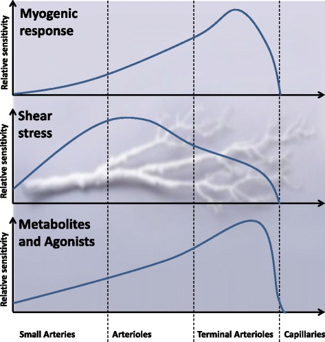
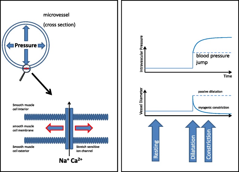
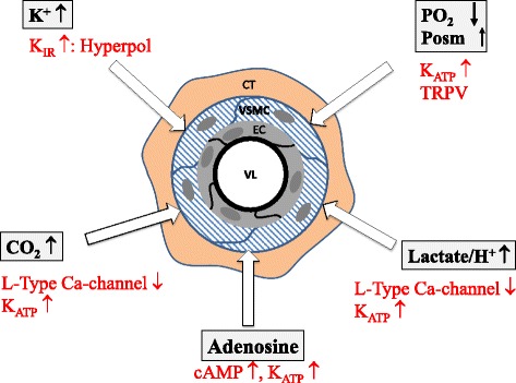
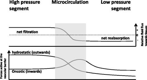
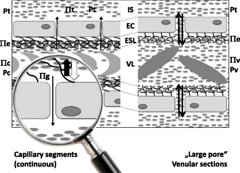
Oxygen delivery to cells is the basic prerequisite of life. Within the human body, an ingenious oxygen delivery system, comprising steps of convection and diffusion from the upper airways via the lungs and the cardiovascular system to the microvascular area, bridges the gap between oxygen in the outside airspace and the interstitial space around the cells. However, the complexity of this evolutionary development makes us prone to pathophysiological problems. While those problems related to respiration and macrohemodynamics have already been successfully addressed by modern medicine, the pathophysiology of the microcirculation is still often a closed book in daily practice. Nevertheless, here as well, profound physiological understanding is the only key to rational therapeutic decisions. The prime guarantor of tissue oxygenation is tissue blood flow. Therefore, on the premise of intact macrohemodynamics, the microcirculation has three major responsibilities: 1) providing access for oxygenated blood to the tissues and appropriate return of volume; 2) maintaining global tissue flood flow, even in the face of changes in central blood pressure; and 3) linking local blood flow to local metabolic needs. It is an intriguing concept of nature to do this mainly by local regulatory mechanisms, impacting primarily on flow resistance, be this via endothelial or direct smooth muscle actions. The final goal of microvascular blood flow per unit of time is to ensure the needed exchange of substances between tissue and blood compartments. The two principle means of accomplishing this are diffusion and filtration. While simple diffusion is the quantitatively most important form of capillary exchange activity for the respiratory gases, water flux across the blood-brain barrier is facilitated via preformed specialized channels, the aquaporines. Beyond that, the vascular barrier is practically nowhere completely tight for water, with paracellular filtration giving rise to generally low but permanent fluid flux outwards into the interstitial space at the microvascular high pressure segment. At the more leaky venular aspect, both filtration and diffusion allow for bidirectional passage of water, nutrients, and waste products. We are just beginning to appreciate that a major factor for maintaining tissue fluid homeostasis appears to be the integrity of the endothelial glycocalyx.

Keywords: Blood flow; Blood vessels; Endothelium; Glycocalyx; Microcirculation; Tissue oxygenation.


Figures

Fig. 1
Fig. 1
Autoregulatory responsiveness of smooth muscle cells to stimuli within the different vessel segments. The reaction of resistance to signals differs between different segments of the increasingly branching vascular tree. It is reasonable that metabolic impact can be found mainly close to the capillary diffusion and exchange area while the influence of hemodynamics is more prominent within the sections next to the large arteries (further explanations in the text)
Fig. 2
Fig. 2
The myogenic response (Bayliss effect) as an example for vascular autoregulation. Dilatation of the microvessel leads to ion influx (Na+, Ca2+) through stretch-sensitive membrane ion channels and, therefore, to contraction of the vessel smooth muscle cells due to depolarisation (left hand panel, a very simple illustration of the reality where the link between stretch and smooth muscle contraction is certainly more complex). The right hand panel shows the impact of an acute increase in blood pressure on intravascular pressure and vessel diameter with (full line) and (potentially) without (dotted line) myogenic response. The Bayliss effect which targets maintaining tissue blood flow in the face of different blood pressure levels can be blocked, e.g., pharmacologically by calcium antagonists
Fig. 3
Fig. 3
Local vasodilatation related to tissue metabolic activity. Local metabolic effects targeting a close relation of regional blood flow to metabolic activity are particularly effective in the terminal arterioles, being elicited foremost by changes in pO2, pCO2, pH, osmolarity, potassium ion concentration, and released catabolites such as adenosine. The respective signaling mechanisms are colored in red. cAMP cyclic adenosine monophosphate; CT connecting tissue, EC endothelial cell, K ATP ATP-dependent potassium ion channel, K IR inward-rectifying potassium ion channel, giving rise to hyperpolarization (Hyperpol), Posm osmotic pressure, TRPV transitory receptor-mediated potential, vallinoid type, VL vascular lumen, VSMC vascular smooth muscle cell
Fig. 4
Fig. 4
The principle of Ernest Starling. The high filtration-high reabsorption scenario proposed by Ernest Starling presumed high filtration in the high-pressure segments due to outweighing hydrostatic forces and reabsorption of a very large part of the filtered volume at the venular aspect owing to prevailing oncotic forces in the lumen. Fluid excess in the interstitial space needs to be drained by the lymphatic system (according to Becker et al. [28])
Fig. 5
Fig. 5
The endothelial surface layer model. Left hand panel: An intact endothelial surface layer, consisting of the endothelial glycocalyx and attached plasma protein molecules, oncotically (thick black arrow) limits hydrostatically driven (thick white arrow) fluid movement across the vascular wall within the microvascular high-pressure segments, which, in addition to narrow interendothelial clefts with high resistance to water flow, allows for hardly any egress of colloidal particles and only very low net rates of fluid extravasation (thin black arrow; Πt and Πc are in equal ranges, but irrelevant because Πe (high) and Πg (low) count). Right hand panel: At the venular aspect, relatively free and easy exchange of colloidal particles is allowed in both directions across the vascular wall (black arrows). This is feasible, because the interstitial space of most organs and tissues is now known to possess oncotic and hydrostatic pressures close to those existing in the end- and post-capillary vessel segments (Πv – Πt is small, but Pv – Pt is also small). There is no need for largescale reabsorption, as suggested by Ernest Starling (according to Jacob et al. [41] and Becker et al. [28]). EC endothelial cell, ESL endothelial surface layer, IS interstitial space, Πc, e, g, t, and v oncotic pressure in capillary plasma, ESL, below the ESL, in the tissue, and venular space, respectively, Pc, t, and v hydrostatic pressure in the capillary, tissue, and venule, respectively

Comment in

References

    1. Ganong WF. Review of medical physiology. 19. Stamford: Appleton & Lange; 1999.
    1. Suetrong B, Walley KR. Lactic acidosis in sepsis: it’s not all anaerobic: implications for diagnosis and management. Chest. 2016;149:252–61. doi: 10.1378/chest.15-1703. - DOI - PubMed
    1. Kimmoun A, Novy E, Auchet T, Ducrocq N, Levy B. Hemodynamic consequences of severe lactic acidosis in shock states: from bench to bedside. Crit Care. 2015;19:175. doi: 10.1186/s13054-015-0896-7. - DOI - PMC - PubMed
    1. Fick A. Ueber diffusion. Ann Phys. 1855;170.
    1. Rizzoni D, De Ciuceis C, Salvetti M, Paini A, Rossini C, Agabiti-Rosei C, et al. Interactions between macro- and micro-circulation: are they relevant? High Blood Press Cardiovasc Prev. 2015;22:119–28. doi: 10.1007/s40292-015-0086-3. - DOI - PubMed