. 2016 Oct 20;17(1):218.
doi: 10.1186/s13059-016-1070-5.

Estimating the population abundance of tissue-infiltrating immune and stromal cell populations using gene expression

Affiliations

Estimating the population abundance of tissue-infiltrating immune and stromal cell populations using gene expression

Etienne Becht et al. Genome Biol. .

Erratum in

Abstract

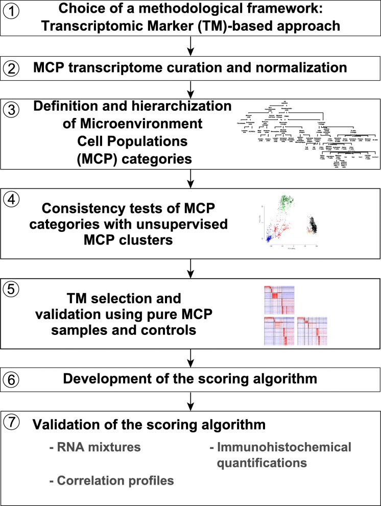
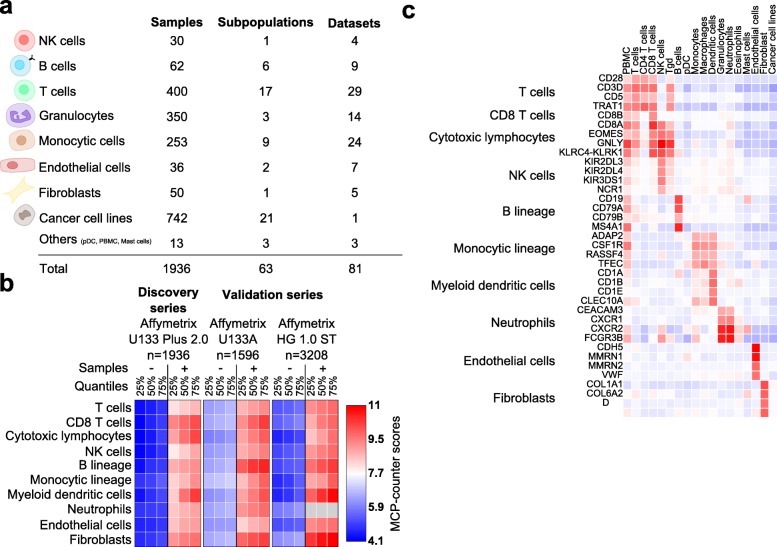
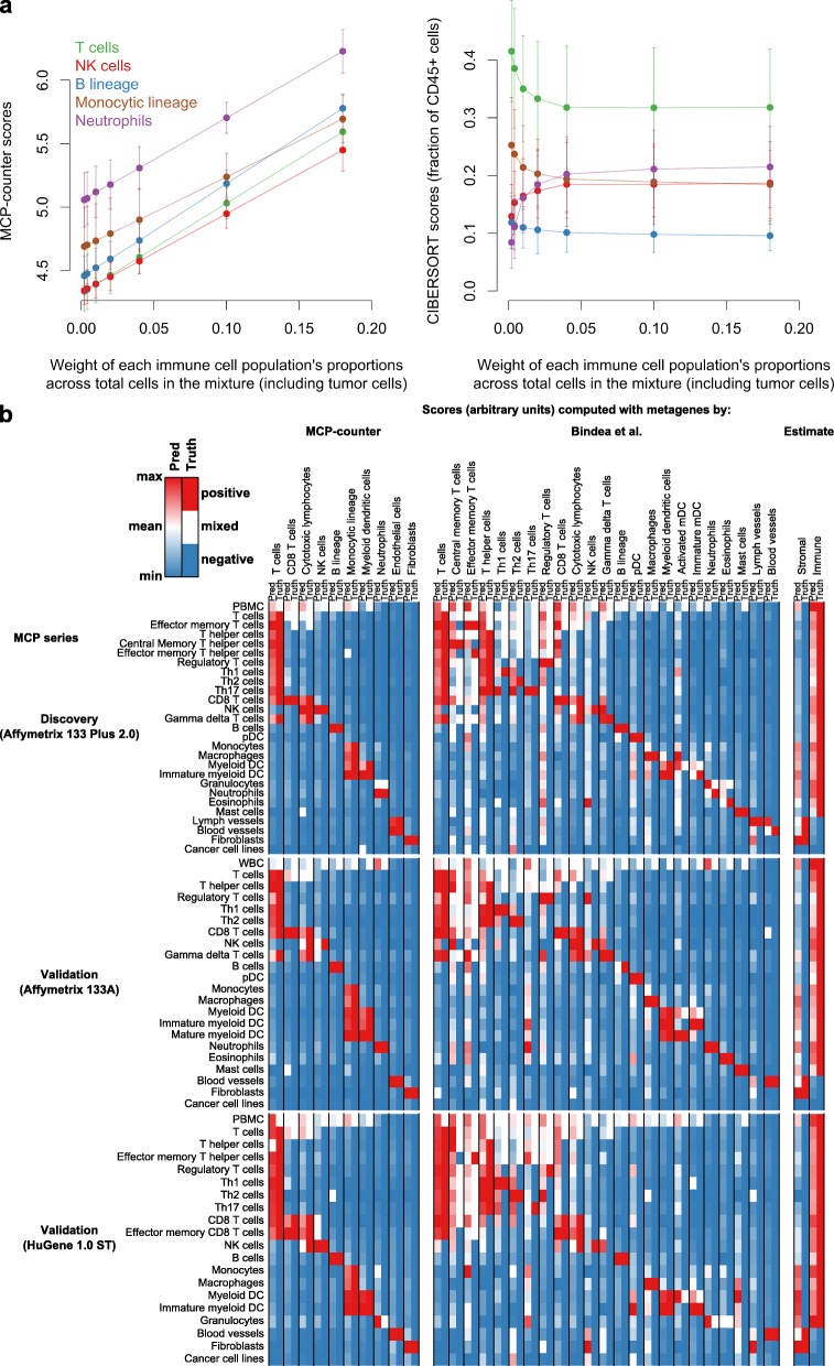
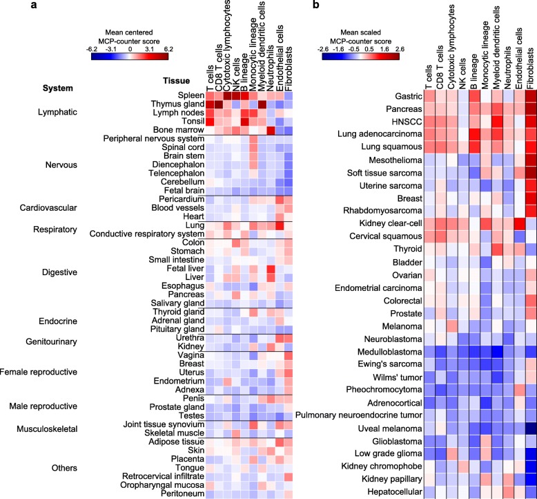
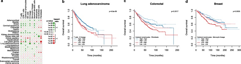
We introduce the Microenvironment Cell Populations-counter (MCP-counter) method, which allows the robust quantification of the absolute abundance of eight immune and two stromal cell populations in heterogeneous tissues from transcriptomic data. We present in vitro mRNA mixture and ex vivo immunohistochemical data that quantitatively support the validity of our method's estimates. Additionally, we demonstrate that MCP-counter overcomes several limitations or weaknesses of previously proposed computational approaches. MCP-counter is applied to draw a global picture of immune infiltrates across human healthy tissues and non-hematopoietic human tumors and recapitulates microenvironment-based patient stratifications associated with overall survival in lung adenocarcinoma and colorectal and breast cancer.

Keywords: Deconvolution; Gene signatures; Transcriptomic markers; Tumor microenvironment.


Figures

Fig. 1
Fig. 1
Purpose and development of the MCP-counter method. a Comparison of MCP-counter estimates and CIBERSORT-estimated leukocytic fractions. b Framework of MCP-counter’s development and validation
Fig. 2
Fig. 2
Identification and qualitative validation of transcriptomic markers. a The MCP discovery series. pDC plasmacytoid dendritic cell, PBMC peripheral blood mononuclear cell. b Quartiles of MCP-counter scores on positive and control samples in the discovery and validation microenvironment series. Gray indicates missing values. c Representative transcriptomic markers and their corresponding expression patterns in the MCP discovery series
Fig. 3
Fig. 3
Quantitative validation of MCP-counter estimates. a Design of the in vitro RNA mixture validation experiment. b Correlation of MCP-counter scores to samples’ known RNA proportions in RNA mixture samples. The dashed line represents the least-square regression line. The red dots correspond to limits of detection (average score of non-hematopoietic discovery MCP samples on the x-axis and corresponding mRNA fraction predicted by this linear regression on the y-axis). c Endothelial cells and fibroblasts were tested in the same fashion as in b but using qPCR data. HUVEC human umbilical vein endothelial cell. d Three-dimensional scatterplot showing the relationship between the cytotoxic lymphocyte MCP-counter score and T and NK cell proportions in the mixtures. e Correlation of MCP-counter scores with corresponding cell densities measured by immunohistochemistry
Fig. 4
Fig. 4
Comparison of MCP-counter with previously published methods. a MCP-counter scores (left) and CIBERSORT estimates (right) on simulated mRNA mixtures where five immune populations are introduced in equal proportions, with a varying proportion of tumor cells (x-axis). Error bars represent standard error estimates. b Heat maps showing scores computed with TM sets from three sources (Pred columns). The Truth columns indicate the status of the sample for each TM set. Thus, the performance of a TM set is indicated by the concordance between its Pred and Truth columns. The complete sets of markers, which do not use information from validation series, were used for MCP-counter
Fig. 5
Fig. 5
Estimation of the abundance of infiltrating immune and stromal cells across healthy tissues and non-hematopoietic human tumors. a MCP-counter scores across healthy tissues. In the case of multiple samples originating from the same type of non-diseased tissue, the resulting MCP-counter scores were averaged. b Means of MCP-counter scores across malignant tissues and three transcriptomic platforms. Rows are ordered using a hierarchical clustering procedure, with Ward’s aggregation criterion and Euclidean distance. HNSCC head and neck squamous cell carcinoma
Fig. 6
Fig. 6
Prognostic value associated with MCP-counter scores in non-hematopoietic human cancers. a Meta-analysis for the univariate prognostic value for overall survival of MCP-counter scores in human cancers. Bigger circles represent lower p values. Green represents hazard ratios lower than 1 (favorable prognostic impact) and red hazard ratios higher than 1 (poor prognostic impact). Dull colors represent p values higher than 0.05. HNSCC head and neck squamous cell carcinoma. bd Tumor microenvironment classifications of b lung adenocarcinomas based on the abundance of infiltrating T and B cells, c colorectal cancer based on the abundance of cytotoxic lymphocytes and fibroblasts, and d breast cancer based on the abundance of cytotoxic lymphocytes and cells of monocytic origin

References

    1. Becht E, Giraldo NA, Germain C, de Reyniès A, Laurent-Puig P, Zucman-Rossi J, Dieu-Nosjean M, Sautès-Fridman C, Fridman WH. Immune contexture, immunoscore, and malignant cell molecular subgroups for prognostic and theranostic classifications of cancers. Adv Immunol. 2016;130:95–190. doi: 10.1016/bs.ai.2015.12.002. - DOI - PubMed
    1. Pagès F, Berger A, Camus M, Sanchez-Cabo F, Costes A, Molidor R, Mlecnik B, Kirilovsky A, Nilsson M, Damotte D, Meatchi T, Bruneval P, Cugnenc P, Trajanoski Z, Fridman W, Galon J. Effector memory T cells, early metastasis, and survival in colorectal cancer. N Engl J Med. 2005;353:2654–66. doi: 10.1056/NEJMoa051424. - DOI - PubMed
    1. Galon J, Costes A, Sanchez-Cabo F, Kirilovsky A, Mlecnik B, Lagorce-Pagès C, Tosolini M, Camus M, Berger A, Wind P, Zinzindohoué F, Bruneval P, Cugnenc P, Trajanoski Z, Fridman W, Pagès F. Type, density, and location of immune cells within human colorectal tumors predict clinical outcome. Science. 2006;313:1960–4. doi: 10.1126/science.1129139. - DOI - PubMed
    1. Fridman WH, Pagès F, Sautès-Fridman C, Galon J. The immune contexture in human tumours: impact on clinical outcome. Nat Rev Cancer. 2012;12:298–306. doi: 10.1038/nrc3245. - DOI - PubMed
    1. Giraldo NA, Becht E, Pagès F, Skliris G, Verkarre V, Vano Y, Mejean A, Saint-Aubert N, Lacroix L, Natario I, Lupo A, Alifano M, Damotte D, Cazes A, Triebel F, Freeman GJ, Dieu-Nosjean M, Oudard S, Fridman WH, Sautès-Fridman C. Orchestration and prognostic significance of immune checkpoints in the microenvironment of primary and metastatic renal cell cancer. Clin Cancer Res. 2015;21:3031–40. doi: 10.1158/1078-0432.CCR-14-2926. - DOI - PubMed

MeSH terms