. 2017 Feb;137(2):377-384.
doi: 10.1016/j.jid.2016.10.008. Epub 2016 Oct 17.

Staphylococcus aureus Induces Increased Serine Protease Activity in Keratinocytes

Affiliations

Staphylococcus aureus Induces Increased Serine Protease Activity in Keratinocytes

Michael R Williams et al. J Invest Dermatol. 2017 Feb.

Abstract

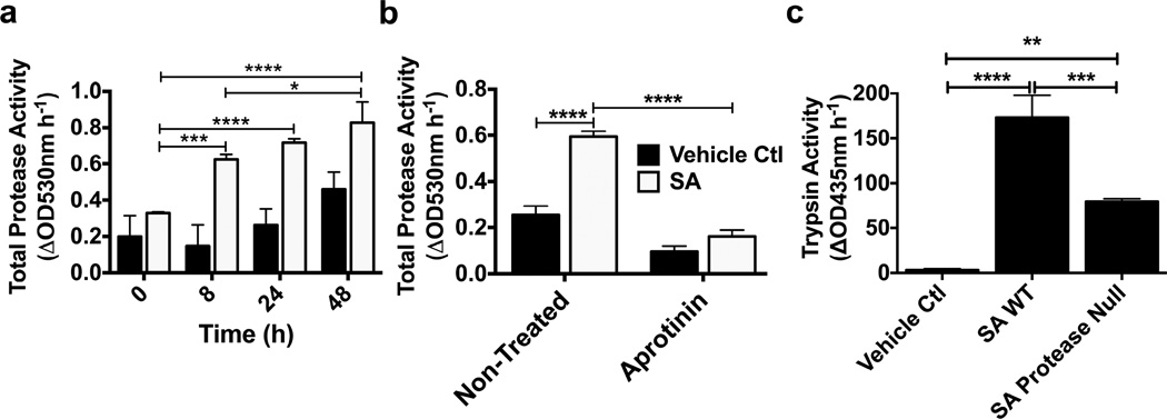
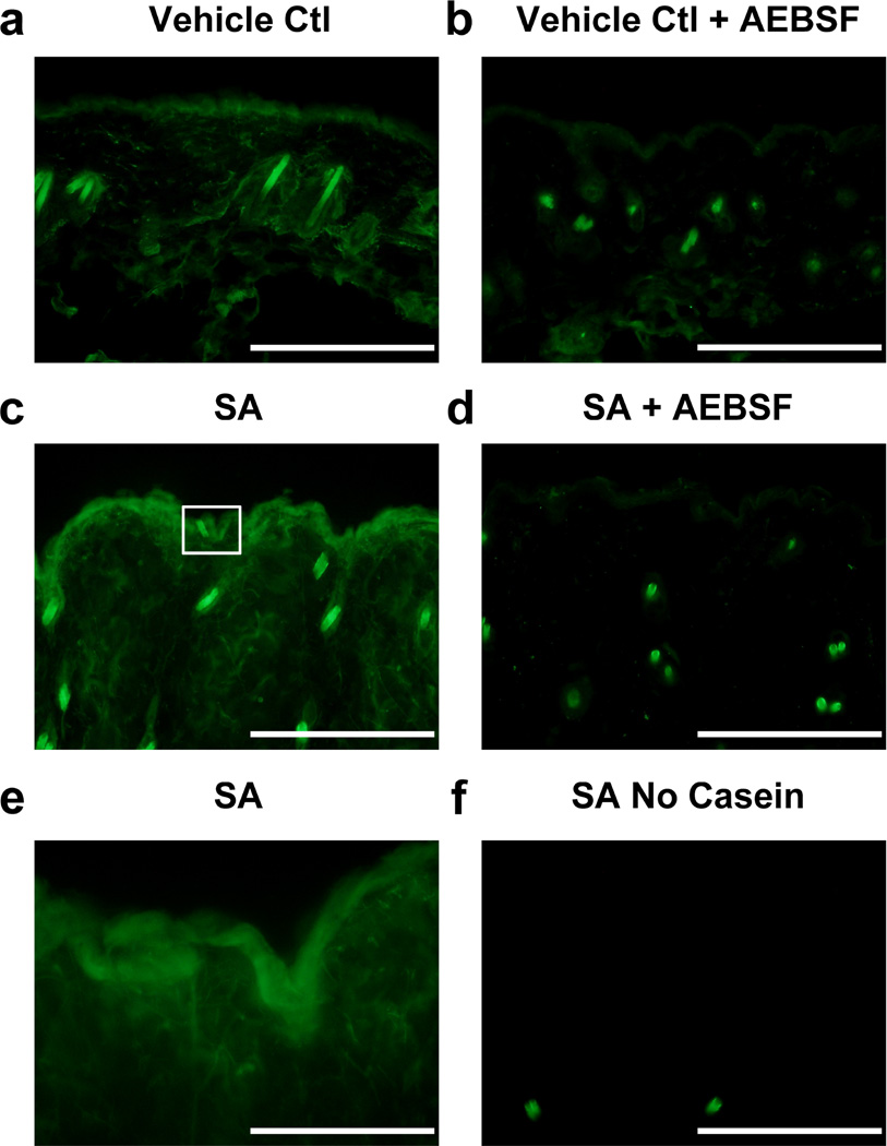
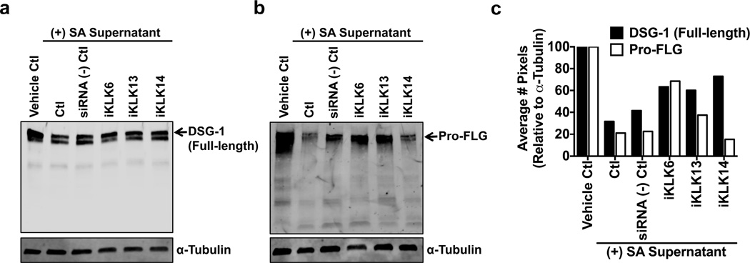
Bacteria that reside on the skin can influence the behavior of the cutaneous immune system, but the mechanisms responsible for these effects are incompletely understood. Colonization of the skin by Staphylococcus aureus (S. aureus) is increased in atopic dermatitis and can result in increased severity of the disease. In this study, we show that S. aureus stimulates human keratinocytes to increase their endogenous protease activity, including specific increases in trypsin activity. This increased protease activity coincided with increased expression of mRNA for kallikreins (KLKs), with KLK6, 13, and 14 showing the greatest induction after exposure to S. aureus. Suppression of mRNA for these KLKs in keratinocytes by targeted small interfering RNA silencing before S. aureus exposure blocked the increase in protease activity. Keratinocytes exposed to S. aureus showed enhanced degradation of desmoglein-1 and filaggrin, whereas small interfering RNA for KLK6, KLK13, and KLK14 partially blocked this degradation. These data illustrate how S. aureus directly influences the skin barrier integrity by stimulating endogenous proteolytic activity and defines a previously unknown mechanism by which S. aureus may influence skin diseases.


Figures

Figure 1
Figure 1. Staphylococci regulate human keratinocyte protease activity
(a–c) NHEKs were treated for 24h with S. aureus (Newman, USA300, 113, SANGER252) and S. epidermidis (ATCC12228, ATCC1457) sterile filtered supernatants and NHEK conditioned medium was analyzed with specific trypsin-like, elastase-like, and MMP protease substrates. (d) S. aureus (Newman) secreted proteases were analyzed for their influence on trypsin activity. Data represent mean ± SEM (n=4) and are representative of at least 3 independent experiments. Oneway ANOVAs (a–c) and two-way ANOVAs (d) were used and significance indicated by: *p<0.05, **p<0.01, ***p<0.001, ****p<0.0001.
Figure 2
Figure 2. S. aureus increases human keratinocyte serine protease activity
(a) Total protease activity (5µg-mL BODIPY FL casein) was measured in NHEK conditioned medium after S. aureus (SA, Newman) supernatant treatment for 0–48h, (b) while the serine protease inhibitor aprotinin (800µg-mL) was applied to 24h post treatment conditioned medium. (c) S. aureus (USA300 LAC) WT and protease null strains were compared for effects on NHEK conditioned medium trypsin activity (Boc-Val-Pro-Arg-AMC, 200µM). Both two-way ANOVAs (a,b) and one-way ANOVAs (c) were used and significance indicated by: *p<0.05, **p<0.01, ***p<0.001, ****p<0.0001.
Figure 3
Figure 3. Murine epidermal serine protease activity is increased by S. aureus
Murine skin colonized with (a,b) TSB vehicle control or (c,d) 2e6 CFU S. aureus (SA, USA300) for 12h was assessed for changes to total protease activity (2µg-mL BODIPY FL casein) with or without the serine protease inhibitor AEBSF (50mM) by in situ zymography; scale bars=200µm. (e) Increased magnification of epidermal total protease activity from panel c as indicated by white box; scale bar=100µm. (f) A 12h S. aureus treated no BODIPY FL casein added control was included to show background staining; scale bar=200µm.
Figure 4
Figure 4. S. aureus increases KLK expression in human keratinocytes
(a) Relative abundance of KLK mRNA expression in NHEKs after 24h S. aureus (SA, Newman) supernatant treatment was analyzed by qPCR. (b–e) KLK5, 6, 13, and 14 were analyzed for fold changes in mRNA expression in NHEKs treated with S. aureus supernatant for 0–48h. All mRNA expression levels were normalized with the housekeeping gene, glyceraldehyde-3-phosphate dehydrogenase (GAPDH). (f) NHEK conditioned medium and cell lysates were analyzed for changes in protein expression of KLK5, 6, 13, and 14 by immunoblotting after a 24h treatment with S. aureus (SA, Newman) supernatant using both published and predicted molecular weights. The housekeeping gene, α-Tubulin, was used as a loading control for cell lysates. Data represent mean ± SEM (n=3) and are representative of at least 3 independent experiments. Twoway ANOVAs (b–e) were used and significance indicated by: *p<0.05, **p<0.01, ***p<0.001, ****p<0.0001.
Figure 5
Figure 5. Multiple KLKs are responsible for S. aureus induced serine protease activity in human keratinocytes
NHEKs were treated with KLK6, 13, or 14 siRNA (15nM) prior to CaCl2 differentiation and the addition of S. aureus (Newman) supernatant. siRNA scrambled (−) controls 1 and 2 were used at 15nM and 45nM respectively. (a) Conditioned medium was analyzed for changes in trypsin activity (Boc-Val-Pro-Arg-AMC, 200µM). (b–d) Transcript levels of KLK6, KLK13, and KLK14 were assessed by qPCR and normalized to the housekeeping gene, GAPDH, to confirm siRNA knockdown efficiency. Data represents mean ± SEM (n=4) and is representative of at least 3 independent experiments. One-way ANOVA (a) was used and significance indicated by: *p<0.05, **p<0.01, ***p<0.001, ****p<0.0001.
Figure 6
Figure 6. Multiple KLKs regulate S. aureus induced DSG-1 and FLG cleavage in human keratinocytes
NHEKs treated with S. aureus (Newman) supernatant for 24h were assessed for changes to (a) desmoglein-1 (DSG-1) and (b) Pro-filaggrin (Pro-FLG) cleavage after siRNA knockdown of KLK6, 13, and 14 (15nM) by immunoblotting. The housekeeping gene, α-Tubulin, was used as a loading control. DSG-1 (full-length) and Pro-FLG are indicated by black arrows. (c) Densitometry analysis of both DSG-1 (full-length) and Pro-FLG represented by the average number of pixels normalized to α-Tubulin (n=1). Immunoblots are representative of at least 3 independent experiments.

References

    1. Bantel H, Sinha B, Domschke W, Peters G, Schulze-Osthoff K, Janicke RU. alpha-Toxin is a mediator of Staphylococcus aureus-induced cell death and activates caspases via the intrinsic death pathway independently of death receptor signaling. J Cell Biol. 2001;155(4):637–648. - PMC - PubMed
    1. Belkaid Y, Segre JA. Dialogue between skin microbiota and immunity. Science. 2014;346(6212):7. - PubMed
    1. Borgono CA, Michael IP, Komatsu N, Jayakumar A, Kapadia R, Clayman GL, et al. A potential role for multiple tissue kallikrein serine proteases in epidermal desquamation. J Biol Chem. 2007;282(6):3640–3652. - PubMed
    1. Caubet C, Jonca N, Brattsand M, Guerrin M, Bernard D, Schmidt R, et al. Degradation of corneodesmosome proteins by two serine proteases of the kallikrein family, SCTE/KLK5/hK5 and SCCE/KLK7/hK7. J Invest Dermatol. 2004;122(5):1235–1244. - PubMed
    1. Chavanas S, Bodemer C, Rochat A, Hamel-Teillac D, Ali M, Irvine AD, et al. Mutations in SPINK5, encoding a serine protease inhibitor, cause Netherton syndrome. Nature Genetics. 2000;25(2):2. - PubMed