. 2016 Dec;15(12):3015-3027.
doi: 10.1158/1535-7163.MCT-16-0017. Epub 2016 Oct 7.

Mutant BRAF Upregulates MCL-1 to Confer Apoptosis Resistance that Is Reversed by MCL-1 Antagonism and Cobimetinib in Colorectal Cancer

Affiliations

Mutant BRAF Upregulates MCL-1 to Confer Apoptosis Resistance that Is Reversed by MCL-1 Antagonism and Cobimetinib in Colorectal Cancer

Hisato Kawakami et al. Mol Cancer Ther. 2016 Dec.

Abstract

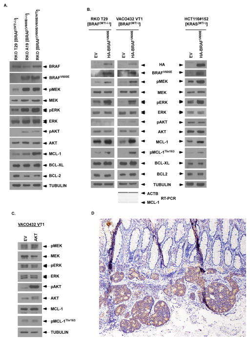
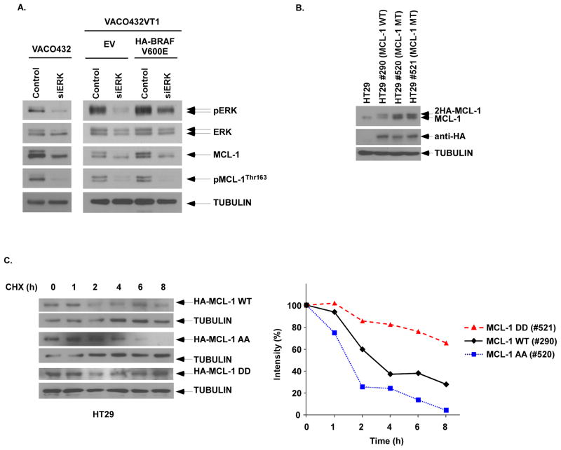
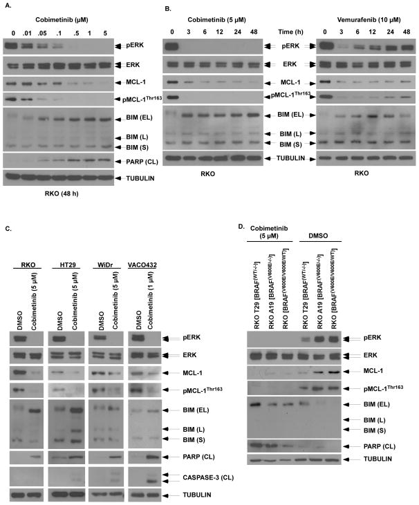
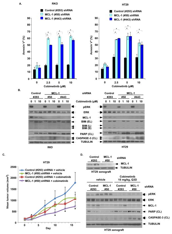
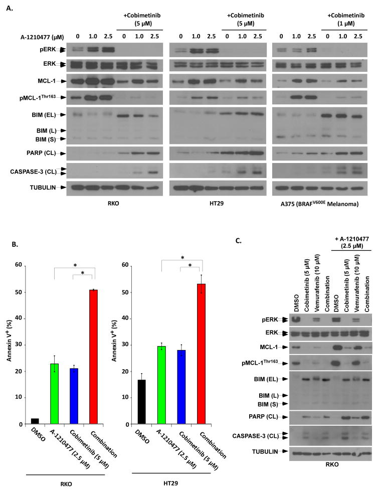
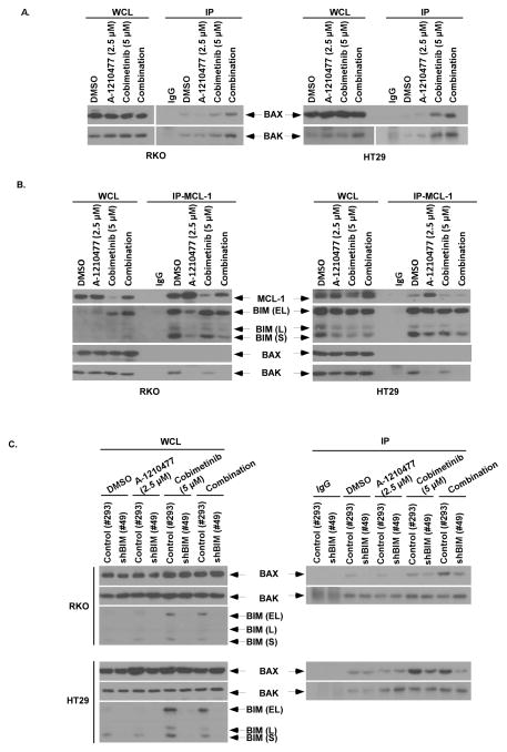
Oncogenic BRAFV600E mutations activate MAPK signaling and are associated with treatment resistance and poor prognosis in patients with colorectal cancer. In BRAFV600E-mutant colorectal cancers, treatment failure may be related to BRAFV600E-mediated apoptosis resistance that occurs by an as yet undefined mechanism. We found that BRAFV600E can upregulate anti-apoptotic MCL-1 in a gene dose-dependent manner using colorectal cancer cell lines isogenic for BRAF BRAFV600E-induced MCL-1 upregulation was confirmed by ectopic BRAFV600E expression that activated MEK/ERK signaling to phosphorylate (MCL-1Thr163) and stabilize MCL-1. Upregulation of MCL-1 was mediated by MEK/ERK shown by the ability of ERK siRNA to suppress MCL-1. Stabilization of MCL-1 by phosphorylation was shown by a phosphorylation-mimicking mutant and an unphosphorylated MCL-1 mutant that decreased or increased MCL-1 protein turnover, respectively. MEK/ERK inhibition by cobimetinib suppressed MCL-1 expression/phosphorylation and induced proapoptotic BIM to a greater extent than did vemurafenib in BRAFV600E cell lines. MCL-1 knockdown versus control shRNA significantly enhanced cobimetinib-induced apoptosis in vitro and in HT29 colon cancer xenografts. The small-molecule MCL-1 inhibitor, A-1210477, also enhanced cobimetinib-induced apoptosis in vitro that was due to disruption of the interaction of MCL-1 with proapoptotic BAK and BIM. Knockdown of BIM attenuated BAX, but not BAK, activation by cobimetinib plus A-1210477. In summary, BRAFV600E-mediated MEK/ERK activation can upregulate MCL-1 by phosphorylation/stabilization to confer apoptosis resistance that can be reversed by MCL-1 antagonism combined with cobimetinib, suggesting a novel therapeutic strategy against BRAFV600E-mutant CRCs. Mol Cancer Ther; 15(12); 3015-27. ©2016 AACR.


Conflict of interest statement

Conflict of Interest. The authors report no conflicts related to the content of the manuscript.

Figures

Figure 1
Figure 1. Mutant BRAF upregulates anti-apoptotic MCL-1 expression
A, Analysis of protein expression by immunoblotting in parental (BRAFV600E/V600E/WT) and isogenic BRAF cell lines including mutant A19 (BRAFV600E/−/−) and wild-type (WT) T29 (BRAF−/−/WT) RKO cells. Tubulin was utilized as control for protein loading. B,C,D, Ectopic expression of lentiviral mutant BRAF (B) or retroviral AKT (C) was performed in isogenic RKO T29 (BRAF−/−/WT) and VACO432 VT1 (BRAFWT/−) cell lines or in isogenic KRAS WT HCT116#152 (KRASWT/−) cells. A competitive RT-PCR assay was performed to quantitate MCL-1 transcripts using β-actin (ACTB) as an internal control (B, bottom). D, Immunohistochemical expression of MCL-1 in a BRAF mutant human colon cancer cells compared to overlying normal colonic epithelium (100X).
Figure 2
Figure 2. MEK/ERK signaling phosphorylates MCL-1 resulting in protein stability
A, ERK knockdown by siRNA was performed in VACO432 (BRAFV600E/WT), as well as isogenic VACO432 VT1 (BRAFWT/−) cells with ectopic BRAFV600E vs empty vector (EV). Protein expression was determined by immunoblotting. B, Ectopic expression of HA-tagged wild type (WT) MCL-1, and phosphorylation-mimicking [T92D/T163D (DD)] or unphosphorylated [T92A/T163A (AA)] MCL-1 mutants was performed in HT29 cells. C, Cell lines were treated with cycloheximide (5 mmol/L) for the indicated times and protein expression against HA-tagged MCL-1 was analyzed by immunoblotting. The level of MCL-1 expression was then quantified by densitometry and normalized using tubulin expression.
Figure 3
Figure 3. Cobimetinib treatment inhibits MCL-1 phosphorylation to downregulate expression and induces BIM
A, RKO cells were treated with increasing doses of cobimetinib for 48 h and protein expression, including BIM isoforms [extra long (EL), long (L) and short (S)] and PARP cleavage (CL), were analyzed by immunboblotting. B, RKO cells were treated with cobimetinib or vemurafenib for the indicated times and expression of pERK/ERK and pMCL-1Thr163/MCL-1 were analyzed by immunoblotting. C, Multiple BRAF mutant CRC cell lines (RKO, HT29, WiDr, VACO432) were treated with cobimetinib at indicated doses for 48 h and protein expression including markers of apoptosis (PARP, caspase-3) were analyzed by immunoblotting. D, RKO BRAF isogenic cell lines with none to two mutant BRAF alleles were treated with cobimetinib vs DMSO and the effect on PARP cleavage was determined.
Figure 4
Figure 4. MCL-1 knockdown by shRNA enhances cobimetinib-induced apoptosis and anti-tumor efficacy in BRAF mutant CRC cells and tumor xenografts
A, RKO and HT29 cells were transduced with lentiviral MCL-1 (#50 or #443) vs control shRNA (#293). Cells with stable expression were then incubated with cobimetinib for 48h at the indicated doses. Apoptosis was analyzed by annexin V+ staining that was quantified using flow cytometry. Mean values were derived from triplicate experiments and bars represent S.D. *p<0.05. B, Apoptosis was also analyzed by expression of cleavage (CL) of PARP and CASPASE-3 by immunoblotting in both cell lines. C, HT29 cells containing stable expression of control (#293) or MCL-1 (#50) shRNA were grown as tumor xenografts in SCID mice. Xenograft-bearing mice with dosed with either vehicle or cobimetinib (15 mg/kg every 3 days by oral gavage) for 14 consecutive days. Xenograft mean tumor volumes are plotted against days of treatment for vehicle- and cobimetinib-treated mice. Error bars represent SEM. Statistical significance(*, P < 0.05) is shown for comparison of MCL-1 (#50) shRNA + cobimetinib vs control (#293) shRNA + cobimetinib. D, Pre-treatment expression of MCL-1 in tumors from xenograft-bearing mice was evaluated by immunoblotting (upper panel). Post-treatment expression of pERK/ERK, MCL-1, cleaved PARP and cleaved caspase-3 in tumor xenografts from the 4 groups of tumor-bearing mice were evaluated by immunoblotting (lower panel).
Figure 5
Figure 5. The combination of a small molecule MCL-1 antagonist, A-1210477, with cobimetinib potently induce apoptosis
A, CRC (RKO, HT29) and melanoma (A375) cell lines were treated with A-1210477 alone or combined with cobimetinib for 48 at the indicated doses. ERK activation, BIM isoform expression, and cleavage of PARP and CASPASE3 were analyzed by immunoblotting. B, RKO and HT29 cell lines were treated with A-1210477 alone or combined with cobimetinib for 48 h, and apoptosis was quantified by annexin V+ staining using flow cytometry. Mean values of triplicate experiments are shown; bars represent S.D; *p<0.05. A synergistic interaction between A-1210477 and cobimetinib (data not shown) was found by calculation of a combination index (CI). C, RKO cells were treated with combimetib, vemurafenib, or their combination in the presence or absence of A-1210477 for 48 h. Protein expression including apoptotic markers was then determined by immunoblotting.
Figure 6
Figure 6. A-1210477 releases BAK from MCL-1 and cobimetinb induces BIM that is required for BAX activation
A, RKO and HT29 cell lines were treated with A-1210477, cobimetinib, or in combination for 16 h. Immunoprecipitation (IP) was performed in whole cell lysates (WCL) using conformation-specific antibodies against BAK or BAX (6A7). Normal rabbit (for BAK) and mouse (for BAX) IgG served as antibody controls. B, Cells were treated with A-1210477, cobimetinib, or their combination for 3 h and IP was then performed in WCL using an anti-MCL-1 antibody. Co-precipitated protein complexes were probed for BIM, BAK or BAX by immunoblotting. Normal rabbit IgG served as an antibody control. C, RKO and HT29 cell lines with stable expression of control (#293) or BIM (#49) shRNA were incubated with A-1210477, cobimetinib. or their combination for 16 h. IP was then performed in WCLs using conformation-specific antibodies against BAK or BAX in these cell lines. The effect of BIM shRNA on BIM protein expression was confirmed by immunoblotting.

References

    1. Hatzivassiliou G, Haling JR, Chen H, Song K, Price S, Heald R, et al. Mechanism of MEK inhibition determines efficacy in mutant KRAS- versus BRAF-driven cancers. Nature. 2013;501:232–6. - PubMed
    1. Davies H, Bignell GR, Cox C, Stephens P, Edkins S, Clegg S, et al. Mutations of the BRAF gene in human cancer. Nature. 2002;417:949–54. - PubMed
    1. Kopetz S, Desai J, Chan E, Hecht JR, O’Dwyer PJ, Maru D, et al. Phase II Pilot Study of Vemurafenib in Patients With Metastatic BRAF-Mutated Colorectal Cancer. J Clin Oncol. 2015;33:4032–8. - PMC - PubMed
    1. Corcoran RB, Atreya CE, Falchook GS, Kwak EL, Ryan DP, Bendell JC, et al. Combined BRAF and MEK Inhibition With Dabrafenib and Trametinib in BRAF V600-Mutant Colorectal Cancer. J Clin Oncol. 2015;33:4023–31. - PMC - PubMed
    1. Roth AD, Tejpar S, Delorenzi M, Yan P, Fiocca R, Klingbiel D, et al. Prognostic role of KRAS and BRAF in stage II and III resected colon cancer: results of the translational study on the PETACC-3, EORTC 40993, SAKK 60-00 trial. J Clin Oncol. 2010;28:466–74. - PubMed

MeSH terms