Review
. 2016 Oct 4;14(Suppl 1):27.
doi: 10.1186/s12959-016-0099-8. eCollection 2016.

New horizon in platelet function: with special reference to a recently-found molecule, CLEC-2

Affiliations
Review

New horizon in platelet function: with special reference to a recently-found molecule, CLEC-2

Yukio Ozaki et al. Thromb J. .

Abstract

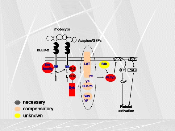
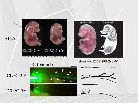
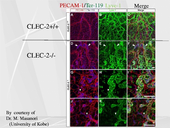
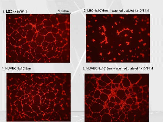
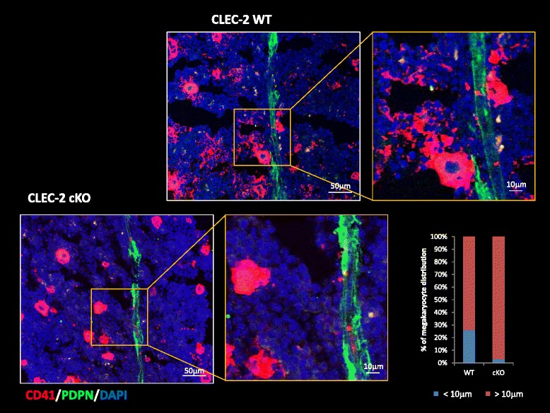
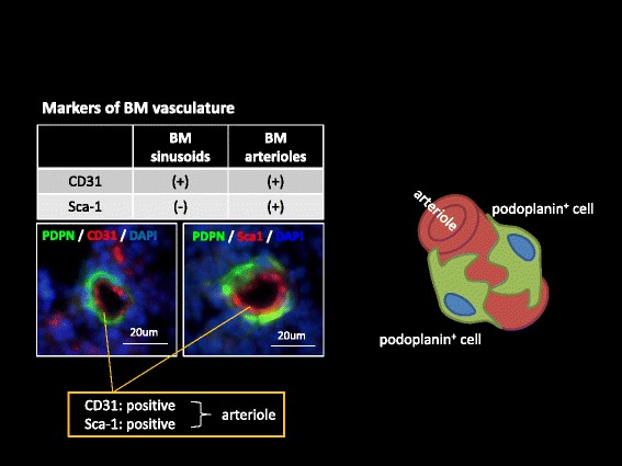
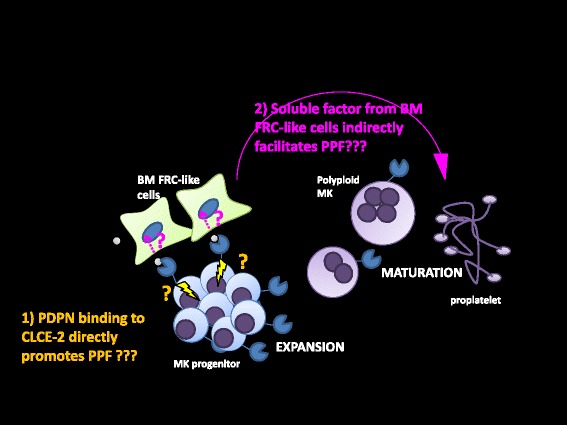
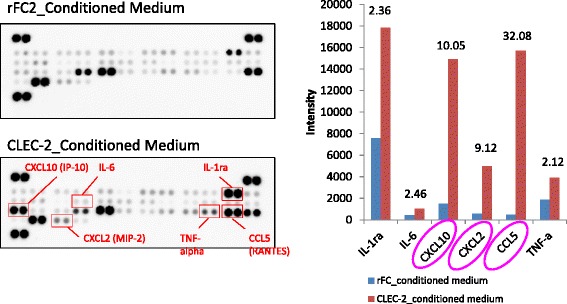
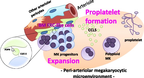
Platelets play a key role in the pathophysiological processes of hemostasis and thrombus formation. However, platelet functions beyond thrombosis and hemostasis have been increasingly identified in recent years. A large body of evidence now exists which suggests that platelets also play a key role in inflammation, immunity, malignancy, and furthermore in organ development and regeneration, such as the liver. We have recently identified CLEC-2 on the platelet membrane, which induces intracellular activation signals upon interaction of a snake venom, rhodocytin. Later we discovered that podoplanin, present in renal podocytes and lymphatic endothelial cells, both of which are not accessible to platelets in blood stream, is an endogenous ligand for CLEC-2. In accord with our expectation, platelet-specific CLEC-2 knockout mice have a phenotype of edema, lymphatic vessel dilatation, and the presence of blood cells in lymphatic vessels. It is suggested that lymphatic/blood vessel separation during the developmental stage is governed by cytokines released from platelets activated by the interaction between platelet CLEC-2 and podoplanin present on lymphatic endothelial cells. Recombinant CLEC-2 bound to early atherosclerotic lesions and normal arterial walls, co-localizing with vascular smooth muscle cells (VSMCs). Flow cytometry and immunocytochemistry showed that recombinant CLEC-2, but not an anti-podoplanin antibody, bound to VSMCs, suggesting that CLEC-2 ligands other than podoplanin are present in VSMCs. Protein arrays and Biacore analysis were used to identify S100A13 as a CLEC-2 ligand in VSMCs. S100A13 was released upon oxidative stress, and expressed in the luminal area of atherosclerotic lesions. Megakaryopoiesis is promoted through the CLEC-2/podoplanin interaction in the vicinity of arterioles, not sinusoids or lymphatic vessels. There exist podoplanin-expressing bone-marrow (BM) arteriolar stromal cells, tentatively termed as BM fibroblastic reticular cell (FRC)-like cells, and megakaryocyte colonies were co-localized with periarteriolar BM FRC-like cells in the BM. CLEC-2/podoplanin interaction induces BM FRC-like cells to secrete CCL5 to facilitate proplatelet formation. These observations indicate that a reciprocal interaction with between CLEC-2 on megakaryocytes and podoplanin on BM FRC-like cells contributes to the periarteriolar megakaryopoietic microenvironment in mouse BM.

Keywords: Beyond hemostasis; CLEC-2; Immunity; Lymphangiogeneis; Megakaryopoiesis; Platelets; Smooth muscle cells; Thrombosis.


Figures

Fig. 1
Fig. 1
Signal transduction pathway mediated through CLEC-2. Overall, the signal transduction pathway is strikingly similar to that of GPVI, involving a number of signaling molecules related to tyrosine kinases. Upon association with its agonists, CLEC-2 assumes multimerization, and Syk and Src family kinases mediates tyrosine phosphorylation of its hemITAM, which is followed by downstream signals culminating in PLCγ2 activation. Signaling molecules required for full activation of platelets are marked in red, those which are partially required are marked in orange, and those which can be spared are marked in yellow
Fig. 2
Fig. 2
CLEC-2 knockout mice have abnormal lymphatic vessels. CLEC-2 knockout mice (upper left inlet) are edematous with dilated vessels, the pattern of which is quite similar to the distribution of lymphatic vessels of porcine fetus (upper right inlet), which was reported previously elsewhere. We found that the injected dye does not run into lymphatic vessels of the CLEC-knockout, suggesting for the presence of lymphatic vessel malformation (lower inlets)
Fig. 3
Fig. 3
CLEC-2-knockout mice have dilated, torturous lymphatic vessels. While blood vessels and lymphatic vessels are distinctly separated in wild-type, they were intermingled with each other with CLEC-2-knockout mice, and the lymphatic vessels are dilated and torturous. PECAM-1 stains blood vessels, and Lyve-1 stains lymphatic endothelial cells. At the sites indicated by arrowheads, blood vessels and lymphatic vessels appear to be connected
Fig. 4
Fig. 4
The supernatant of activated platelets inhibited tube formation of lymphatic endothelial cells (LEC) but not that of human umbilical venous endothelial cells (HUVEC). The upper left inlet shows the pattern of LEC tube formation without platelets, and the upper right inlet shows that LEC tube formation is disturbed in the presence of washed platelets which express CLEC-2. On the other hand, tube formation of HUVEC (lower left inlet) is not affected in the presence of platelets (lower right inlet)
Fig. 5
Fig. 5
S100A13 is present in the normal aorta, and its distribution coincides with that of smooth muscle cells. In figures a, the distribution patterns of smooth muscle actin is similar to that of S100A12. Figures b show that CLEC-2 binding is abundantly present in atherosclerotic aorta, but less so in the normal aorta, and the binding of CLEC-2 colocalizes with that of S100A13. Podoplanin which is a ligand for CLEC-2 is expressed in the atherosclerotic tissues. However, the distribution of podoplanin is distinct from that of S100A13 (figures c), suggesting that CLEC-2 binding in the normal and atherosclerotic aorta is attributed to its binding to S100A13, but not to podoplanin
Fig. 6
Fig. 6
CD41+ clusters were formed adjacent to the podoplanin+ stromal cells in the BM CD41+ clusters which represent megakaryocytes were observed, lying close to the podoplanin+ stromal cells lining vasculature in the bone marrow. However, this phenomenon is not present with CLEC-2 knockout mice. The right inlet shows the quantitative distribution of megakaryocytes within 10 μm of vasculature in wild-type mice vs. CLEC-2 knockout
Fig. 7
Fig. 7
BM arteriolar stromal cells are podoplanin-positive. There are three type of vessels in the bone marrow, arterioles, sinusoids and lymphatic vessels. Only the bone marrow (BM) arteriolar stromal cells (CD31- and Sca-1-positive) are podoplanin-positive, and these cells are tentatively termed as BM fibroblastic reticular cell (FRC)-like cells. BM-FRC-like cells surround arterioles as illustrated in the right inlet
Fig. 8
Fig. 8
Hitherto, we have found that podoplain/CLEC-2 interaction induces megakaryocyte expansion. We then asked whether podoplain/CLEC-2 interaction acts on megakaryocytes to induce proplatelet formation, or on BM FCR-like cells which then contributes to proplatelet formation. Two hypotheses are depicted in Fig. 8
Fig. 9
Fig. 9
Proteome Profiler_Cytokine array. We found that some substances released from BM FCR-like cells upon interaction with CLEC-2-positive megakaryocytes serve to induce proplatelet formation. By the use of proteome profile cytokine array, three cytokines were identified, CXCL10, CXCL2, andCCL5. CCL5 was identified to be the most potent molecule released from BM FCR-like cells to induce proplatelet formation in megakaryocytes
Fig. 10
Fig. 10
Microenvironment for megakaryopoiesis related to CLEC-2/podoplanin interaction. Our finding in this study suggest that a reciprocal interaction with between CLEC-2 on megakaryocytes and podoplanin on BM FRC-like cells contributes to the periarteriolar megakaryopoietic microenvironment in mouse BM

Similar articles

Cited by

References

    1. McFadyen JD, Kaplan ZS. Platelets are not just for clots. Transfus Med Rev. 2015;26:110–9. doi: 10.1016/j.tmrv.2014.11.006. - DOI - PubMed
    1. Suzuki-Inoue K, Fuller GL, Garcia A, Eble JA, Pohlmann S, Inoue O, et al. A novel Syk-dependent mechanism of platelet activation by the C-type lectin receptor CLEC-2. Blood. 2006;107:542–9. doi: 10.1182/blood-2005-05-1994. - DOI - PubMed
    1. Suzuki-Inoue K, Kato Y, Inoue O, Kaneko MK, Mishima K, Yatomi Y, et al. Involvement of the snake toxin receptor CLEC-2, in podoplanin-mediated platelet activation, by cancer cells. J Biol Chem. 2007;282:25993–6001. doi: 10.1074/jbc.M702327200. - DOI - PubMed
    1. Bertozzi CC, Schmaier AA, Mericko P, Hess PR, Zou Z, Chen M, et al. Platelets regulate lymphatic vascular development through CLEC-2-SLP-76 signaling. Blood. 2010;116:661–70. doi: 10.1182/blood-2010-02-270876. - DOI - PMC - PubMed
    1. Suzuki-Inoue K, Inoue O, Ding G, Nishimura S, Hokamura K, Eto K, et al. Essential in vivo roles of the C-type lectin receptor CLEC-2: embryonic/neonatal lethalilty of CLEC-2-deficient mice by blood/lymphatic misconnections and impaired thrombus formation of CLEC-2-deficient platelets. J Biol Chem. 2010;285:24494–507. doi: 10.1074/jbc.M110.130575. - DOI - PMC - PubMed

LinkOut - more resources