. 2016 Oct 6:7:411.
doi: 10.3389/fimmu.2016.00411. eCollection 2016.

NFAT2 Regulates Generation of Innate-Like CD8+ T Lymphocytes and CD8+ T Lymphocytes Responses

Affiliations

NFAT2 Regulates Generation of Innate-Like CD8+ T Lymphocytes and CD8+ T Lymphocytes Responses

Emilia Pachulec et al. Front Immunol. .

Abstract

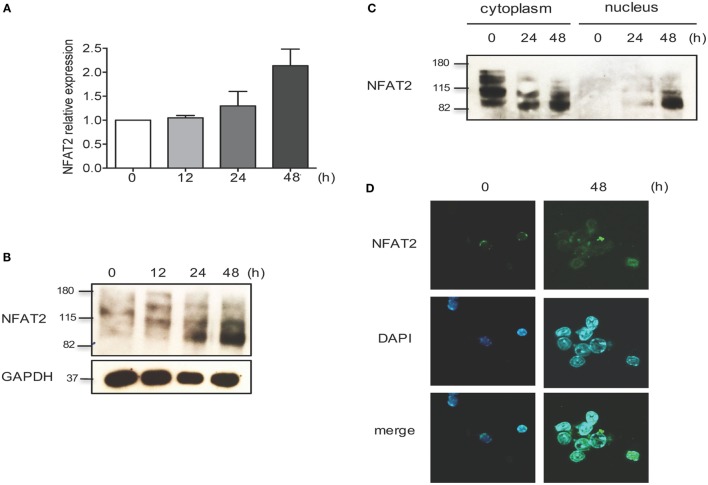
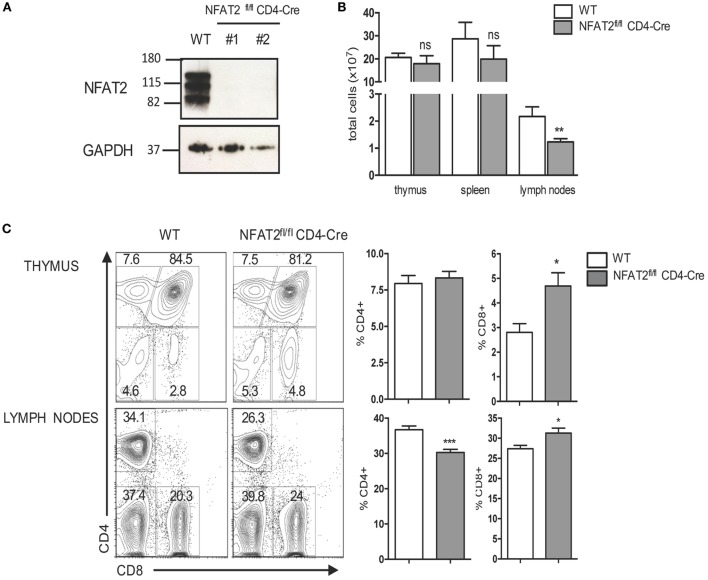
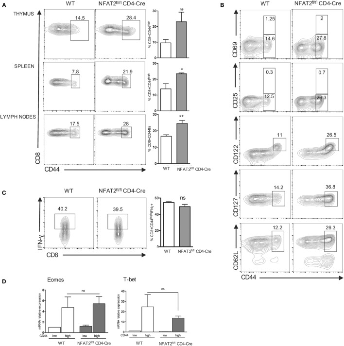
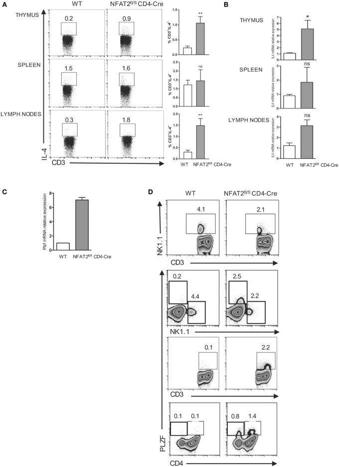
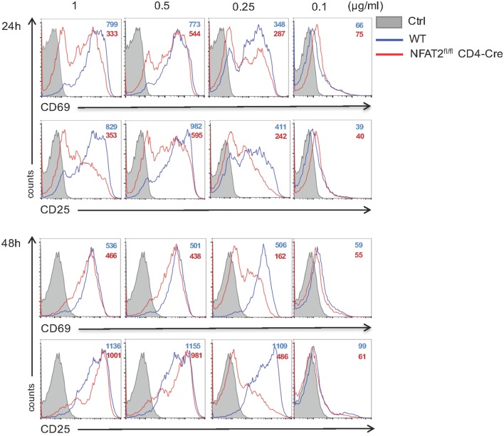
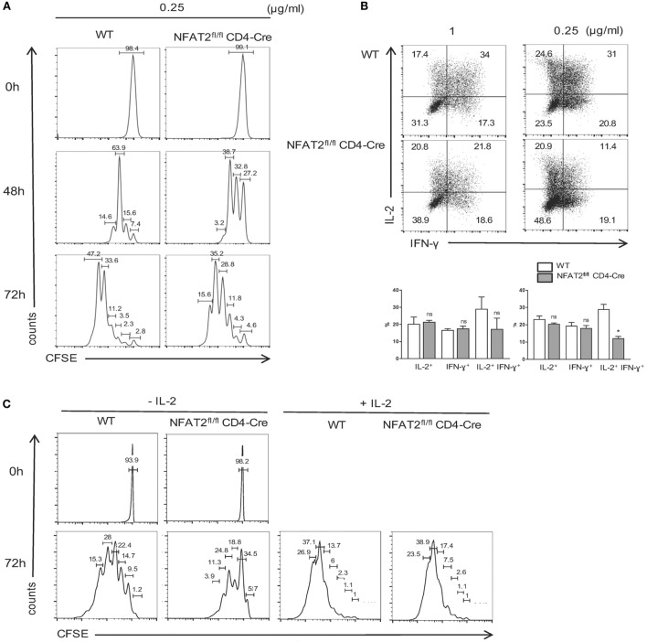
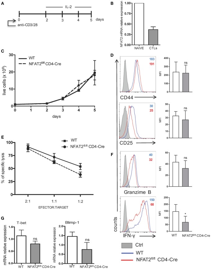
Nuclear factor of activated T cells (NFAT) 2 null mutant mice die in utero of cardiac failure, precluding analysis of the role of NFAT2 in lymphocyte responses. Only the NFAT2-/-/Rag-1-/- chimeric mice model gave insight into the role of NFAT2 transcription factor in T lymphocyte development, activation, and differentiation. As reports are mainly focused on the role of NFAT2 in CD4+ T lymphocytes activation and differentiation, we decided to investigate NFAT2's impact on CD8+ T lymphocyte responses. We report that NFAT2 is phosphorylated and inactive in the cytoplasm of naive CD8+ T cells, and upon TCR stimulation, it is dephosphorylated and translocated into the nucleus. To study the role of NFAT2 in CD8+ T responses, we employed NFAT2fl/flCD4-Cre mice with NFAT2 deletion specifically in T cells. Interestingly, the absence of NFAT2 in T cells resulted in increased percentage of non-conventional innate-like CD8+ T cells. These cells were CD122+, rapid producer of interferon gamma (IFN-γ) and had characteristics of conventional memory CD8+ T cells. We also observed an expansion of PLZF+ expressing CD3+ thymocyte population in the absence of NFAT2 and increased IL-4 production. Furthermore, we found that CD8+ T lymphocytes deficient in NFAT2 had reduced activation, proliferation, and IFN-γ and IL-2 production at suboptimal TCR strength. NFAT2 absence did not significantly influence differentiation of CD8+ T cells into cytotoxic effector cells but reduced their IFN-γ production. This work documents NFAT2 as a negative regulator of innate-like CD8+ T cells development. NFAT2 is required for complete CD8+ T cell responses at suboptimal TCR stimulation and regulates IFN-γ production by cytotoxic CD8+ T cells in vitro.

Keywords: CD8+ T lymphocytes; IFN-γ; NFAT2; PLZF; innate-like CD8+ T cells.


Figures

Figure 1
Figure 1
NFAT2 is present and functional in CD8+ T lymphocytes. CD8+ T lymphocytes were purified from naive C57BL/6 mice, as described, and then left unstimulated (0) or stimulated in vitro with anti-CD3 plus anti-CD28 (both at 1 μg/ml) for indicated times. (A) Total RNA was isolated, and NFAT2 mRNA levels were analyzed by real-time RT-PCR assay using SYBR green master mix. The data are normalized to the β-actin levels. Data are shown as mean ± SD of three independent experiments. Detection of NFAT2 transcription factor in CD8+ T cells in total lysates (B) and in cytoplasmic and nuclear fractions (C) by Western blot. (D) Cellular localization of NFAT2 protein in CD8+ T cells by immunofluorescence staining. All data are representative of at least two independent experiments.
Figure 2
Figure 2
NFAT2 deficiency increases the frequency of CD8+ T cells. (A) Detection of NFAT2 transcription factor in CD8+ T cells’ total protein lysates from WT and NFAT2fl/flCD4-Cre mice by Western blot. (B) Thymus, spleens, and lymph nodes were harvested from WT or NFAT2fl/flCD4-Cre mice, and the total number of cells were counted using Trypan blue exclusion. (C) Flow cytometric analysis of thymocytes and total lymph nodes cells from WT or NFAT2fl/fl CD4-Cre mice. Shown is the frequency of CD4- and CD8-expressing cells. Right, the percentage of CD4+ and CD8+ cells for all the mice. All results are representative of at least two independent experiments. All data are shown as mean ± SD. The ns indicates not significant, *indicates p < 0.05, and **indicates p < 0.01 compared to WT mice.
Figure 3
Figure 3
NFAT2 deficiency leads to the development of innate-like CD8+ T cells. (A) Flow cytometric analysis CD8+ T cells for CD44 expression. Right, the percentage of CD8+CD44high cells for all the mice. (B) Total CD8+ T cells were analyzed for the expression of CD69, CD25, CD122, CD127, and CD62L. Shown are the percentages of T CD8+CD44high populations expressing or not specific surface markers. (C) Total CD8+ T cells were stimulated with PMA plus ionomycin for 6 h, and cells in CD8+CD44high gate were analyzed for IFN-γ production. Right, the percentage of CD8+CD44highIFN-γ+ cells for all the mice. (D) Total RNA was isolated from CD8+CD44high and CD8+CD44low populations from lymph nodes of WT and NFAT2fl/flCD4-Cre mice, and Tbx21 and Eomes mRNA levels were analyzed by real-time RT-PCR assay using Taqman probe. The data are normalized to the HPRT levels. All results are representative of at least two independent experiments. All data are shown as mean ± SD. The ns indicates not significant, *indicates p < 0.05, and **indicates p < 0.01 compared to WT mice.
Figure 4
Figure 4
Expansion of PLZF expressing cells in the absence of NFAT2. Total thymocytes, splenocytes, and lymph nodes cells were stimulated with PMA plus ionomycin for 6 h. IL-4-producing cells were analyzed in CD3+ gate. Right, the percentage of CD3+IL-4+ cells for all the mice (A). Total RNA was isolated from thymus, spleen, and lymph nodes from WT and NFAT2fl/flCD4-Cre mice, and IL-4 mRNA levels were analyzed by real-time RT-PCR assay using Taqman probe. The data are normalized to the HPRT levels (B). Total RNA was isolated from CD4+ thymocytes and analyzed for PLZF mRNA level by real-time RT-PCR using SyberGreen. The data are normalized to the actin levels (C). Flow cytometric analysis of CD3+, CD4+, and NK1.1+ thymocytes for PLZF expression (D). All results are representative of at least two independent experiments. All data are shown as mean ± SD. The ns indicates not significant, *indicates p < 0.05, and **indicates p < 0.01 compared to WT mice.
Figure 5
Figure 5
NFAT2 absence increases the threshold of CD8+ T cells activation. Total lymph nodes cells from WT and NFAT2fl/flCD4-Cre mice were activated for 24 and 48 h with different doses of anti-CD3 and 1 μg/ml of anti-CD28. Cells in CD8+ gate were analyzed for the expression of CD69 and CD25. All data are representative of at least three independent experiments.
Figure 6
Figure 6
NFAT2 deficiency increases the threshold of CD8+ T cells proliferation and cytokines production. (A) CD8+ T cells from lymph nodes of WT and NFAT2fl/flCD4-Cre mice were purified, stained with CFSE as described, and activated with 0.25 μg/ml of anti-CD3 plus 1 μg/ml of anti-CD28 for indicated times. At each time point, CFSE dye dilution was analyzed by flow cytometry. (B) CD8+ T cells from lymph nodes of WT and NFAT2fl/flCD4-Cre mice were purified and activated for 48 h with either 1 or 0.25 μg/ml of anti-CD3 plus 1 μg/ml of anti-CD28. Then, cells were restimulated with PMA plus ionomycin for 6 h, and intracellular IL-2 and IFN-γ production was analyzed by flow cytometry. Shown is the percentage of IL-2- and IFN-γ-expressing cells. (C) CD8+ T cells from lymph nodes of WT and NFAT2fl/flCD4-Cre mice were purified, stained with CFSE as described, and activated with 0.25 μg/ml of anti-CD3 plus 1 μg/ml of anti-CD28 in the absence or presence of 200 U/ml IL-2 for indicated times. At each time point, CFSE dye dilution was analyzed by flow cytometry. All data are representative of at least two independent experiments.
Figure 7
Figure 7
IFN-γ production by cytotoxic CD8+ T cells is regulated by NFAT2. (A) Schematic representation of cytotoxic CD8+ T cell differentiation in vitro. Purified CD8+ T cells from WT and NFAT2fl/flCD4-Cre mice were differentiated in vitro into cytotoxic CD8+ T lymphocytes as described. (B) Total RNA isolated from naive CD8+ T cells (NAIVE) or from in vitro differentiated cytotoxic CD8+ T lymphocytes (CTLs) were analyzed for NFAT2 mRNA levels by real-time RT-PCR assay using SYBR green master mix. The data are normalized to the β-actin levels. (C) Total number of live WT and NFAT2-deficient CD8+ T lymphocytes was recorded daily during in vitro differentiation using Trypan blue exclusion method. (D) Flow cytometric analysis of CD44 and CD25 expression on differentiated cytotoxic WT and NFAT2-deficient CD8+ T cells at day 5. The analysis of MFI from three independent experiments on the right. (E) At day 5, differentiated cytotoxic CD8+ T cells were tested in cytotoxicity assay against P815 cells as described at indicated efector:target ratios or restimulated with PMA plus ionomycin for 6 h, and intracellular granzime B and IFN-γ production was analyzed by flow cytometry. The analysis of MFI from three independent experiments on the right (F). (G) Total RNA was isolated from differentiated in vitro WT and NFAT2-deficient cytotoxic CD8+ T lymphocytes, and T-bet and Blimp-1 mRNA levels were analyzed by real-time RT-PCR assay using Taqman probes. The data are normalized to the HPRT RNA levels. All data are shown as mean ± SD of three independent experiments. The ns indicates not significant and *indicates p < 0.05 compared to WT mice.

Similar articles

Cited by

References

    1. Zhang N, Bevan MJ. CD8(+) T cells: foot soldiers of the immune system. Immunity (2011) 35:161–8.10.1016/j.immuni.2011.07.010 - DOI - PMC - PubMed
    1. Sallusto F, Geginat J, Lanzavecchia A. Central memory and effector memory T cell subsets: function, generation, and maintenance. Annu Rev Immunol (2004) 22:745–63.10.1146/annurev.immunol.22.012703.104702 - DOI - PubMed
    1. Takemoto N, Intlekofer AM, Northrup JT, Wherry EJ, Reiner SL. Cutting edge: IL-12 inversely regulates T-bet and eomesodermin expression during pathogen-induced CD8+ T cell differentiation. J Immunol (2006) 177:7515–9.10.4049/jimmunol.177.11.7515 - DOI - PubMed
    1. Prlic M, Williams MA, Bevan MJ. Requirements for CD8 T-cell priming, memory generation and maintenance. Curr Opin Immunol (2007) 19:315–9.10.1016/j.coi.2007.04.010 - DOI - PubMed
    1. Prince AL, Yin CC, Enos ME, Felices M, Berg LJ. The Tec kinases Itk and Rlk regulate conventional versus innate T-cell development. Immunol Rev (2009) 228:115–31.10.1111/j.1600-065X.2008.00746.x - DOI - PMC - PubMed