. 2016 Oct 12;3(6):e289.
doi: 10.1212/NXI.0000000000000289. eCollection 2016 Dec.

Alemtuzumab treatment alters circulating innate immune cells in multiple sclerosis

Affiliations

Alemtuzumab treatment alters circulating innate immune cells in multiple sclerosis

Catharina C Gross et al. Neurol Neuroimmunol Neuroinflamm. .

Abstract

Objective: To characterize changes in myeloid and lymphoid innate immune cells in patients with relapsing-remitting multiple sclerosis (MS) during a 6-month follow-up after alemtuzumab treatment.

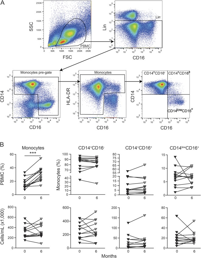
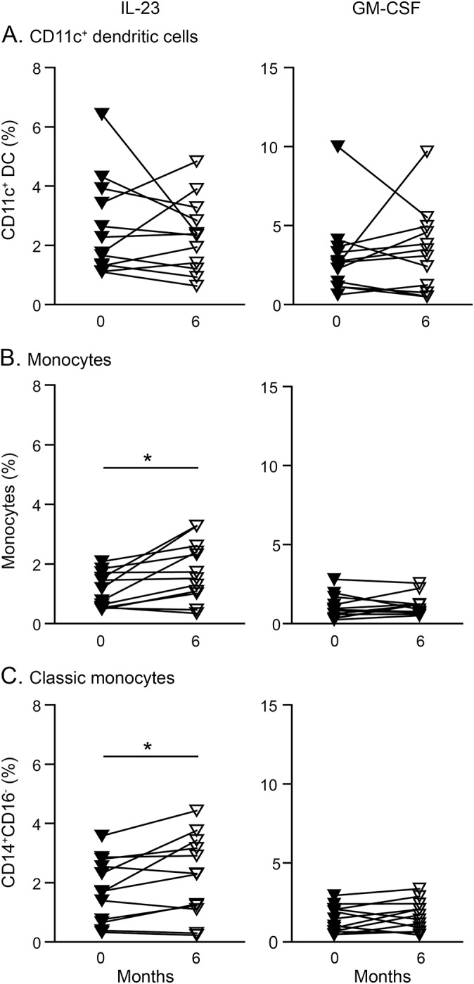
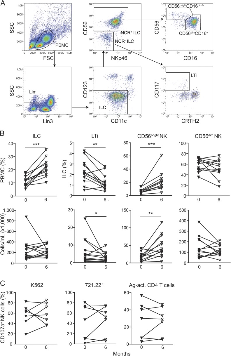
Methods: Circulating innate immune cells including myeloid cells and innate lymphoid cells (ILCs) were analyzed before and 6 and 12 months after onset of alemtuzumab treatment. Furthermore, a potential effect on granulocyte-macrophage colony-stimulating factor (GM-CSF) and interleukin (IL)-23 production by myeloid cells and natural killer (NK) cell cytolytic activity was determined.

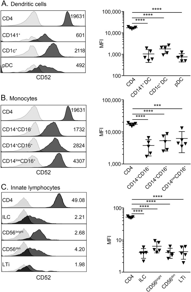
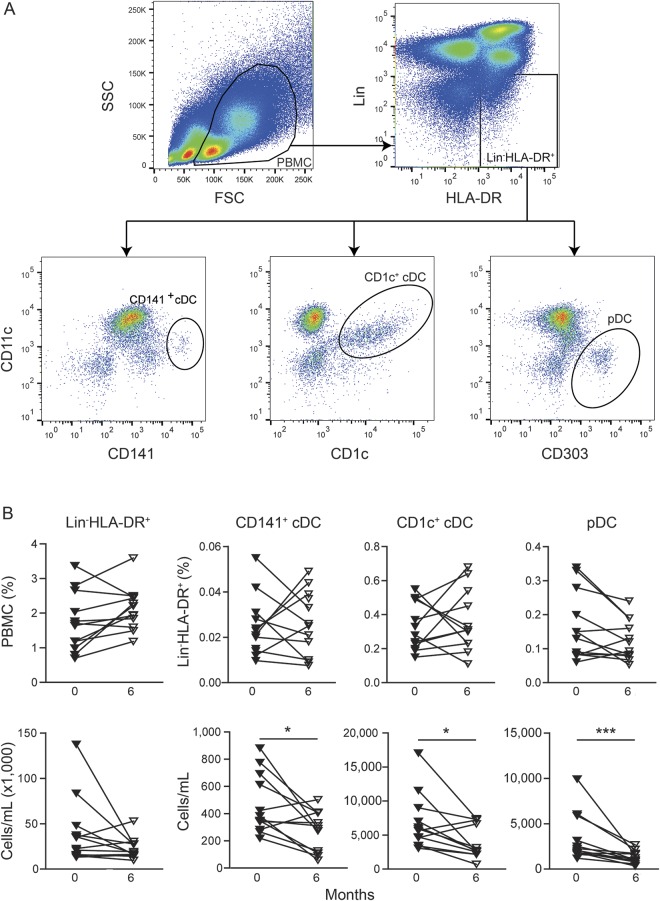
Results: In comparison to CD4+ T lymphocytes, myeloid and lymphoid innate cell subsets of patients with MS expressed significantly lower amounts of CD52 on their cell surface. Six months after CD52 depletion, numbers of circulating plasmacytoid dendritic cells (DCs) and conventional DCs were reduced compared to baseline. GM-CSF and IL-23 production in DCs remained unchanged. Within the ILC compartment, the subset of CD56bright NK cells specifically expanded under alemtuzumab treatment, but their cytolytic activity did not change.

Conclusions: Our findings demonstrate that 6 months after alemtuzumab treatment, specific DC subsets are reduced, while CD56bright NK cells expanded in patients with MS. Thus, alemtuzumab specifically restricts the DC compartment and expands the CD56bright NK cell subset with potential immunoregulatory properties in MS. We suggest that remodeling of the innate immune compartment may promote long-term efficacy of alemtuzumab and preserve immunocompetence in patients with MS.


Figures

Figure 1
Figure 1. CD52 expression on innate myeloid and lymphoid cell subsets
Peripheral blood mononuclear cells derived from treatment-naive patients with relapsing-remitting multiple sclerosis (n = 5, down triangles) were stained with fluorochrome-conjugated antibodies specific for (A) dendritic cell subsets, (B) monocyte subsets, and (C) innate lymphoid cell subsets as well as for CD52. CD52 expression on CD4+ T cells served as a positive control. Left: histograms of one representative donor show CD52 expression of the respective cell subset (dark gray) in comparison to the isotype control (light gray). Right: graphs display the median fluorescence intensities (MFI) of the respective cell subsets. Error bars indicate the SD. p Values were calculated by unpaired Student t test, ***p < 0.001, ****p < 0.0001. ILC = innate lymphoid cell; LTi = lymphoid tissue inducer cell.
Figure 2
Figure 2. Alemtuzumab-induced changes in the dendritic cell compartment
(A) Gating strategy for dendritic cells. Peripheral blood mononuclear cells (PBMCs) were gated by forward scatter vs side scatter characteristics. Lineage (CD3, CD14, CD19) negative cells expressing human leukocyte antigen–DR (HLA-DR) were further categorized into CD141+ conventional DCs, CD1c+ conventional DCs, and CD303+ plasmacytoid dendritic cells (pDCs). (B) Graphs display proportions of LinHLA-DR+ cells within PBMCs or the respective dendritic cell (DC) subsets within LinHLA-DR+ (upper row) and total cell numbers (lower row) of DC subsets derived from alemtuzumab-treated patients with relapsing-remitting multiple sclerosis (n = 12) at baseline (filled triangles) and 6-month follow-up (open triangles). p Values were calculated by paired Student t test or Wilcoxon matched-pairs signed rank test, respectively, *p < 0.05, **p < 0.01, ***p < 0.001.
Figure 3
Figure 3. Alemtuzumab-induced changes in monocytes
(A) Gating strategy for monocytes. Monocytes and their subsets were separated from the total lineage (CD3, CD19) negative compartment based on the expression of CD14, CD16, and human leukocyte antigen–DR (HLA-DR), as previously described. (B) Graphs display proportions of monocytes within peripheral blood mononuclear cells or the respective subsets within monocytes (upper row) and total cell numbers (lower row) of monocyte subsets derived from alemtuzumab-treated patients with relapsing-remitting multiple sclerosis (n = 12) at baseline (filled triangles) and 6-month follow-up (open triangles). p Values were calculated by paired Student t test or Wilcoxon matched-pairs signed rank test, respectively, *p < 0.05, **p < 0.01, ***p < 0.001.
Figure 4
Figure 4. Alemtuzumab-mediated effects on interleukin (IL)–23 and granulocyte-macrophage colony-stimulating factor (GM-CSF) production in innate myeloid cells
Graphs display proportions of IL-23 and GM-CSF producing (A) CD11c+ dendritic cells (DCs), (B) monocytes, and (C) CD14+CD16 classical monocytes derived from alemtuzumab-treated patients with relapsing-remitting multiple sclerosis (n = 12) at baseline (filled triangles) and 6-month follow-up. p Values were calculated by paired Student t test or Wilcoxon matched-pairs signed rank test, respectively, *p < 0.05.
Figure 5
Figure 5. Alemtuzumab-induced changes in the innate lymphoid cell (ILC) compartment
(A) Gating strategy for ILCs. ILCs were defined as lineage (CD3, CD14, CD19, CD20)–negative peripheral blood mononuclear cells (PBMCs) devoid for dendritic cell (DC) markers CD123 and CD11c. CD56brightCD16dim/− natural killer (NK) cells were gated from CD56+NKp46+ ILC, whereas CD117+CRTH2 lymphoid tissue inducer cells (LTis) were gated from CD56NKp46 ILCs. (B) Graphs display proportions of ILCs within PBMCs or ILC subsets within ILCs (upper row) and total cell numbers (lower row) of ILC subsets derived from alemtuzumab-treated patients with relapsing-remitting multiple sclerosis (n = 12) at baseline (filled triangles) and 6-month follow-up (open triangles). (C) Release of cytolytic granules by NK cells at baseline and 6 months after alemtuzumab therapy (n = 7) in response to K562 (left), 721.221 (middle), or antigen-activated allogenic CD4+ T cells. p Values were calculated by paired Student t test or Wilcoxon matched-pairs signed rank test, respectively, *p < 0.05, **p < 0.01, ***p < 0.001.

References

    1. Hale G, Xia MQ, Tighe HP, Dyer MJ, Waldmann H. The CAMPATH-1 antigen (CDw52). Tissue Antigens 1990;35:118–127. - PubMed
    1. Rao SP, Sancho J, Campos-Rivera J, et al. . Human peripheral blood mononuclear cells exhibit heterogeneous CD52 expression levels and show differential sensitivity to alemtuzumab mediated cytolysis. PLoS One 2012;7:e39416. - PMC - PubMed
    1. Hu Y, Turner MJ, Shields J, et al. . Investigation of the mechanism of action of alemtuzumab in a human CD52 transgenic mouse model. Immunology 2009;128:260–270. - PMC - PubMed
    1. Mone AP, Cheney C, Banks AL, et al. . Alemtuzumab induces caspase-independent cell death in human chronic lymphocytic leukemia cells through a lipid raft-dependent mechanism. Leukemia 2006;20:272–279. - PubMed
    1. Geissmann F, Manz MG, Jung S, Sieweke MH, Merad M, Ley K. Development of monocytes, macrophages, and dendritic cells. Science 2010;327:656–661. - PMC - PubMed