Review
. 2016 Apr 25;1(3):131-142..
doi: 10.1016/j.jacbts.2016.03.008. eCollection 2016 Apr.

Cross-Linking GPVI-Fc by Anti-Fc Antibodies Potentiates Its Inhibition of Atherosclerotic Plaque- and Collagen-Induced Platelet Activation

Affiliations
Review

Cross-Linking GPVI-Fc by Anti-Fc Antibodies Potentiates Its Inhibition of Atherosclerotic Plaque- and Collagen-Induced Platelet Activation

Janina Jamasbi et al. JACC Basic Transl Sci. .

Abstract

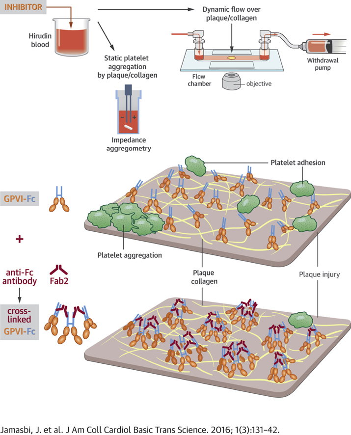
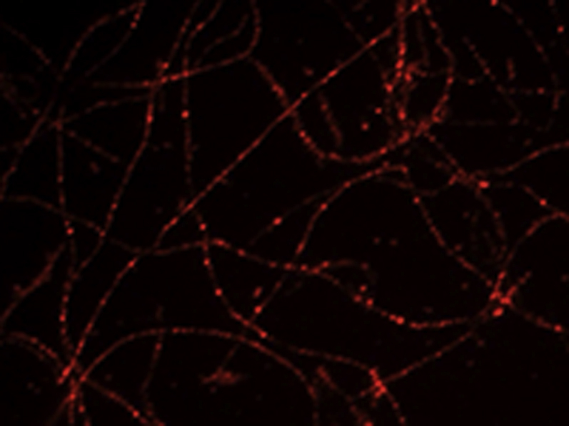
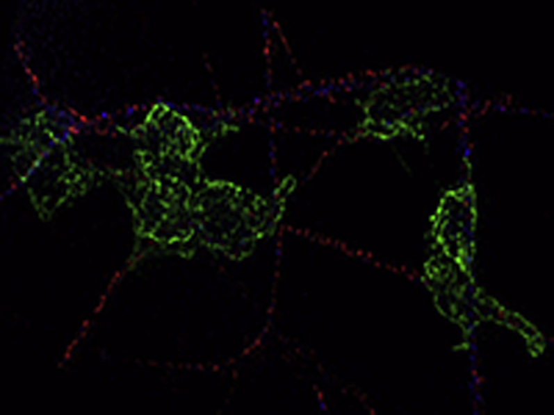
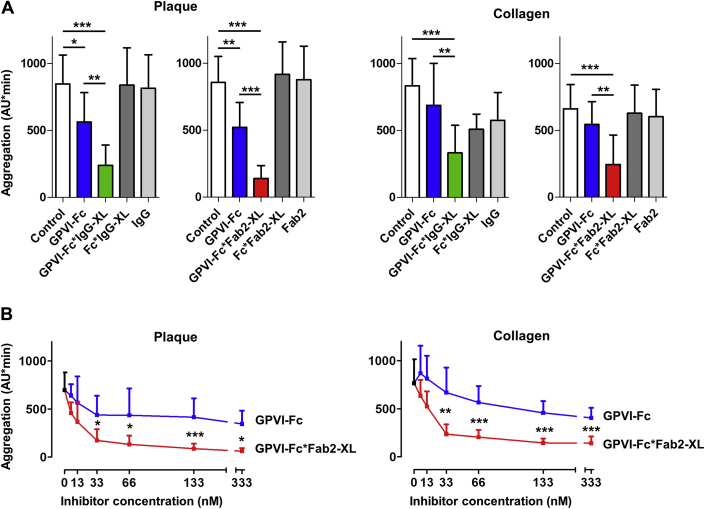
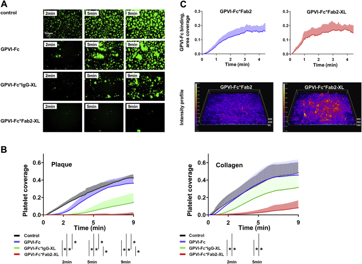
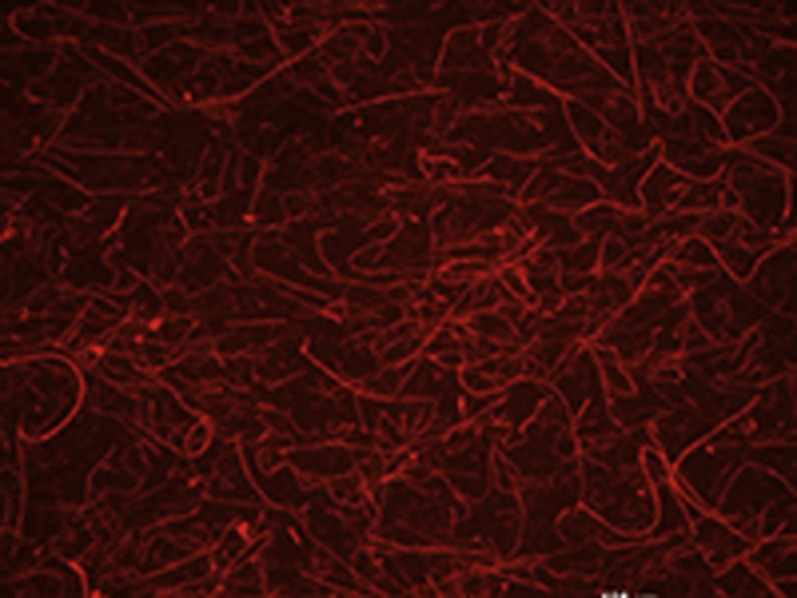
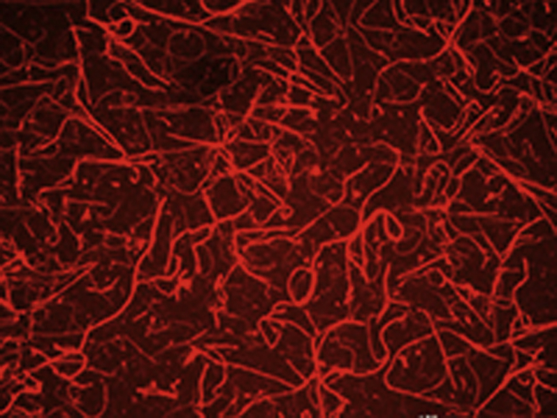
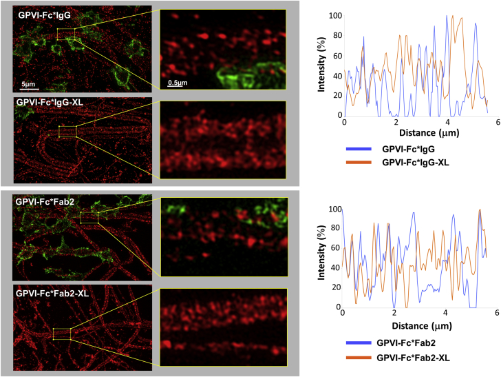
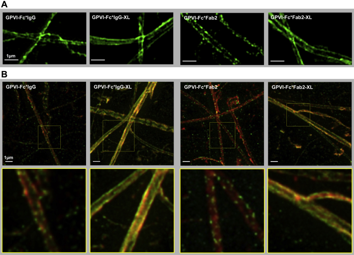
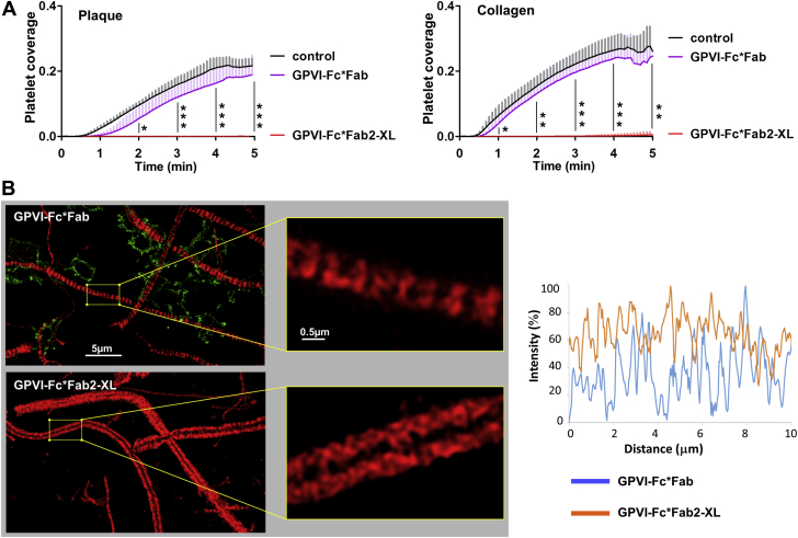
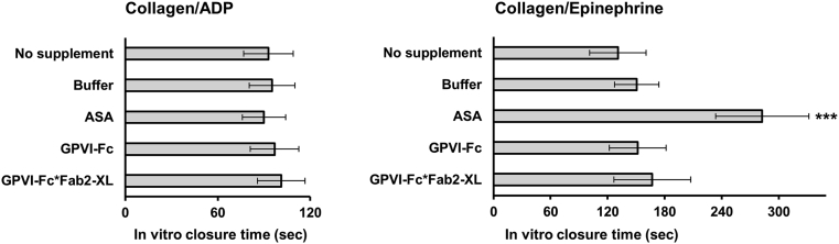
To enhance the antithrombotic properties of recombinant glycoprotein VI fragment crystallizable (GPVI-Fc), the authors incubated GPVI-Fc with anti-human Fc antibodies to cross-link the Fc tails of GPVI-Fc. Cross-linking potentiated the inhibition of human plaque- and collagen-induced platelet aggregation by GPVI-Fc under static and flow conditions without increasing bleeding time in vitro. Cross-linking with anti-human-Fc Fab2 was even superior to anti-human-Fc immunoglobulin G (IgG). Advanced optical imaging revealed a continuous sheath-like coverage of collagen fibers by cross-linked GPVI-Fc complexes. Cross-linking of GPVI into oligomeric complexes provides a new, highly effective, and probably safe antithrombotic treatment as it suppresses platelet GPVI-plaque interaction selectively at the site of acute atherothrombosis.

Keywords: Fc, fragment crystallizable; GPVI, glycoprotein VI; IgG, immunoglobulin G; PE, phycoerythrin; SIM, structured illumination microscopy; STED, stimulated emission depletion; XL, cross-linked; antithrombotic; atherothrombosis; glycoprotein VI; plaque rupture.


Figures

None
Graphical abstract
Figure 1
Figure 1
Dynamics of GPVI-Fc Binding and Platelet Adhesion to Collagen Under Flow Glycoprotein VI–fragment crystallizable (GPVI-Fc) (50 μg/ml final concentration; 333 nM) labeled with phycoerythrin (PE)-conjugated anti–human-Fc antibody (red) was added to blood containing abciximab (to inhibit platelet aggregation and allow only platelet adhesion) before perfusion over collagen (550/s). Differential interference contrast (DIC) and fluorescence images were taken by video microscopy (1 frame/2 s) using a 100× NA1.4 oil objective. Images are representative for 6 individual experiments. Upper rows: overlay of DIC (collagen/platelets) and fluorescence (PE-labeled GPVI-Fc) images. Bottom rows: fluorescence images of PE-labeled GPVI-Fc. See also Video 1. (A) A single platelet (thick arrow) rolls over PE-labeled GPVI-Fc (thin arrows in row below) bound to collagen. Two platelets (asterisks) adhere to segments of collagen fibers that contain less fluorescent GPVI-Fc. (B) A single platelet (arrow, top row) attaching downstream of the fiber and moving back against the blood flow displaces GPVI-Fc-PE from collagen (arrows in bottom row). (C) Structured illumination microscopy imaging. Platelets adhere to collagen segments carrying little GPVI-Fc. Collagen coated onto glass coverslips was stained with anti–collagen type I and type III antibody (Ab) and AlexaFluor405-conjugated secondary Ab (blue). PE-labeled GPVI-Fc (50 μg/ml) was added to blood containing Arg-Gly-Asp-Ser (to inhibit platelet aggregation and allow only platelet adhesion) before the start of perfusion (shear rate 550/s). After 4 min of flow, platelets were fixed and stained with anti-CD41 Ab and DyLight 488-conjugated secondary Ab (green). The fluorescence micrograph shows a maximum intensity projection of 0.15 μm z sections (total z 2.5 μm) of a structured illumination microscopy image (ELYRA PS.1; Carl Zeiss MicroImaging GmbH, Jena, Germany). i and ii, Identical pictures. i, Binding of GPVI-Fc (red) onto collagen fibers (blue). Green channel (platelets) was omitted. ii, Picture as in i but with platelets (green). Image is representative of 5 others from different experiments. Video 2 presents a 3-dimensional representation.
None
Online Video 1 GPVI-Fc binding to collagen GPVI-Fc (50μg/ml final concentration; 333nM) labeled with PE-conjugated anti-human-Fc antibody (red) was added to blood containing abciximab before perfusion over collagen (600/s). PE-labeled GPVI-Fc was detected by fluorescence microscopy (excitation 560nm, emission 605nm) using a 100x NA 1.4 oil objective. Flow direction: from right to left.
None
Online Video 2 Platelets adhere mainly to collagen segments carrying little GPVI-Fc Three-dimensional animation of the SIM image in Fig. 1 C processed with Huygens software. Platelet attachment (green) to collagen (blue) in the presence of GPVI-Fc (red).
Figure 2
Figure 2
Cross-Linking of GPVI-Fc With Anti–Human-Fc IgG and Anti–Human-Fc Fab2 Increases Inhibition of Static Platelet Aggregation Induced by Collagen or Plaque Compared With GPVI-Fc (A) Bar diagrams show the effects of glycoprotein VI–fragment crystallizable (GPVI-Fc) (20 μg/ml; 133 nM), GPVI-Fc, and Fc cross-linked (XL) with anti–human-Fc immunoglobulin G (IgG) (GPVI-Fc*IgG-XL, Fc*IgG-XL; 133 nM) (left panels) or with anti–human-Fc Fab2 (GPVI-Fc*Fab2-XL, Fc*Fab2-XL; 133 nM) (right panels) on plaque- or collagen-induced platelet aggregation. Further controls were anti–human-Fc IgG (133 nM) (left panels) and anti–human-Fc Fab2 (133 nM) (right panels). Cumulative platelet aggregation (AU*min) was measured by using multiple electrode aggregometry. Mean ± SD; n = 4. Repeated measures analysis of variance (overall p < 0.001) with secondary pair-wise comparisons (as indicated by bars) by Tukey correction. (B) Concentration dependency of the effects of GPVI-Fc*Fab2-XL on plaque- and collagen-induced platelet aggregation compared with GPVI-Fc. Mean ± SD; n = 4 to 6. Comparisons were made by using the Student t test or, if inappropriate, by using the Mann-Whitney U test (at 333 nM with plaque and at 33 nM with collagen). ∗p < 0.05; ∗∗p < 0.01; ∗∗∗p < 0.001.
Figure 3
Figure 3
Cross-Linking of GPVI-Fc With Anti–Human-Fc IgG or Anti–Human-Fc Fab2 Increases Its Inhibition of Platelet Aggregation Stimulated by Collagen or Plaque Under Flow Buffer (control), GPVI-Fc, or GPVI-Fc (50 μg/ml; 333 nM) premixed with equimolar anti–human-Fc IgG or anti–human-Fc Fab2 antibodies for crosslinking was added to blood containing DiOC6 for platelet visualization and perfused over plaque homogenate at a shear rate of 600/s. (A) Representative micrographs display platelet coverage of plaque at 2, 5, and 9 min after start of blood flow. (B) Effect of buffer, GPVI-Fc, GPVI-Fc*IgG-XL, or GPVI-Fc*Fab2-XL on the kinetics of platelet deposition from flowing blood onto plaque and collagen. Measurements are each second. Mean (solid line) ± SD (shaded area); n = 4 to 6. Comparison at 2, 5, and 9 min by using repeated measures analysis of variance or if inappropriate by repeated measures analysis of variance on ranks (only 2 min). Significance of secondary pair-wise comparisons by Tukey correction is indicated by bars. ∗p < 0.05. (C) Kinetics of not cross-linked and cross-linked GPVI-Fc binding to collagen. (Videos 3 and 4 present additional details.) GPVI-Fc pre-incubated with PE-labeled anti–human-Fc Fab2 (20:1 mol/mol) (GPVI-Fc*Fab2) or with an equimolar mixture of PE-labeled and unlabeled anti–human-Fc Fab2 (1:10) for cross-linking (GPVI-Fc*Fab2-XL), was added to blood (333 nM GPVI-Fc final concentration) containing abciximab and perfused over collagen at a shear rate of 600/s. Binding of PE-labeled GPVI-Fc (cross-linked or not cross-linked) to collagen was quantified every second by fluorescence microscopy using a 10× objective. Top: Time course of GPVI-Fc*Fab2 and GPVI-Fc*Fab2-XL binding to collagen (area coverage). Mean (solid line) ± SD (shaded area); n = 4. Bottom: Fluorescence intensity surface plots of GPVI-Fc*Fab2 and GPVI-Fc*Fab2-XL bound to collagen at 3 min after start of blood flow. Color gradients indicate increase of fluorescence intensity. Abbreviations as in Figures 1 and 2.
None
Online Video 3 Kinetics of not cross-linked GPVI-Fc binding to collagen under flow GPVI-Fc was incubated with PE-labeled anti-human-Fc Fab2 antibodies in a 20:1 molar ratio. The mixtures were added to blood (333nM GPVI-Fc f.c.) containing abciximab before perfusion over collagen at a shear rate of 600/sec for 3min. Binding of PE-labeled GPVI-Fc*Fab2 to collagen was recorded by fluorescence video microscopy (1frame/sec) using a 10x objective.
None
Online Video 4 Kinetics of cross-linked GPVI-Fc binding to collagen under flow GPVI-Fc was incubated with 10% PE-labeled and 90% unlabeled anti-human-Fc Fab2 antibodies in a 1:1 molar ratio for GPVI-Fc cross-linking. The mixtures were added to blood (333nM GPVI-Fc f.c.) containing abciximab before perfusion over collagen at a shear rate of 600/sec for 3min. Binding of PE-labeled GPVI-Fc*Fab2-XL to collagen was recorded by fluorescence video microscopy (1frame/sec) using a 10x objective.
Figure 4
Figure 4
SIM Imaging of Binding of GPVI-Fc (Not Cross-Linked and Cross-Linked) and Platelets to Collagen GPVI-Fc was incubated with anti–human-Fc antibodies in a 20:1 molar ratio (GPVI-Fc*IgG, GPVI-Fc*Fab2) or in a 1:1 molar ratio for crosslinking (GPVI-Fc*IgG-XL, GPVI-Fc*Fab2-XL). The antibodies contained 10% PE-conjugated anti–human-Fc IgG or anti–human-Fc Fab2. The mixtures were added to blood (333 nM GPVI-Fc) containing abciximab before perfusion over collagen at a shear rate of 600/s. After 4 min of flow, platelets were fixed and stained with anti-CD41 antibody and DyLight 488-conjugated secondary Ab. (Left) Structured illumination microscopy (SIM) images showing binding of PE-labeled GPVI-Fc (not cross-linked and cross-linked) (red) and platelets (green) to collagen with enlarged sections (×800) on the right. Maximum intensity projections of 0.15 μm z sections (total z 2.5 μm) are shown. (Right) Line intensity profiles (maximum intensity 100%) drawn along comparable, platelet-free segments of collagen fibers in the designated images (left). Images are representative of 5 different experiments. Binding of not cross-linked GPVI-Fc to collagen is discontinuous, showing fiber segments that are not occupied by GPVI-Fc (zero intensity in line profiles). Abbreviations as in Figures 1 and 2.
Figure 5
Figure 5
SIM and STED Imaging of Collagen Stained With Not Cross-Linked GPVI-Fc (GPVI-Fc*IgG, GPVI-Fc*Fab2) and Cross-Linked GPVI-Fc (GPVI-Fc*IgG-XL, GPVI-Fc*Fab2-XL) GPVI-Fc was incubated with Alexa Fluor 488-conjugated anti–human-Fc IgG or anti–human-Fc Fab2 antibodies in either a 10:1 molar ratio (not cross-linked GPVI-Fc) or a 1:1 molar ratio (cross-linked GPVI-Fc). The mixtures were then incubated with collagen coated glass coverslips. (A) SIM imaging. The SIM micrographs are maximum intensity projections of 0.15 μm z sections (total z 1.5 μm). The image is representative of 5 others from different experiments. (B) Stimulated emission depletion (STED) microscopy imaging. Collagen coated glass coverslips were stained simultaneously with AlexaFluor594-labeled anti-collagen type I and type III antibodies (red) in addition to GPVI-Fc, which was either labeled or cross-linked with anti–human-Fc antibodies. Yellow indicates co-localization of cross-linked GPVI-Fc with anti-collagen antibodies. Abbreviations as in Figures 1, 2, and 4.
Figure 6
Figure 6
GPVI-Fc*Fab in Contrast to GPVI-Fc*Fab2-XL Does Not Inhibit Platelet Aggregation Induced by Plaque and Collagen Under Flow: Functional and SIM Imaging Studies GPVI-Fc (100 μg/ml; 666 nM) was incubated with 3-fold molar excess of anti–human-Fc Fab-AlexaFluor594 (GPVI-Fc*Fab) or equimolar concentrations of anti–human-Fc Fab2 or anti-human-Fc Fab2–PE (GPVI-Fc*Fab2-XL). (A) Effect of GPVI-Fc*Fab and GPVI-Fc*Fab2-XL on the kinetic of platelet deposition onto plaque and collagen in flowing blood. Plaque homogenate and collagen coated onto coverslips were incubated with GPVI-Fc*Fab and GPVI-Fc*Fab2-XL complexes before perfusion with blood at a shear rate of 600/s. Fluorescence images of platelet deposition were continuously recorded (1 frame/5 s) and analyzed as detailed in the Methods section. Mean ± SD; n = 4. Comparison by using analysis of variance for repeated measures at full 2 to 5 min and secondary pair-wise comparisons by using Tukey correction. Significance of pair-wise comparison of GPVI-Fc*Fab2-XL to control and to GPVI-Fc*Fab was equal at all time points and is only indicated by a single line with asterisks. ∗p < 0.05; ∗∗p < 0.01; ∗∗∗p < 0.001. (B) SIM imaging of GPVI-Fc*Fab and GPVI-Fc*Fab2-XL binding and platelet adhesion to collagen fibers. Collagen-coated coverslips were incubated with GPVI-Fc*Fab-AlexaFluor594 or GPVI-Fc*Fab2-PE-XL (red) before perfusion with blood containing abciximab at a shear rate of 600/s. After 4 min of flow, platelets were fixed and stained with anti-CD41 antibody and DyLight 488-conjugated second Ab (green). (Left) SIM micrographs (maximum intensity projections of 0.15 μm z sections (total z 2.5 μm) with enlarged sections (×800) showing the different pattern of GPVI-Fc*Fab and GPVI-Fc*Fab2-XL binding (red) to collagen. Platelet adhesion (green) to collagen is observed in GPVI-Fc*Fab but not GPVI-Fc*Fab2-XL treated samples (left). Images are representative of 5 different experiments. (Right) Line intensity profiles (maximum intensity 100%) along comparable, platelet-free sections of collagen fibers in the designated images (left). Binding of GPVI-Fc*Fab to collagen is discontinuous with segments of fibers lacking significant binding (<10% in line profiles). Abbreviations as in Figures 1, 2, and 4.
Figure 7
Figure 7
GPVI-Fc*Fab2-XL Does Not Increase In Vitro Bleeding Blood was either pre-incubated with no supplement, buffer, aspirin (ASA) (300 μg/ml), GPVI-Fc (333 nM), or GPVI-Fc*Fab2-XL (333 nM) before transfer to collagen/adenosine diphosphate (ADP) or collagen/epinephrine cartridges and determination of the in vitro closure time with the platelet function analyzer PFA-200. Mean ± SD; n = 6. ***p < 0.001 by repeated measures analysis of variance and secondary pair-wise comparison of ASA versus all other conditions. Abbreviations as in Figure 2.
Figure 8
Figure 8
Model of GPVI-Fc and Cross-Linked GPVI-Fc Binding to Plaque Collagen Cross-linking of dimeric glycoprotein VI–fragment crystallizable (GPVI-Fc) fusion protein (GPVI, brown; Fc, blue) by anti–Fc Fab2 creates oligomeric (n = 2 to 4) GPVI-Fc molecules, which bind to their corresponding motifs on collagen fibers more densely (bottom) than GPVI-Fc (top).

References

    1. Fuster V., Moreno P.R., Fayad Z.A., Corti R., Badimon J.J. Atherothrombosis and high-risk plaque: part I: evolving concepts. J Am Coll Cardiol. 2005;46:937–954. - PubMed
    1. Badimon L., Vilahur G. Thrombosis formation on atherosclerotic lesions and plaque rupture. J Intern Med. 2014;276:618–632. - PubMed
    1. van Zanten G.H., de Graaf S., Slootweg P.J. Increased platelet deposition on atherosclerotic coronary arteries. J Clin Invest. 1994;93:615–632. - PMC - PubMed
    1. Penz S., Reininger A.J., Brandl R. Human atheromatous plaques stimulate thrombus formation by activating platelet glycoprotein VI. FASEB J. 2005;19:898–909. - PubMed
    1. Reininger A.J., Bernlochner I., Penz S.M. A 2-step mechanism of arterial thrombus formation induced by human atherosclerotic plaques. J Am Coll Cardiol. 2010;55:1147–1158. - PubMed