Review
. 2017 Feb;133(2):223-244.
doi: 10.1007/s00401-016-1631-4. Epub 2016 Oct 20.

Multiple sclerosis: experimental models and reality

Affiliations
Review

Multiple sclerosis: experimental models and reality

Hans Lassmann et al. Acta Neuropathol. 2017 Feb.

Abstract

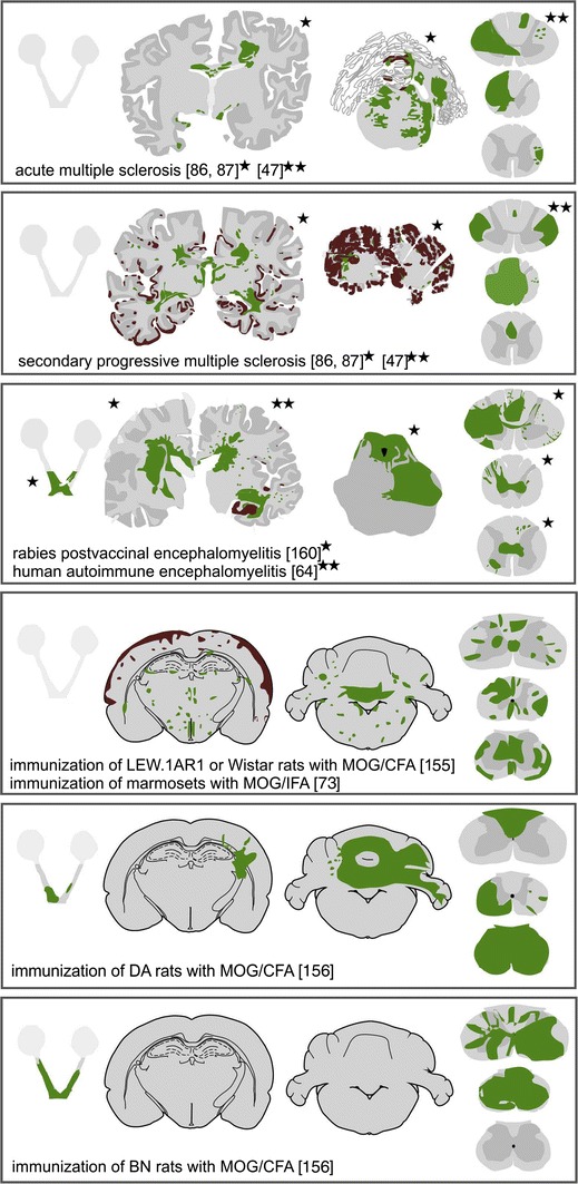
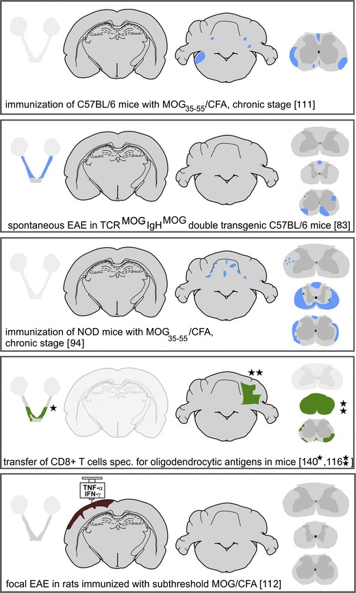
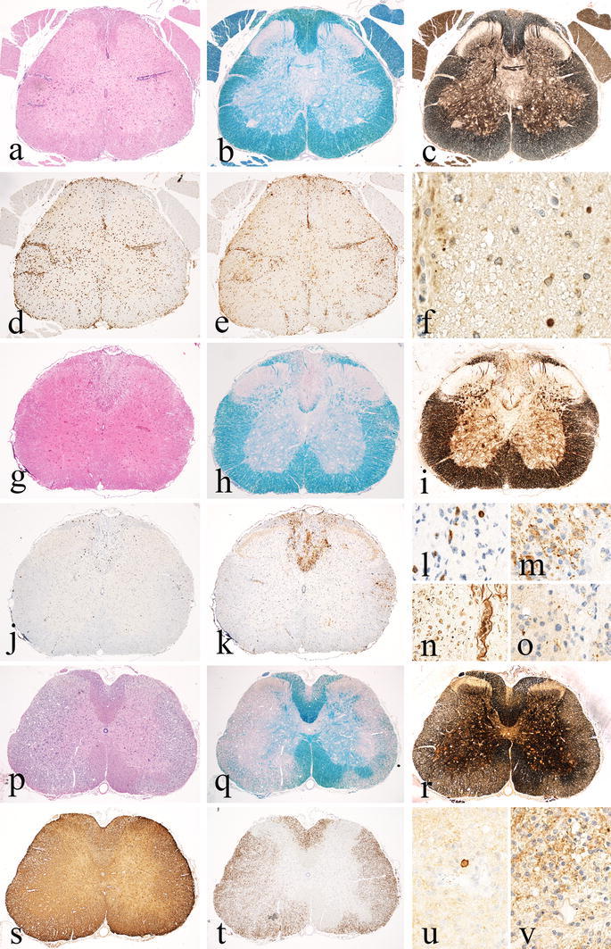
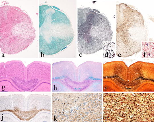
One of the most frequent statements, provided in different variations in the introduction of experimental studies on multiple sclerosis (MS), is that "Multiple sclerosis is a demyelinating autoimmune disease and experimental autoimmune encephalomyelitis (EAE) is a suitable model to study its pathogenesis". However, so far, no single experimental model covers the entire spectrum of the clinical, pathological, or immunological features of the disease. Many different models are available, which proved to be highly useful for studying different aspects of inflammation, demyelination, remyelination, and neurodegeneration in the central nervous system. However, the relevance of results from such models for MS pathogenesis has to be critically validated. Current EAE models are mainly based on inflammation, induced by auto-reactive CD4+ T-cells, and these models reflect important aspects of MS. However, pathological data and results from clinical trials in MS indicate that CD8+ T-cells and B-lymphocytes may play an important role in propagating inflammation and tissue damage in established MS. Viral models may reflect key features of MS-like inflammatory demyelination, but are difficult to use due to their very complex pathogenesis, involving direct virus-induced and immune-mediated mechanisms. Furthermore, evidence for a role of viruses in MS pathogenesis is indirect and limited, and an MS-specific virus infection has not been identified so far. Toxic models are highly useful to unravel mechanisms of de- and remyelination, but do not reflect other important aspects of MS pathology and pathogenesis. For all these reasons, it is important to select the right experimental model to answer specific questions in MS research.


Figures

Fig. 1
Fig. 1
Distribution of demyelinating lesions in MS and different EAE-based models. The sites of demyelinated lesions were shown in camera lucida drawings of human brain sections, were projected into schemes redrawn after Paxinos and Watson [124] for rat and murine brain sections, or were outlined in optic nerve and spinal cord schemes. Areas of primary demyelination are shown in green, lesions with dominant axonal loss, and secondarily demyelinated areas in blue, and cortical demyelination in brown. Shaded schemes indicate lack of sufficient information for lesion distribution
Fig. 1
Fig. 1
Distribution of demyelinating lesions in MS and different EAE-based models. The sites of demyelinated lesions were shown in camera lucida drawings of human brain sections, were projected into schemes redrawn after Paxinos and Watson [124] for rat and murine brain sections, or were outlined in optic nerve and spinal cord schemes. Areas of primary demyelination are shown in green, lesions with dominant axonal loss, and secondarily demyelinated areas in blue, and cortical demyelination in brown. Shaded schemes indicate lack of sufficient information for lesion distribution
Fig. 2
Fig. 2
Basic patterns of pathology in different MS Models Part 1. Pure inflammatory models exemplified by passive transfer of CD4+ T-cells directed against myelin basic protein (MBP) in the Lewis rat. Spinal cord with massive inflammation reflected by the presence of perivenous inflammatory cuffs and diffuse infiltration of the tissue by T-cells and macrophages (a H&E; d CD3; e macrophage marker ED1). Sections stained for myelin (b, Luxol fast blue) or axons (c Bielschowsky silver impregnation) do not show demyelination or axonal loss, but there are some axons with accumulation of amyloid precursor protein (f APP), indicating a mild-to-moderate degree of (in part reversible) axonal injury. Models with chronic inflammatory axonopathy leading to focal lesions with secondary demyelination. As an example, spinal cord pathology of a NOD mouse with chronic EAE, 90 days after active sensitization with myelin oligodendrocyte glycoprotein peptide (MOG35–55), is shown. A confluent inflammatory demyelinated lesion is present in the dorsal column of the spinal cord (g H&E, h Luxol fast blue). There is nearly complete axonal loss within the lesion (i, n Bielschowsky silver impregnation); the lesion is infiltrated by a moderate number of T-cells (j, l CD3) and shows a broad rim of activated macrophages at the lesion edges (k Mac3); ongoing tissue destruction is shown by the presence of myelin protein reactive degradation products in macrophages (m PLP) and by the presence of numerous axons with disturbed fast axonal transport (intra-axonal accumulation of amyloid precursor protein; o APP). Chronic EAE in the DA rat 60 days after active immunization with full-length recombinant myelin oligodendrocyte glycoprotein as a model for extensive inflammatory demyelinating disease; profound inflammation (p H&E) and widespread confluent demyelination (q Luxol fast blue), but nearly complete axonal preservation (r Bielschowsky silver impregnation) and pronounced astrogliosis (s GFAP); the areas of active demyelination are highly infiltrated by macrophages (t ED1), but contain only very few T-lymphocytes (u CD3); and myelin sheaths and myelin degradation products in macrophages are decorated by activated complement (v C9neo antigen)
Fig. 3
Fig. 3
Basic patterns of pathology in different MS models Part 2. MHV-induced spinal cord pathology as an example for inflammatory demyelinating lesions with extensive oligodendrocyte and astrocyte loss; large confluent demyelinated lesions in the lateral column of the spinal cord with inflammation and tissue edema (a H&E), complete demyelination (b Luxol fast blue), and nearly complete axonal preservation (c, d Bodian silver impregnation), but nearly complete loss of astrocytes (e GFAP); some of the astrocytes at the lesion edge contain virus antigen (f). The cuprizone model as an example for toxic demyelination; large hyper-cellular demyelinating lesion in the corpus callosum after 6 weeks of cuprizone exposure (g H&E). The lesion shows complete demyelination (h Luxol fast blue) and only mild or moderate axonal loss (i, l Bielschowsky silver impregnation); the site of active demyelination is highly infiltrated by macrophages and activated microglia (j Mac3); oligodendrocytes are lost within the areas of active demyelination and only preserved in the areas with intact myelin (k CNPase)

References

    1. Alvord EC. Acute disseminated encephalomyelitis and “allergic” neuroencephalopathies. In: Vinken PY, Bruyn GW, editors. Handbook of clinical neurology. New York: Elsevier; 1970. pp. 500–571.
    1. Anderson AC, Chandvaskar R, Lee DH, Sullivan JM, Solomon A, Rodriguez-Manzanet R, Greve B, Sobel RA, Kuchroo VK. A transgenic model of central nervous system autoimmunity mediated by CD4+ and CD8+ T and B cells. J Immunol. 2012;188:2084–2092. doi: 10.4049/jimmunol.1102186. - DOI - PMC - PubMed
    1. Anderson JM, Hampton DW, Patani R, Pryce G, Crowther RA, Reynolds RA, Franklin RJ, Giovannoni G, Compston DA, Baker D, Spillantini MG, Chandran S. Abnormally phosphorylated tau is associated with neuronal and axonal loss in experimental autoimmune encephalomyelitis and multiple sclerosis. Brain. 2008;131:1736–1748. doi: 10.1093/brain/awn119. - DOI - PubMed
    1. Ascherio A, Munger KL. Epidemiology of multiple sclerosis. From risk factor to prevention—an update. Semin Neurol. 2016;36:103–114. doi: 10.1055/s-0036-1579693. - DOI - PubMed
    1. Bailey OT, Pappenheimer AM, Sargent F, Cheever MD, Daniels JB. A murine virus (JHM) causing disseminated encephalomyelitis with extensive destruction of myelin. II. Pathology. J Exp Med. 1949;90:195–212. doi: 10.1084/jem.90.3.195. - DOI - PMC - PubMed

Publication types