

Lung adenocarcinoma with anomalous bronchi and pulmonary veins preoperatively identified by computed tomography
- PMID: 27766780
- PMCID: PMC5130314
- DOI: 10.1111/1759-7714.12362
Lung adenocarcinoma with anomalous bronchi and pulmonary veins preoperatively identified by computed tomography
Abstract
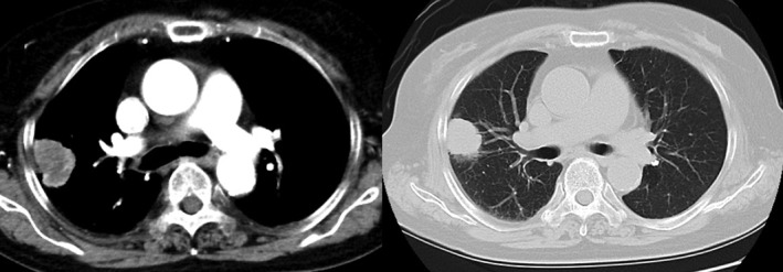
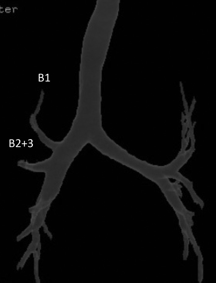
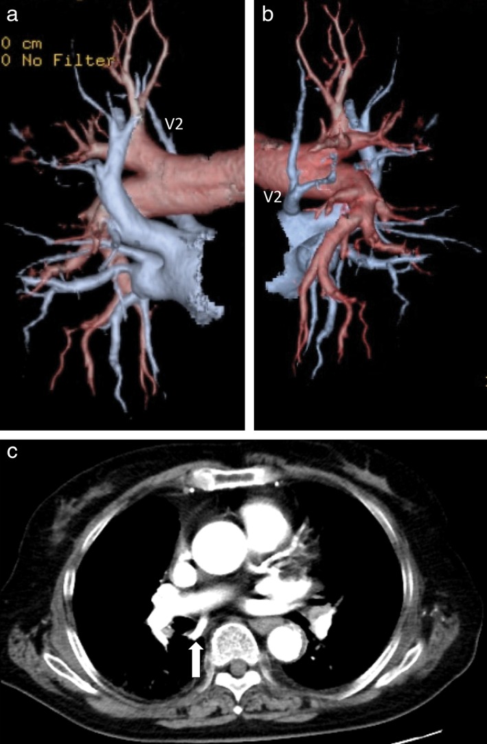
A 69-year-old woman visited our hospital complaining of right chest pain. Chest computed tomography showed a 55 × 45 mm tumor in the right upper lobe. Bronchoscopy revealed displaced anomalous B 1 and B 2+3 arising from the right main bronchus, and the patient was diagnosed with lung adenocarcinoma by transbronchial lung biopsy from the displaced B 2+3 . Three-dimensional computed tomography with multiplanar reconstruction revealed a displaced anomalous B 1 and B 2+3 branching directly from the right main bronchus, respectively, and abnormal distribution of the aberrant pulmonary vein (V 2 ) descended dorsally to the right main bronchus and emptied into the left atrium. Video-assisted right upper lobectomy with nodal dissection was successfully performed. Attention should be paid to the anomalous bronchus and pulmonary vessels for safer lung cancer operations, especially for video-assisted thoracic surgery.
Keywords: Abnormal distribution of the pulmonary vein; bronchial anomaly; lung cancer.
© 2016 The Authors. Thoracic Cancer published by China Lung Oncology Group and John Wiley & Sons Australia, Ltd.
Figures
References
-
- Asai K, Urabe N, Yajima K, Suzuki K, Kazui T. Right upper lobe venous drainage posterior to the bronchus intermedius: Preoperative identification by computed tomography. Ann Thorac Surg 2005; 79: 1866–71. - PubMed
-
- Yamashita S, Goto T, Mori T et al. Video‐assisted thoracic surgery for lung cancer: Republication of a systematic review and a proposal by the guidelines committee of the Japanese Association for Chest Surgery 2014. Gen Thorac Cardiovasc Surg 2014; 62: 701–5. - PubMed
-
- Tsuboi M, Asamura H, Naruke H, Nakayama H, Kondo H, Tsuchiya R. A VATS lobectomy for lung cancer in a patient with an anomalous pulmonary vein: Report of a case. Surg Today 1997; 27: 1074–6. - PubMed
-
- Spaggiari L, Solli P, Leo F, Veronesi G, Pastorino U. Anomalous segmental vein for right upper lobe: An unusual anatomical variation. Ann Thorac Surg 2002; 74: 267. - PubMed
-
- Yurugi Y, Nakamura H, Taniguchi Y et al. Case of thoracoscopic right upper lobectomy for lung cancer with tracheal bronchus and pulmonary vein variation. Asian J Endosc Surg 2012; 5: 93–5. - PubMed
Publication types
MeSH terms
LinkOut - more resources
Full Text Sources
Other Literature Sources
Medical
