. 2016 Nov;26(11):1212-1225.
doi: 10.1038/cr.2016.119. Epub 2016 Oct 21.

Inhibitory phosphorylation of Cdk1 mediates prolonged prophase I arrest in female germ cells and is essential for female reproductive lifespan

Affiliations

Inhibitory phosphorylation of Cdk1 mediates prolonged prophase I arrest in female germ cells and is essential for female reproductive lifespan

Deepak Adhikari et al. Cell Res. 2016 Nov.

Abstract

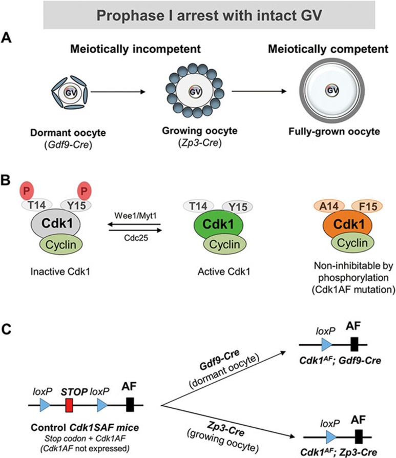
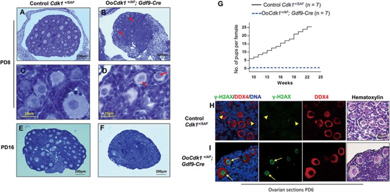
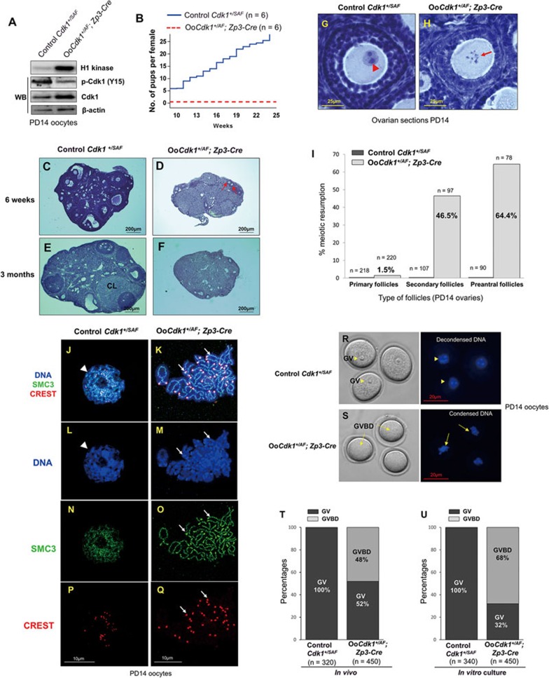
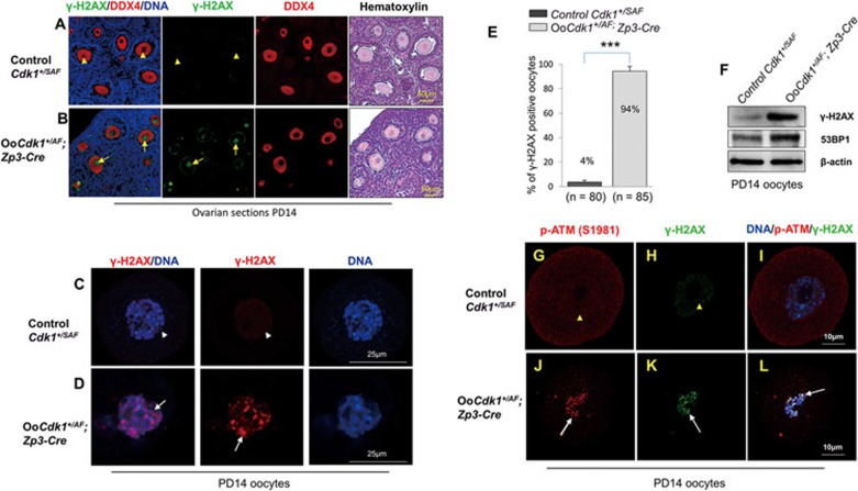
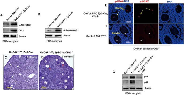
A unique feature of female germ cell development in mammals is their remarkably long arrest at the prophase of meiosis I, which lasts up to 50 years in humans. Both dormant and growing oocytes are arrested at prophase I and completely lack the ability to resume meiosis. Here, we show that the prolonged meiotic arrest of female germ cells is largely achieved via the inhibitory phosphorylation of Cdk1 (cyclin-dependent kinase 1). In two mouse models where we have introduced mutant Cdk1T14AY15F which cannot be inhibited by phosphorylation (Cdk1AF) in small meiotically incompetent oocytes, the prophase I arrest is interrupted, leading to a premature loss of female germ cells. We show that in growing oocytes, Cdk1AF leads to premature resumption of meiosis with condensed chromosomes and germinal vesicle breakdown followed by oocyte death, whereas in dormant oocytes, Cdk1AF leads to oocyte death directly, and both situations damage the ovarian reserve that maintains the female reproductive lifespan, which should be around 1 year in mice. Furthermore, interruption of the inhibitory phosphorylation of Cdk1 results in DNA damage, which is accompanied by induction of the Chk2 (checkpoint kinase 2)-p53/p63-dependent cell death pathway, which eventually causes global oocyte death. Together, our data demonstrate that the phosphorylation-mediated suppression of Cdk1 activity is one of the crucial factors that maintain the lengthy prophase arrest in mammalian female germ cells, which is essential for preserving the germ cell pool and reproductive lifespan in female mammals.


Figures

Figure 1
Figure 1
Illustrations of oocyte prophase I arrest and the strategy for expressing the Cdk1AF allele in dormant and growing oocytes. (A) All of the dormant and growing oocytes remain in prophase I arrest with GV and are meiotically incompetent. Only the fully grown oocytes are meiotically competent and have the capability of resuming meiosis. Gdf9-Cre recombinase starts to be expressed in dormant oocytes in primordial follicles, and Zp3-Cre starts to be expressed only in growing oocytes in developing follicles. GV, germinal vesicle. (B) Cdk1 remains in an inactive state due to its phosphorylation at T14 and Y15 by Wee1/Myt1. Cdk1 activation requires dephosphorylation of T14 and Y15 by Cdc25. Cdk1 with mutation of T14 and Y15 to A14 and F15 cannot be phosphorylated and therefore Cdk1 activity cannot be inhibited by Wee1/Myt1. (C) Oocyte-specific expression of the Cdk1AF allele starting from dormant oocytes as mediated by Gdf9-Cre or starting from growing oocytes as mediated by Zp3-Cre.
Figure 2
Figure 2
Expression of Cdk1AF in dormant oocytes causes DNA damage and oocyte depletion in OoCdk1+/AF; Gdf9-Cre mice. (A-F) Hematoxylin-stained sections of mouse ovary at PD8 and PD16. By PD8, only a few follicles were left in OoCdk1+/AF; Gdf9-Cre ovaries (B arrows), and the oocytes of surviving primordial follicles remained arrested at GV stage (D, arrows). The follicular structures had mostly disappeared in the mutant ovaries by PD16 (F). As controls, normal ovaries of PD8 (A and C) and PD16 (E) Cdk1+/SAF mice containing healthy follicles are shown. The experiments were repeated more than three times each, and for each time and each age ovaries from one mouse of each genotype were used. (G) Comparison of the average cumulative number of pups per OoCdk1+/AF; Gdf9-Cre female (dotted blue line) and per Cdk1+/SAF female (solid black line), indicating that the OoCdk1+/AF; Gdf9-Cre females were infertile. The numbers of females used are shown as n. (H and I) Prominent γ-H2AX staining was observed in dormant oocytes enclosed in primordial follicles of PD6 OoCdk1+/AF; Gdf9-Cre ovaries (I, arrows) but not in the control oocytes (H, arrowheads). DDX4 was used to label the oocytes and the same sections were counterstained with hematoxylin to visualize ovarian histology. The experiments were repeated more than three times each.
Figure 3
Figure 3
Premature in vivo resumption of meiosis in small growing oocytes in OoCdk1+/AF; Zp3-Cre mice. (A) Elevated Cdk1 kinase activity and total Cdk1 level but decreased inhibitory phosphorylation of Cdk1 (p-Cdk1, Y15) were observed in OoCdk1+/AF;Zp3-Cre oocytes, suggesting that Cdk1AF was successfully introduced into the growing oocytes in the mutant mice. β-Actin was used as the loading control. For the Cdk1 kinase assay, 10 oocytes per reaction were used. For Western blots (WB), lysate from 200 oocytes was loaded in each lane. The experiments were repeated more than three times each. (B) Comparison of the average cumulative number of pups per OoCdk1+/AF; Zp3-Cre female (red dotted line) and per Cdk1+/SAF female (solid blue line). All OoCdk1+/AF; Zp3-Cre females were infertile. The numbers of females are shown as n. (C-F) Hematoxylin-stained sections from mouse ovary at 6 weeks and 3 months. By 6 weeks of age, only a few follicles were observed in OoCdk1+/AF; Zp3-Cre ovaries (D, arrows), and healthy follicular structures had mostly disappeared in 3-month-old OoCdk1+/AF;Zp3-Cre ovaries (F). Control Cdk1+/SAF ovaries (C and E) contained healthy oocytes and follicles. CL, corpus luteum. The experiments were repeated more than three times each, and for each time and each age; ovaries from one mouse of each genotype were used. (G, H) Premature in vivo GVBD and chromosome condensation in small primary oocytes of OoCdk1+/AF; Zp3-Cre mice. Ovaries from PD14 OoCdk1+/AF; Zp3-Cre and Cdk1+/SAF females were embedded in paraffin, and serial sections of 8-μm thickness were prepared and stained with hematoxylin. (G) A wild-type primary oocyte in a secondary follicle showing the GV (G, arrowhead) from a PD14 Cdk1+/SAF ovary. (H) A representative growing primary oocyte from a PD14 OoCdk1+/AF; Zp3-Cre ovary where GVBD and chromosome condensation had already occurred (H, arrow). The experiments were repeated more than three times each, and for each time and each age ovaries from one mouse of each genotype were used. (I) Quantification of the percentages of oocytes that resumed meiosis in vivo in PD14 ovaries within primary, secondary, and preantral follicles. Numbers of each type of follicles included (n) are shown. (J-Q) Condensed chromosomes with cohesin localization at the chromosome axis in PD14 OoCdk1+/AF; Zp3-Cre oocytes. Staining with DAPI for DNA (L, M, blue), SMC3 for cohesin (N, O, green), CREST for kinetochores (P, Q, red), and merge (J, K) in OoCdk1+/AF; Zp3-Cre and Cdk1+/SAF oocytes. The experiments were repeated more than three times each, and the representative images from one experiment are shown. (R, S) Increased GVBD in mutant OoCdk1+/AF; Zp3-Cre oocytes upon release from the follicular environment. In wild-type PD14 Cdk1+/SAF oocytes, GV was maintained even if the oocytes were released from the follicles by enzymatic digestion (R, arrowheads). However, an increased rate of GVBD was seen in PD14 OoCdk1+/AF; Zp3-Cre oocytes upon release from the follicles (S, arrows) by enzymatic digestion of ovaries from PD14 mice. Representative Hoechst-stained oocyte nuclei are shown to the right. Quantification of GV and GVBD rates of oocytes immediately after their isolation from follicles in PD14 Cdk1+/SAF and OoCdk1+/AF; Zp3-Cre mice (T) and after further in vitro culture (U). The numbers of oocytes analyzed (n) are shown.
Figure 4
Figure 4
Expression of Cdk1AF leads to DNA damage and ATM activation in growing OoCdk1+/AF; Zp3-Cre oocytes. (A) Absence of γ-H2AX staining in the oocytes of control PD14 Cdk1+/SAF ovarian sections (arrowheads). (B) Prominent γ-H2AX staining in the oocytes of PD14 OoCdk1+/AF; Zp3-Cre ovaries (arrows). DDX4 was used to label the oocytes and the same sections were counterstained with hematoxylin to visualize ovarian histology. The experiments were repeated more than three times each, and representative images are shown. (C) Absence of γ-H2AX staining in isolated oocytes of control PD14 Cdk1+/SAF mice (arrowhead). (D) Strong γ-H2AX staining in isolated oocytes from PD14 OoCdk1+/AF; Zp3-Cre ovaries (arrow). (E) Quantification of γ-H2AX-positive oocytes that were isolated from PD14 Cdk1+/SAF and OoCdk1+/AF; Zp3-Cre ovaries. Numbers of each genotype of oocytes checked (n) are shown. (F) Western blot for γ-H2AX and 53BP1 levels in oocytes isolated from PD14 Cdk1+/SAF and OoCdk1+/AF; Zp3-Cre ovaries. β-actin was used as the loading control. Lysate from 200 oocytes was loaded in each lane. The experiments were repeated more than three times. (G-L) High level of p-ATM (S1981) accompanied by increased γ-H2AX staining in PD14 OoCdk1+/AF; Zp3-Cre oocytes (J-L, arrows), in comparison with PD14 control Cdk1+/SAF oocytes where there was almost no signal for p-ATM (S1981) or γ-H2AX (G-I, arrowheads). The experiments were repeated more than three times each, and representative images are shown.
Figure 5
Figure 5
Cdk1AF triggers the Chk2-p63/p53 and cell death pathways in OoCdk1+/AF; Zp3-Cre oocytes. (A) Western blot for p-Chk2 (T68) and Chk2 in oocytes isolated from PD14 Cdk1+/SAF and OoCdk1+/AF; Zp3-Cre ovaries. (B) Western blot for active caspase 3 in oocytes isolated from PD14 Cdk1+/SAF and OoCdk1+/AF; Zp3-Cre ovaries. β-actin was used as the loading control. Lysate from 200 oocytes was loaded in each lane. The experiments were repeated more than three times each. (C, D) Hematoxylin-stained sections of mouse ovary at 2 month of age. Rescue of follicle depletion by the concomitant deletion of Chk2 in OoCdk1+/AF; Zp3-Cre ovaries. By 2 months of age, although healthy follicular structures and oocytes had mostly disappeared in mutant OoCdk1+/AF; Zp3-Cre ovaries (C), concomitant deletion of Chk2 largely prevented the death of follicles in double-mutant OoCdk1+/AF; Zp3-Cre ; Chk2−/− ovaries, and many surviving oocytes could be observed (D, arrows). The experiments were repeated more than three times each, and for each time and each age ovaries from one mouse of each genotype were used. (E) Surviving oocytes in PD60 double-mutant OoCdk1+/AF; Zp3-Cre ; Chk2−/− ovaries stained positive for γ-H2AX (arrows) indicating that DNA damage is still present. (F) Control Cdk1+/SAF ovaries with γ-H2AX-negative oocytes. The experiments were repeated more than three times, and representative images are shown. (G) Western blot for p63 and p53 levels in oocytes isolated from PD14 Cdk1+/SAF, OoCdk1+/AF; Zp3-Cre, and OoCdk1+/AF; Zp3-Cre ; Chk2−/− ovaries. β-actin was used as the loading control, and lysate from 200 oocytes was loaded in each lane. The experiments were repeated more than three times.

Similar articles

Cited by

References

    1. Nagaoka SI, Hassold TJ, Hunt PA. Human aneuploidy: mechanisms and new insights into an age-old problem. Nat Rev Genet 2012; 13:493–504. - PMC - PubMed
    1. Zheng W, Zhang H, Gorre N, Risal S, Shen Y, Liu K. Two classes of ovarian primordial follicles exhibit distinct developmental dynamics and physiological functions. Hum Mol Genet 2014; 23:920–928. - PMC - PubMed
    1. McGee EA, Hsueh AJ. Initial and cyclic recruitment of ovarian follicles. Endocr Rev 2000; 21:200–214. - PubMed
    1. Adhikari D, Liu K. Molecular mechanisms underlying the activation of mammalian primordial follicles. Endocr Rev 2009; 30:438–464. - PubMed
    1. Eppig JJ, Viveiros MM, Bivens CM, De La Fuente R. Regulation of mammalian oocyte maturation. In: Leung PC, Adashi EY, eds. The Ovary. Academic Press: Amsterdam 2004:113–129.

MeSH terms