. 2017 Feb 15;595(4):1143-1158.
doi: 10.1113/JP273097. Epub 2016 Dec 12.

FoxO-dependent atrogenes vary among catabolic conditions and play a key role in muscle atrophy induced by hindlimb suspension

Affiliations

FoxO-dependent atrogenes vary among catabolic conditions and play a key role in muscle atrophy induced by hindlimb suspension

Lorenza Brocca et al. J Physiol. .

Abstract

Key points: Muscle atrophy is a debilitating condition that affects a high percentage of the population with a negative impact on quality of life. Dissecting the molecular level of the atrophy process, and the similarities/dissimilarities among different catabolic conditions, is a necessary step for designing specific countermeasures to attenuate/prevent muscle loss. The FoxO family transcription factors represent one of the most important regulators of atrophy programme stimulating the expression of many atrophy-related genes. The findings of the present study clearly indicate that the signalling network controlling the atrophy programme is specific for each catabolic condition.

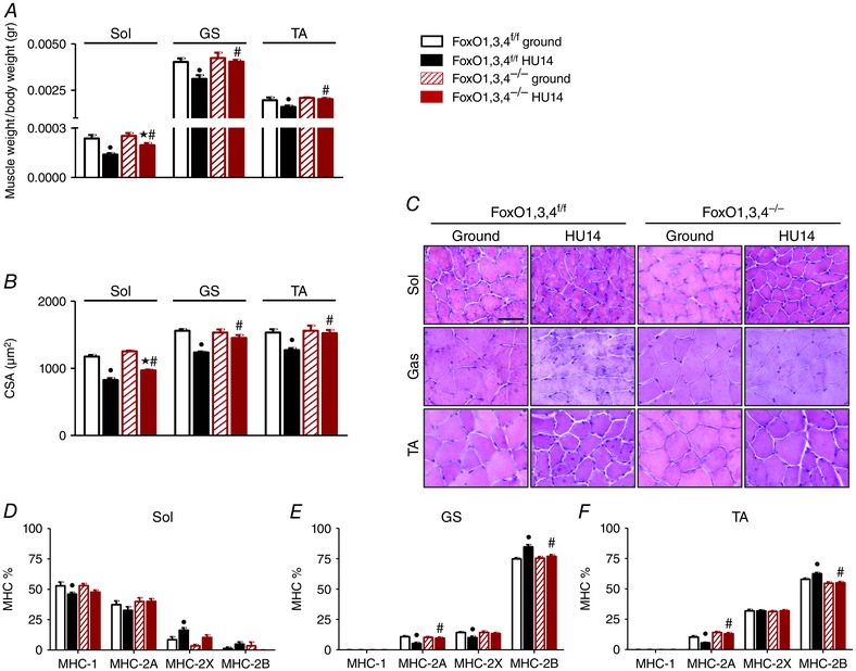
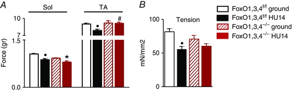
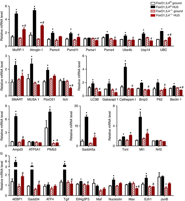
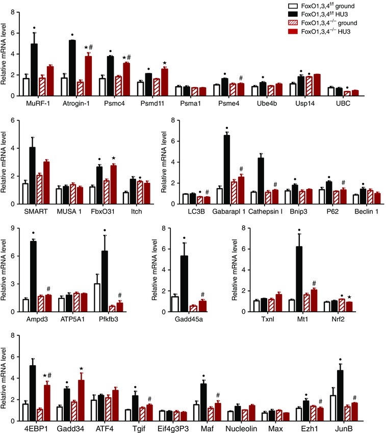
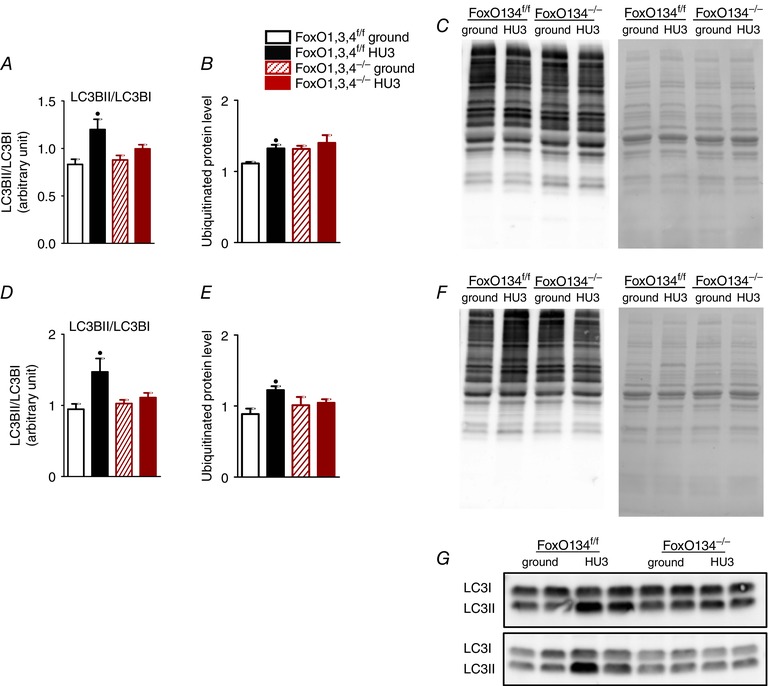
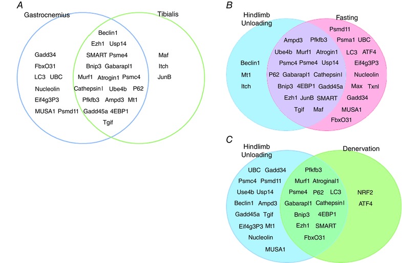
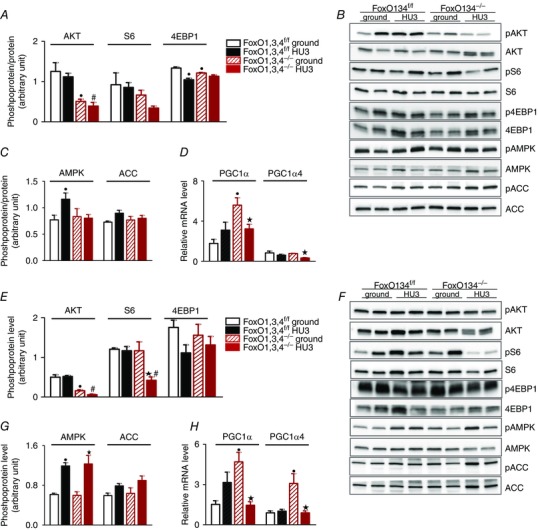
Abstract: Muscle atrophy is a complex process that is in common with many different catabolic diseases including disuse/inactivity and ageing. The signalling pathways that control the atrophy programme in the different disuse/inactivity conditions have not yet been completely dissected. The inhibition of FoxO is considered to only partially spare muscle mass after denervation. The present study aimed: (i) to determine the involvement of FoxOs in hindlimb suspension disuse model; (ii) to define whether the molecular events of protein breakdown are shared among different unloaded muscles; and finally (iii) to compare the data obtained in this model with another model of inactivity such as denervation. Both wild-type and muscle-specific FoxO1,3,4 knockout (FoxO1,3,4-/- ) mice were unloaded for 3 and 14 days and muscles were characterized by functional, morphological, biochemical and molecular assays. The data obtained show that FoxOs are required for muscle loss and force drop during unloading. Moreover, we found that FoxO-dependent atrogenes vary in different unloaded muscles and that they diverge from denervation. The findings of the present study clearly indicate that the signalling network that controls the atrophy programme is specific for each catabolic condition.

Keywords: atrogenes regulation; muscle atrophy; muscle disuse.


Figures

Figure 1
Figure 1. FoxOs deletion preserves muscle mass and prevents fibre‐type switch during HU
A, determination of muscle weight/body weight in Sol, GS and TA from FoxO1,3,4f/f and FoxO1,3,4−/− mice in ground condition and after 14 days of unloading (HU14). B, quantification of average fibre area (CSA) of Sol, GS and TA from FoxO1,3,4 f/f and FoxO1,3,4−/− in ground condition and after 14 days of unloading (HU14). C, representative cryosections stained with haematoxylin and eosin; scale bar = 100 μm. D, E and F, MHC isoforms distribution of Sol, GS and TA from FoxO1,3,4f/f and FoxO1,3,4−/− in ground condition and after 14 days of unloading (HU14) (n = 4 in each group). Significantly different from WT‐g (P < 0.05). Significantly different from KO‐g (P < 0.05). #Significantly different from WT‐HU14 (P < 0.05). Data are presented as the mean ± SEM.
Figure 2
Figure 2. FoxOs deletion prevents muscle weakness induced by HU
A, force measured in Sol and TA ex vivo muscles from FoxO1,3,4f/f and FoxO1,3,4−/− mice in ground condition and after 14 days of unloading (HU14). B, tension of GS single muscle fibres from FoxO1,3,4f/f and FoxO1,3,4−/− mice in ground condition and after 14 days of unloading (HU14) (n = 4 in each group). Significantly different from WT‐g (P < 0.05). #Significantly different from WT‐HU14 (P < 0.05). Data are presented as the mean ± SEM.
Figure 3
Figure 3. FoxOs inhibition prevents induction of atrogenes during HU in GS
Quantitative RT‐PCR of the indicated atrogenes in GS from FoxO1,3,4f/f and FoxO1,3,4−/− mice in ground condition and after 3 days of unloading (HU3) (gene functions are reported in Table 3). Data are normalized to GAPDH (n = 4 in each group). Significantly different from WT‐g (P < 0.05). Significantly different from KO‐g (P < 0.05). #Significantly different from WT‐HU3 (P < 0.05). Data are presented as the mean ± SEM.
Figure 4
Figure 4. FoxOs inhibition prevents atrogenes induction during HU in tibialis
Quantitative RT‐PCR of the indicated atrogenes in tibialis from FoxO1,3,4f/f and FoxO1,3,4−/− mice in ground condition and after 3 days of unloading (HU3) (gene functions are reported in Table 3). Data are normalized to GAPDH (n = 4 in each group). Significantly different from WT‐g (P < 0.05). Significantly different from KO‐g (P < 0.05). #Significantly different from WT‐HU3 (P < 0.05). Data are presented as the mean ± SEM.
Figure 5
Figure 5. FoxOs deletion blocks the ubiquitin proteasome and autophagy lysosome systems
A, quantification of protein levels of LC3BII in gastrocnemius from FoxO1,3,4f/f and FoxO1,3,4−/− mice in ground condition and after 3 days of unloading (HU3) based on the ratio between the content in forms II and I of LC3 by western blotting. B, relative content and representative blot (C) of polyubiquitinated proteins in GS muscle. D, quantification of protein levels of LC3BII in TA. E, relative content and representative blot (F) of polyubiquitinated proteins in TA. G, representative blot of LC3 in GS (upper) and TA (bottom) n = 4 in each group. Significantly different from WT‐g (P < 0.05). #Significantly different from WT‐HU3 (P < 0.05). Data are presented as the mean ± SEM.
Figure 6
Figure 6. FoxO‐dependent atrogenes vary in different muscles and catabolic conditions
A, FoxO‐dependent atrogenes common in gastrocnemius and tibialis muscle following hindlimb suspension. B, FoxO‐dependent atrogenes common in HU and fasting condition (TA muscle). C, FoxO‐dependent atrogenes common in HU and denervation condition (GS muscle).
Figure 7
Figure 7. Pathways involved in maintenance of muscle mass
Determination of p‐AKT, p‐S6 and p‐4EBP1 levels in GS (A) and TA (E) muscle from FoxO1,3,4f/f and FoxO1,3,4−/− mice in ground condition and after 3 days of HU (HU3) by western blot analysis. Representative western blot of synthetic factors in GS (B) and TA (F). Determination of p‐AMPK and p‐ACC levels in GS (C) and TA (G) muscle from FoxO1,3,4f/f and FoxO1,3,4−/− mice in ground condition and following HU3 by western blotting. Quantification of PGC1α and PGC1α4 mRNA levels in GS (D) and TA (H) from FoxO1,3,4f/f and FoxO1,3,4−/− mice in ground condition and following HU3 by RT‐PCR. Data are normalized to GAPDH (RT‐PCR). The activity levels of AKT, S6R and 4EBP1 were calculated as the ratio between the content in the phosphorylated (p) and total forms (n = 4 in each group). Significantly different from WT‐g (P < 0.05). Significantly different from KO‐g (P < 0.05). #Significantly different from WT‐HU3 (P < 0.05). Data are presented as the mean ± SEM.

Comment in

References

    1. Blaauw B, Canato M, Agatea L, Toniolo L, Mammucari C, Masiero E, Abraham R, Sandri M, Schiaffino S & Reggiani C (2009). Inducible activation of Akt increases skeletal muscle mass and force without satellite cell activation. FASEB J 23, 3896–3905. - PubMed
    1. Bodine SC & Baehr LM (2014). Skeletal muscle atrophy and the E3 ubiquitin ligases MuRF1 and MAFbx/atrogin‐1. Am J Physiol Endocrinol Metab 307, E469–E484. - PMC - PubMed
    1. Bodine SC, Latres E, Baumhueter S, Lai VK, Nunez L, Clarke BA, Poueymirou WT, Panaro FJ, Na E, Dharmarajan K, Pan ZQ, Valenzuela DM, DeChiara TM, Stitt TN, Yancopoulos GD & Glass DJ (2001). Identification of ubiquitin ligases required for skeletal muscle atrophy. Science 294, 1704–1708. - PubMed
    1. Bonaldo P & Sandri M (2013). Cellular and molecular mechanisms of muscle atrophy. Dis Model Mech 6, 25–39. - PMC - PubMed
    1. Bottinelli R & Reggiani C (2000). Human skeletal muscle fibres: molecular and functional diversity. Prog Biophys Mol Biol 73, 195–262. - PubMed

Publication types

MeSH terms

Substances