. 2017;55(3):1069-1082.
doi: 10.3233/JAD-160834.

Association of Kidney Function Biomarkers with Brain MRI Findings: The BRINK Study

Affiliations

Association of Kidney Function Biomarkers with Brain MRI Findings: The BRINK Study

Prashanthi Vemuri et al. J Alzheimers Dis. 2017.

Abstract

Background: Chronic kidney disease (CKD) studies have reported variable prevalence of brain pathologies, in part due to low inclusion of participants with moderate to severe CKD.

Objective: To measure the association between kidney function biomarkers and brain MRI findings in CKD.

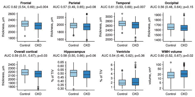
Methods: In the BRINK (BRain IN Kidney Disease) study, MRI was used to measure gray matter volumes, cerebrovascular pathologies (white matter hyperintensity (WMH), infarctions, microhemorrhages), and microstructural changes using diffusion tensor imaging (DTI). We performed regression analyses with estimated glomerular filtration rate (eGFR) and urine albumin to creatinine ratio (UACR) as primary predictors, and joint models that included both predictors, adjusted for vascular risk factors.

Results: We obtained 240 baseline MRI scans (150 CKD with eGFR <45 in ml/min/1.73 m2; 16 mild CKD: eGFR 45-59; 74 controls: eGFR≥60). Lower eGFR was associated with greater WMH burden, increased odds of cortical infarctions, and worsening diffusion changes throughout the brain. In eGFR models adjusted for UACR, only cortical infarction associations persisted. However, after adjusting for eGFR, higher UACR provided additional information related to temporal lobe atrophy, increased WMH, and whole brain microstructural changes as measured by increased DTI mean diffusivity.

Conclusions: Biomarkers of kidney disease (eGFR and UACR) were associated with MRI brain changes, even after accounting for vascular risk factors. UACR adds unique additional information to eGFR regarding brain structural and diffusion biomarkers. There was a greater impact of kidney function biomarkers on cerebrovascular pathologies and microstructural brain changes, suggesting that cerebrovascular etiology may be the primary driver of cognitive impairment in CKD.

Keywords: Cerebrovascular disease; chronic kidney disease; infarctions; magnetic resonance imaging.


Figures

Fig. 1
Fig. 1
Box plots of regional and overall thicknesses, hippocampal and ventricle volumes divided by total intracranial volume (TIV), and white matter hyperintensity (WMH) volumes. WMH volume is on the log scale to account for skew in the data. Group-wise differences are summarized by the non-parametric area under the receiver operating characteristic curve (AUC) and p-value based on the Wilcoxon rank-sum/Mann-Whitney U test.
Fig. 2
Fig. 2
Percentage change (95% confidence interval, CI) in MRI outcome measures thickness (A), volume (B), and fractional anisotropy (FA) and MD (C) for a 10% decrease in eGFR or 10% increase in UACR. Estimates are from linear regression models adjusting for age, sex, education, race, diabetes, hypertension, cholesterol, systolic blood pressure, diastolic blood pressure, stroke/TIA, AFIB, CVD, smoking, and alcohol use. We fit models with GFR alone and UACR alone, and finally both GFR and UACR in the same model. The left panels show the effect of eGFR alone (black square) and the effect of eGFR after adjusting for UACR from the joint model (light blue triangle). The right panels show the effect of UACR alone (black square) and the effect of UACR after adjusting for eGFR from the joint model (light blue triangle).
Fig. 3
Fig. 3
RR and OR (95% CI) for microhemorrhage (MCH) and infarctions (respectively) for a 10% decrease in eGFR or 10% increase in UACR after adjusting for age, sex, education, race, diabetes, hypertension, cholesterol, systolic blood pressure, diastolic blood pressure, stroke/TIA, AFIB, CVD, smoking, and alcohol use. We fit models with GFR alone and UACR alone, and finally both GFR and UACR in the same model. The left panel shows the effect of eGFR alone and the effect of eGFR after adjusting for UACR from the joint model (black square). The right panels show the effect of UACR alone and the effect of UACR after adjusting for eGFR from the joint model (light blue triangle).

References

    1. Knopman DS, Mosley TH, Jr, Bailey KR, Jack CR, Jr, Schwartz GL, Turner ST. Associations of microal-buminuria with brain atrophy and white matter hyperintensities in hypertensive sibships. J Neurol Sci. 2008;271:53–60. - PMC - PubMed
    1. Kurella Tamura M, Wadley V, Yaffe K, McClure LA, Howard G, Go R, Allman RM, Warnock DG, McClellan W. Kidney function and cognitive impairment in US adults: The Reasons for Geographic and Racial Differences in Stroke (REGARDS) Study. Am J Kidney Dis. 2008;52:227–234. - PMC - PubMed
    1. Murray AM. Cognitive impairment in the aging dialysis and chronic kidney disease populations: An occult burden. Adv Chronic Kidney Dis. 2008;15:123–132. - PMC - PubMed
    1. Seliger SL, Siscovick DS, Stehman-Breen CO, Gillen DL, Fitzpatrick A, Bleyer A, Kuller LH. Moderate renal impairment and risk of dementia among older adults: The Cardiovascular Health Cognition Study. J Am Soc Nephrol. 2004;15:1904–1911. - PubMed
    1. Yaffe K, Lindquist K, Shlipak MG, Simonsick E, Fried L, Rosano C, Satterfield S, Atkinson H, Windham BG, Kurella-Tamura M. Cystatin C as a marker of cognitive function in elders: Findings from the health ABC study. Ann Neurol. 2008;63:798–802. - PMC - PubMed

Publication types

MeSH terms