. 2017 Feb 15;123(4):617-628.
doi: 10.1002/cncr.30385. Epub 2016 Oct 21.

An independent validation of a gene expression signature to differentiate malignant melanoma from benign melanocytic nevi

Affiliations

An independent validation of a gene expression signature to differentiate malignant melanoma from benign melanocytic nevi

Loren E Clarke et al. Cancer. .

Abstract

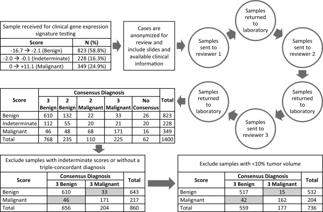
Background: Recently, a 23-gene signature was developed to produce a melanoma diagnostic score capable of differentiating malignant and benign melanocytic lesions. The primary objective of this study was to independently assess the ability of the gene signature to differentiate melanoma from benign nevi in clinically relevant lesions.

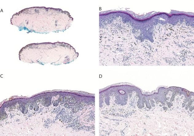
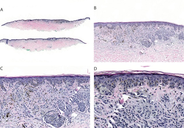
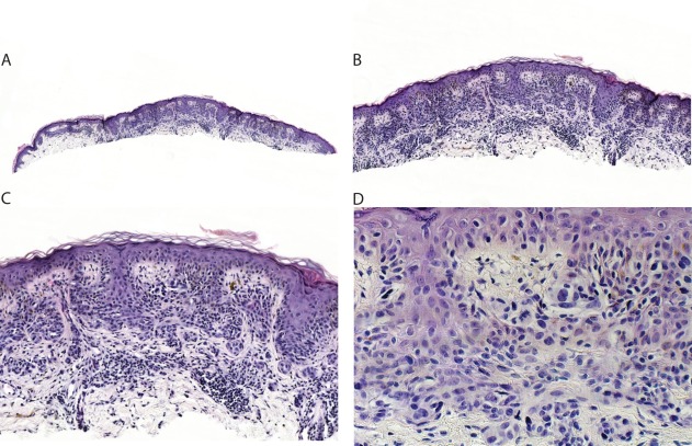
Methods: A set of 1400 melanocytic lesions was selected from samples prospectively submitted for gene expression testing at a clinical laboratory. Each sample was tested and subjected to an independent histopathologic evaluation by 3 experienced dermatopathologists. A primary diagnosis (benign or malignant) was assigned to each sample, and diagnostic concordance among the 3 dermatopathologists was required for inclusion in analyses. The sensitivity and specificity of the score in differentiating benign and malignant melanocytic lesions were calculated to assess the association between the score and the pathologic diagnosis.

Results: The gene expression signature differentiated benign nevi from malignant melanoma with a sensitivity of 91.5% and a specificity of 92.5%.

Conclusions: These results reflect the performance of the gene signature in a diverse array of samples encountered in routine clinical practice. Cancer 2017;123:617-628. © 2016 American Cancer Society.

Keywords: clinical validation; gene expression; melanoma; molecular diagnosis; reverse transcription-polymerase chain reaction.


Figures

Figure 1
Figure 1
Schematic of the gene expression testing, case review, tumor volume evaluation, and validation. False‐positives and false‐negatives are highlighted in gray.
Figure 2
Figure 2
This lesion from the back of a 72‐year‐old man was submitted with a pretest diagnosis of “favor junctional dysplastic nevus with moderate atypia.” Each of the 3 reviewing dermatopathologists classified the lesion as malignant melanoma (either in situ melanoma or lentigo maligna). The score was + 4.6. After the test, the submitting dermatopathologist changed the diagnosis to lentigo maligna and recommended excision with appropriate margins. Hematoxylin‐eosin; original magnification x20 (A), x100 (B), x200 (C), and x400 (D).
Figure 3
Figure 3
This lesion from the abdomen of a 36‐year‐old female was submitted with a pretest diagnosis of “favor traumatized atypical nevus.” All 3 of the reviewing dermatopathologists classified the lesion as melanoma. The score was + 1.1. The submitting dermatopathologist changed the diagnosis to atypical melanocytic proliferation. Hematoxylin‐eosin; original magnification x20 (A), x100 (B), x200 (C), and x400 (D).
Figure 4
Figure 4
This lesion from a 74‐year‐old male (anatomic site unknown) was submitted with a pretest diagnosis of “indeterminate; favor irritated compound nevus with moderate to severe atypia.” All 3 reviewing dermatopathologists classified the lesion as melanoma. The score was + 2.4. After the test, the submitting dermatopathologist changed the diagnosis to superficial spreading melanoma. Hematoxylin‐eosin; original magnification x20 (A), x100 (B), x200 (C), and x400 (D).
Figure 5
Figure 5
This lesion from the back of a 58‐year‐old male was submitted with a pretest diagnosis of “benign; favor severely dysplastic nevus.” All 3 reviewing dermatopathologists classified the lesion as melanoma. The score was + 2.2. After the test, the submitting dermatopathologist changed the diagnosis to melanoma in situ. Hematoxylin‐eosin; original magnification x20 (A), x100 (B), x200 (C), and x400 (D).
Figure 6
Figure 6
This lesion from the back of a 26‐year‐old female was submitted as “indeterminate; favor atypical Spitz tumor.” All 3 reviewing dermatopathologists classified the lesion as a benign Spitz nevus. The score was –7.2. Hematoxylin‐eosin; original magnification x20 (A), x100 (B), x200 (C), and x400 (D).
Figure 7
Figure 7
This lesion from the neck of a 71‐year‐old female was submitted as “indeterminate; favor dysplastic nevus with severe dysplasia versus evolving melanoma.” The 3 reviewing dermatopathologists all favored a benign diagnosis, but 2 of the 3 dermatopathologists noted that they could not entirely exclude early melanoma and would recommend re‐excision. The score was + 3.1. This case was counted as a false‐positive. However, note the atypical mitotic figure within a melanocyte in the papillary dermis. On the basis of the score, the submitting dermatopathologist changed his diagnosis to superficial spreading melanoma. Hematoxylin‐eosin; original magnification x20 (A), x100 (B), x200 (C), and x400 (D).
Figure 8
Figure 8
This lesion from the scalp of a 52‐year‐old male was submitted as lentigo maligna. The 3 reviewing dermatopathologists all categorized the lesion as malignant melanoma in situ. The score was –2.3. This case is an example of a false‐negative. Hematoxylin‐eosin; original magnification x20 (A), x100 (B), x200 (C), and x400 (D).

References

    1. American Cancer Society . Cancer Facts & Figures 2015. Atlanta, GA: American Cancer Society; 2015.
    1. American Cancer Society . What are the survival rates for melanoma skin cancer by stage? http://www.cancer.org/cancer/skincancer-melanoma/detailedguide/melanoma-.... Accessed January 21, 2016.
    1. Shoo BA, Sagebiel RW, Kashani‐Sabet M. Discordance in the histopathologic diagnosis of melanoma at a melanoma referral center. J Am Acad Dermatol. 2010;62:751–756. - PubMed
    1. Hawryluk EB, Sober AJ, Piris A, et al. Histologically challenging melanocytic tumors referred to a tertiary care pigmented lesion clinic. J Am Acad Dermatol. 2012;67:727–735. - PubMed
    1. Veenhuizen KC, De Wit PE, Mooi WJ, Scheffer E, Verbeek AL, Ruiter DJ. Quality assessment by expert opinion in melanoma pathology: experience of the pathology panel of the Dutch Melanoma Working Party. J Pathol. 1997;182:266–272. - PubMed

Publication types

Substances