. 2016 Dec;590(23):4381-4392.
doi: 10.1002/1873-3468.12467. Epub 2016 Nov 3.

X-linked inhibitor of apoptosis-associated factor 1 regulates TNF receptor 1 complex stability

Affiliations

X-linked inhibitor of apoptosis-associated factor 1 regulates TNF receptor 1 complex stability

Boren Lin et al. FEBS Lett. 2016 Dec.

Abstract

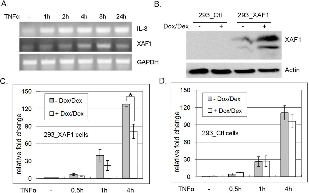
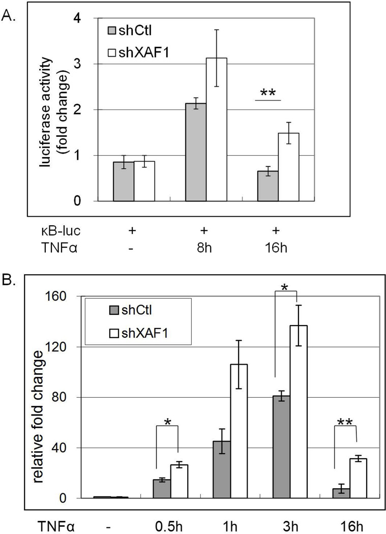
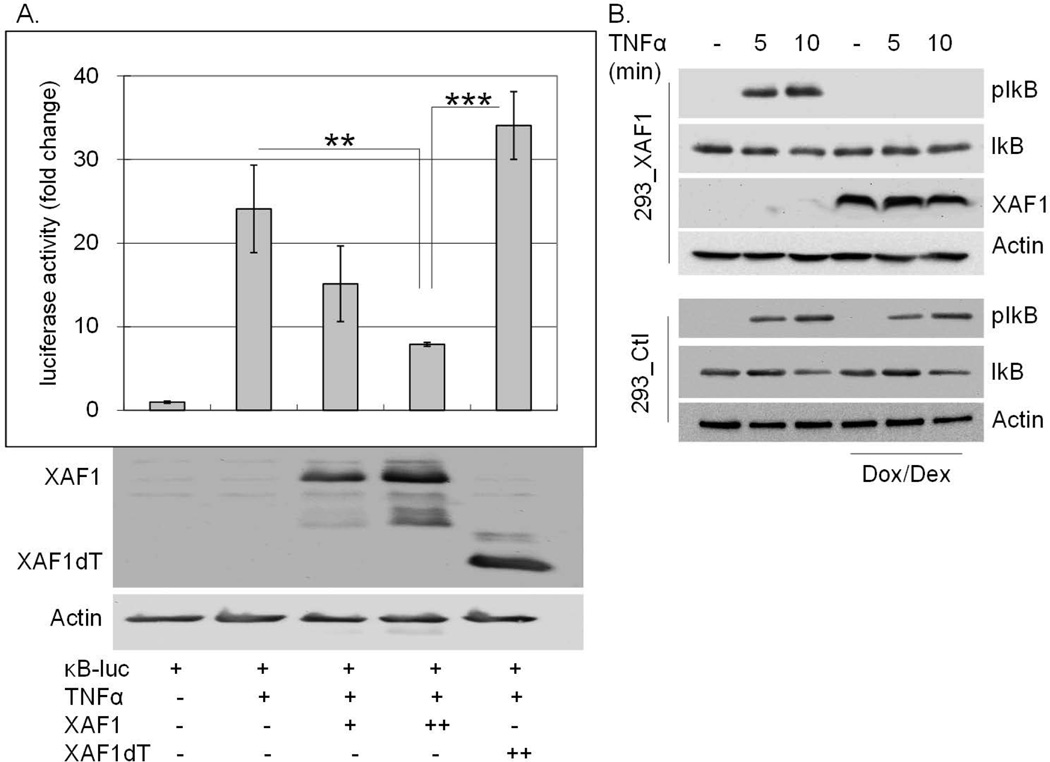
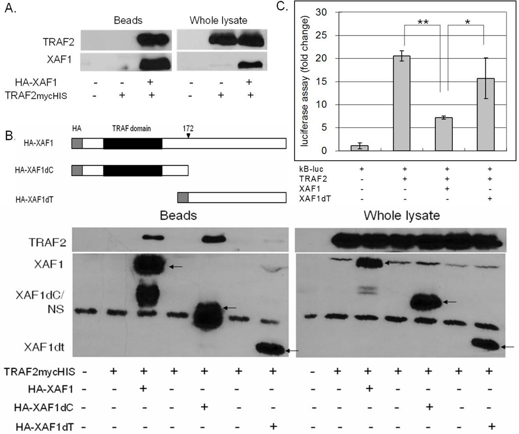
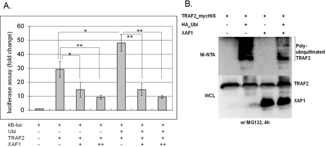
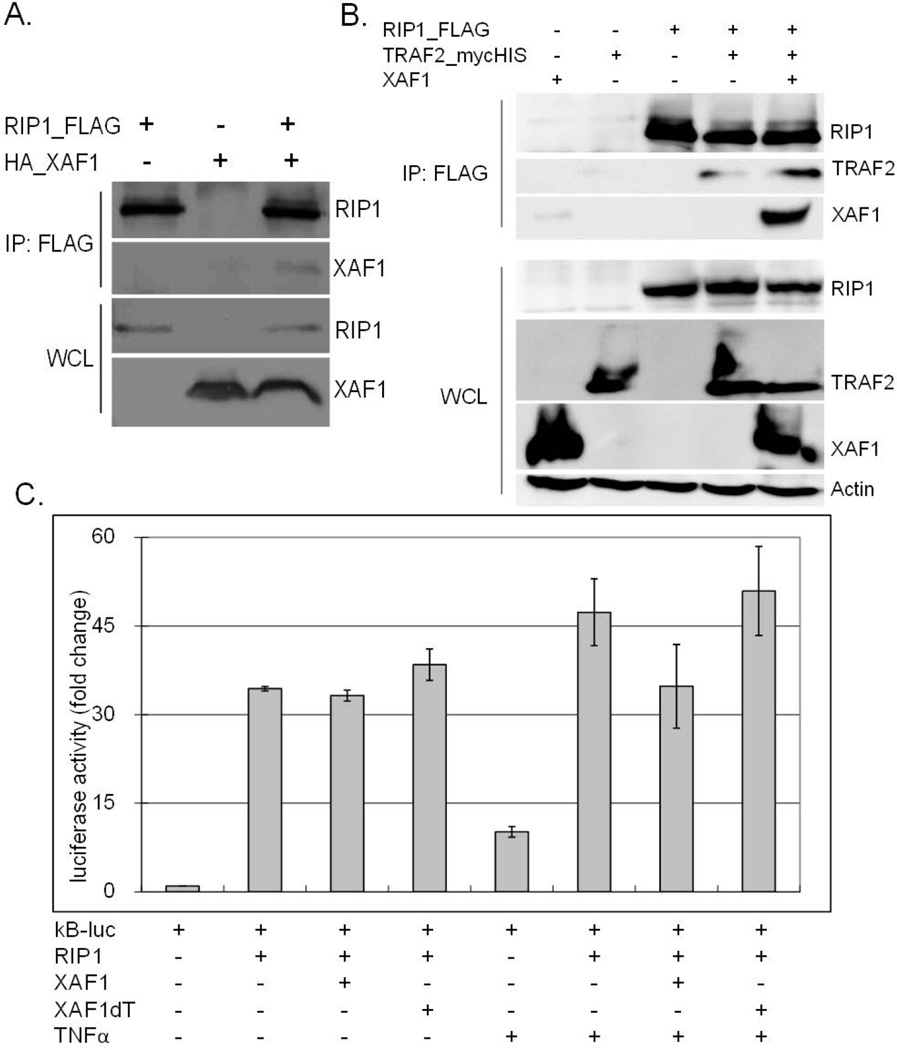
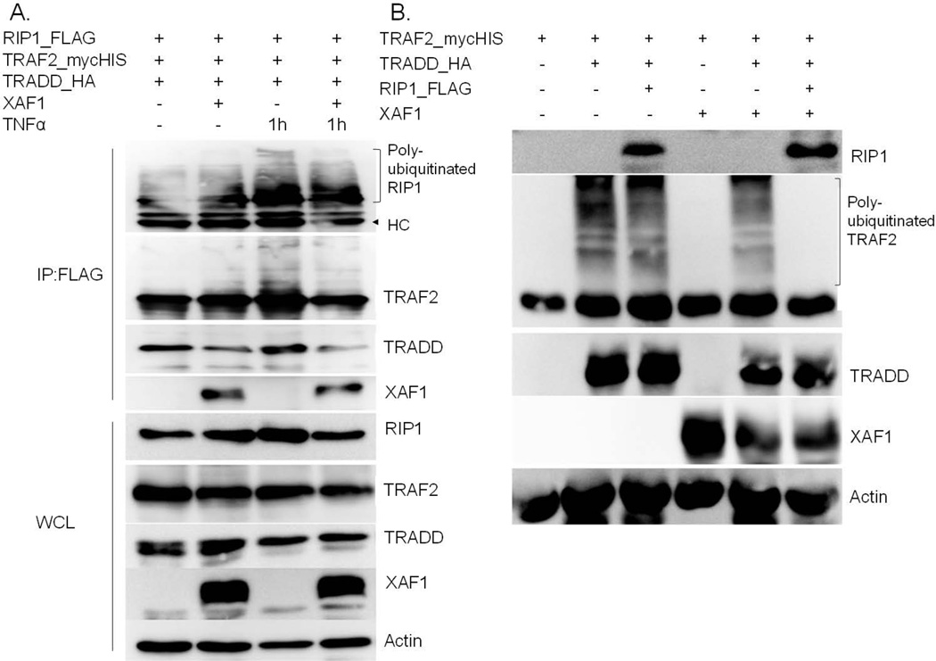
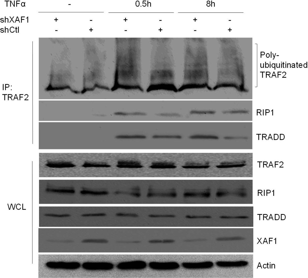
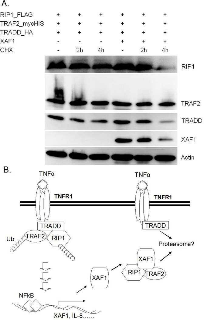
X-linked inhibitor of apoptosis (XIAP)-associated factor 1 (XAF1) is a cytokine-regulated, tumor necrosis factor (TNF) receptor-associated factor (TRAF) domain-containing protein that has a poorly defined cellular function. Here, we show that ectopically expressed XAF1 inhibits TNF-ɑ-induced NF-κB activation, whereas shRNA silencing of endogenous XAF1 augments it. Our data suggest that XAF1 may inhibit TNF-ɑ-induced NF-κB activation by disrupting the assembly of the TRADD/TRAF2/RIP1 complex (complex I) downstream of TNF receptor activation. XAF1 interacts with TRAF2 and inhibits TRAF2-dependent NF-κB activation, in part, by blocking TRAF2 polyubiquitination. Our findings also indicate that although XAF1 does not directly inhibit RIP1-dependent NF-κB activation, it binds RIP1 and disrupts RIP1/TRADD association. Our data suggest that XAF1 acts as a feedback regulator of the TNF receptor signaling pathway to suppress NF-κB activation.

Keywords: Receptor interacting protein kinase 1; XIAP associated factor 1; inflammatory response; nuclear factor-kappa B; tumor necrosis factor; tumor necrosis factor receptor-associated factor 2.


Figures

Figure 1
Figure 1. XAF1 inhibits TNF-α-induced IL-8 expression
(A) ACHN cells were treated with 20 ng/ml TNF-α, RNA isolated at the indicated times and reverse transcribed. IL-8, XAF1 and GAPDH mRNAs were amplified by RT PCR. (B) Dox/Dex regulated 293 cells were generated as described in Materials and Methods. XAF1 induction by Dox/Dex in the 293_XAF1 cells, but not empty vector control cells, was confirmed by immunoblot using anti-XAF1 antibody. XAF1-inducible cells (C) or empty vector control cells (D) were treated with Dox/Dex overnight or left untreated and then treated with 20 ng/ml TNF-α for the indicated times. IL8 transcript was quantified by real-time RT PCR using GAPDH as an internal control. Relative IL8 mRNA levels are compared to untreated samples. The mean and SEM were calculated based on data from three independent experiments (* p<0.05).
Figure 2
Figure 2. XAF1 knockdown aμgments TNF-α response
(A) Transient knockdown of XAF1 in ACHN cells was achieved by infection with XAF1 shRNA lentivirus as described in Materials and Methods. XAF1-knockdown cells or cells receiving control virus were cotransfected for 24 h with an NF-κB responsive luciferase plasmid (0.4 µg) and a β-galactosidase-expression plasmid (0.4µg), followed by 20 ng/ml TNF-α treatment for another 8 h or 16 h. Luciferase activities were measured, normalized to β-galactosidase values and results presented as fold change relative to controls without TNF-α treatment. The mean and SEM were calculated based on data from three independent experiments (**p<0.01). (B) XAF1-knockdown cells or control cells were treated with 20 ng/ml TNF-α and RNA isolated at the indicated times and reverse transcribed. Quantitative real-time RT PCR was used to quantify IL-8 mRNA levels, which were normalized to GAPDH mRNA levels. Relative IL-8 mRNA levels were calculated by comparing to untreated controls. The mean and SEM were based on three independent experiments (**p<0.01; * p<0.05).
Figure 3
Figure 3. XAF1 inhibits TNF-α-induced NF-κB activation
(A) 293 cells were cotransfected for 24 h with an NF-κB responsive luciferase plasmid (0.1 µg) and a β-galactosidase-expression plasmid (0.1 µg) with or without plasmids expressing HA-XAF1 (0.2 [+] or 0.4 [++] µg) or HA-XAF1dT (0.4 [++] µg), followed by 20 ng/ml TNF-α treatment for another 8 h. Luciferase activities were measured, normalized to β-galactosidase values and results presented as fold change relative to empty vector controls without TNF-α treatment. The mean and SEM were calculated based on data from three independent experiments (*** p<0.001; ** p<0.01). These cell lysates were also subjected to SDS PAGE separation followed by immunoblot using an anti-HA antibody to confirm the protein expression of HA-XAF1 or HA-XAF1dT. (B) XAF1-inducible (293_XAF1) or control (293_Ctl) cells were left untreated or treated with Dox/Dex overnight followed by 20 ng/ml of TNF-α for 0, 5 or 10 min. Cell lysates were separated by SDS PAGE and immunoblotted with anti-phospho-IκB, XAF1 and Actin antibodies. Data are representative of at least three independent experiments.
Figure 4
Figure 4. XAF1 interacts with TRAF2
(A) 293 cells were transiently transfected with plasmids expressing TRAF2mycHis and/or HA-XAF1. 24 h after transfection, XAF1 was captured on anti-HA-agarose and then eluted proteins assessed by immunoblot. Input cell lysates were immunoblotted in parallel for comparison. (B) 293 cells were transfected with plasmids expressing TRAF2mycHis and HA-XAF1, C-terminus deleted XAF1 (HA-XAF1dC) or TRAF domain deleted XAF1 (HA-XAF1dT). After 24 h XAF1 variants were captured on anti-HA-agarose, and proteins in the whole cell lysate and eluted complexes assessed by immunoblot analysis. (C) 293 cells were cotransfected with an NF-κB responsive luciferase plasmid (0.1 µg), a β-galactosidase-expressing plasmid (0.1 µg) and plasmids expressing TRAF2mycHis (0.2 µg), HA-XAF1 (0.2 µg) or HA-XAF1dT (0.2 µg). After 24 h, luciferase activities were measured and normalized to β-galactosidase activity. Results are presented as a fold change in normalized luciferase activity as compared to cells transfected with the empty vector. The mean and SEM were calculated based on data from three independent experiments (** p<0.01; * p<0.05).
Figure 5
Figure 5. XAF1 inhibited ubiquitin-enhanced TRAF2 activity
(A) 293 cells were cotransfected with a NF-κB responsive luciferase plasmid (0.1 µg), a β-galactosidaseexpressing plasmid (0.1 µg) and plasmids expressing XAF1 (0.2 [+] or 0.4 [++] µg) or TRAF2 (0.2 µg) or ubiquitin (0.2 µg). After 24 h, Luciferase activities were measured and normalized to β-galactosidase values. Results are presented as fold change relative to empty vector controls without TNF-α treatment. The mean and SEM were calculated based on data from three independent experiments (** p<0.01; * p<0.05). (B) 293 cells were transiently transfected with TRAF2mycHis, HA-ubiquitin, and/or untagged XAF1 expression plasmids. 24 h after transfection, TRAF2mycHis was pulled down by Ni-NTA beads followed by SDS PAGE and immunoblotting with an anti-HA antibody to detect ubiquitinated TRAF2 (top panel). Total XAF1 and TRAF2 were detected in immunoblots of the total cell lysates with antibodies for XAF1 and myc, respectively. Data are representative of at least three independent experiments.
Figure 6
Figure 6. XAF1 interacted with RIP1
(A) 293 cells were transiently transfected with plasmids expressing FLAG tagged RIP1 with or without HA tagged XAF1 or an empty vector. After 24 h, RIP1 was precipitated by anti-FLAG affinity gel, and eluted proteins separated by SDS PAGE and assessed by immunoblot with the indicated antibodies. Whole cell lysates were immunoblotted in parallel to verify XAF1 and RIP1 expression. (B) 293 cells ectopically expressing FLAG-RIP1, TRAF2mycHis, and/or untagged XAF1 were lysed and FLAG-RIP1 immunoprecipitated using anti-FLAG affinity gel. The proteins in the immune complex and whole cell lysates were separated by SDS PAGE and immunoblotted with epitope tag-specific, XAF1-specific or actin-specific antibodies. Data are representative of at least three independent experiments. (C) 293 cells were cotransfected with a NF-κB response element-regulated luciferase plasmid (0.1 µg), along with a β-galactosidase-expressing plasmid (0.1 µg) and plasmid expressing FLAG-RIP1 (0.2 µg), HA-XAF1 (0.2 µg) or HA-XAF1dT (0.4 µg). After 24 h, cells were left untreated or treated with TNF-α for 8 h, luciferase activities measured and normalized to β-galactosidase values. Results are presented as fold change relative to empty vector controls without TNF-α treatment. The mean and SEM were calculated based on data from three independent experiments.
Figure 7
Figure 7. XAF1 interfered with RIP1-TRADD association
(A) 293 cells were transfected with FLAG-RIP1, TRAF2mycHis and HA-TRADD with or without untagged XAF1 cotransfection for 24 h and then left untreated or treated with TNF-α for 1h before lysate preparation. RIP1- associated proteins were captured on anti-FLAG beads and proteins in the immune complexes or whole cell lysates separated by SDS PAGE and transfected proteins detected by immunoblotting with epitope tagspecific antibodies. XAF1 and actin were detected by anti-XAF1 and anti-actin antibodies, respectively. Data are representative of at least three independent experiments. (B) 293 cells were transfected as in (A) with the indicated expression plasmids. After 24 h, whole cell lysates were collected for SDS PAGE and immunoblot analysis with the same antibodies as (A). Data are representative of at least three independent experiments.
Figure 8
Figure 8
XAF1 knockdown prolonged TNF-α-induced TRAF2 ubiquitination and TRAF2/RIP1/TRADD complex. XAF1-knockdown cells or control cells (generated as described in Materials and Methods) were treated with 20 ng/ml TNF-α and cell lysates collected after 0.5 h or 8 h. TRAF2-associated proteins were captured on anti-TRAF2 beads. The proteins in the immune complexes or whole cell lysates were separated by SDS PAGE and detected by immunoblotting with antibodies against the proteins of interest. Data are representative of at least three independent experiments.
Figure 9
Figure 9. XAF1 regulates TRADD and RIP1 half-life
(A) 293 cells were transfected with FLAG-RIP1, TRAF2mycHis, and HA-TRADD with or without untagged XAF1. After 24 h, some cells received 100 μgmL −1 of cycloheximide for 2 or 4 h to block protein synthesis, after which time cell lysates were collected and subjected to SDS/PAGE and immunoblot analysis using epitope tag-specific antibodies or anti-XAF1 and anti-actin antibodies. Data are representative of at least three independent experiments. (B) A model of XAF1 action based on the collected data. XAF1 is induced by TNF-a and feeds back to inhibit the signaling complex. By associating with both TRAF2 and RIP1, XAF1 appears to disrupt interaction between these proteins and TRADD, thereby disrupting the TNFR signaling complex and leading to suppression of NF- jB signaling. Data in (A) suggested that components of the disrupted complex may be targeted for degradation.

Similar articles

Cited by

References

    1. Gruss HJ, Dower SK. Tumor necrosis factor ligand superfamily: involvement in the pathology of malignant lymphomas. Blood. 1995;85:3378–3404. - PubMed
    1. Tartaglia LA, Goeddel DV. Two TNF receptors. Immunol Today. 1992;13:151–153. - PubMed
    1. Tracey KJ, Cerami A. Tumor necrosis factor: a pleiotropic cytokine and therapeutic target. Annu Rev Med. 1994;45:491–503. - PubMed
    1. Chen G, Goeddel DV. TNF-R1 signaling: a beautiful pathway. Science. 2002;296:1634–1635. - PubMed
    1. Hsu H, Shu HB, Pan MG, Goeddel DV. TRADD-TRAF2 and TRADD-FADD interactions define two distinct TNF receptor 1 signal transduction pathways. Cell. 1996;84:299–308. - PubMed

MeSH terms

Substances