. 2016 Oct 20;167(3):789-802.e12.
doi: 10.1016/j.cell.2016.10.003.

Toxic PR Poly-Dipeptides Encoded by the C9orf72 Repeat Expansion Target LC Domain Polymers

Affiliations

Toxic PR Poly-Dipeptides Encoded by the C9orf72 Repeat Expansion Target LC Domain Polymers

Yi Lin et al. Cell. .

Abstract

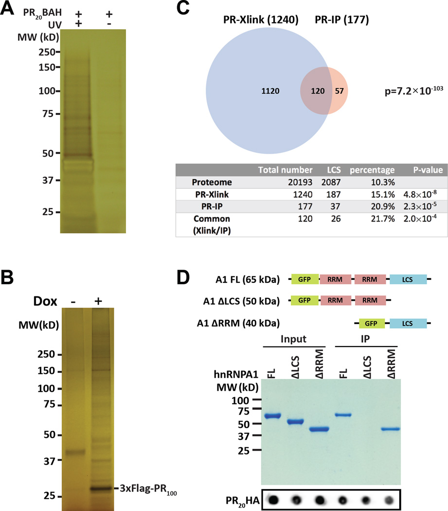
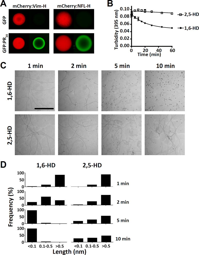
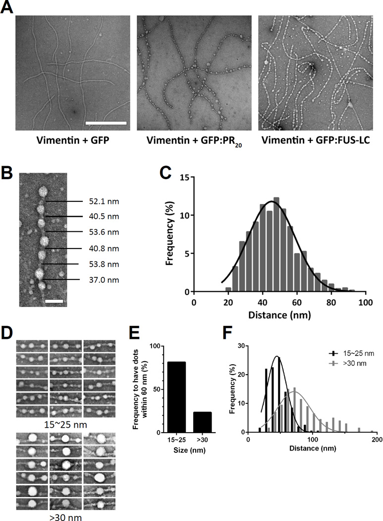
Two complementary approaches were used in search of the intracellular targets of the toxic PR poly-dipeptide encoded by the repeat sequences expanded in the C9orf72 form of amyotrophic lateral sclerosis. The top categories of PRn-bound proteins include constituents of non-membrane invested cellular organelles and intermediate filaments. PRn targets are enriched for the inclusion of low complexity (LC) sequences. Evidence is presented indicating that LC sequences represent the direct target of PRn binding and that interaction between the PRn poly-dipeptide and LC domains is polymer-dependent. These studies indicate that PRn-mediated toxicity may result from broad impediments to the dynamics of cell structure and information flow from gene to message to protein.

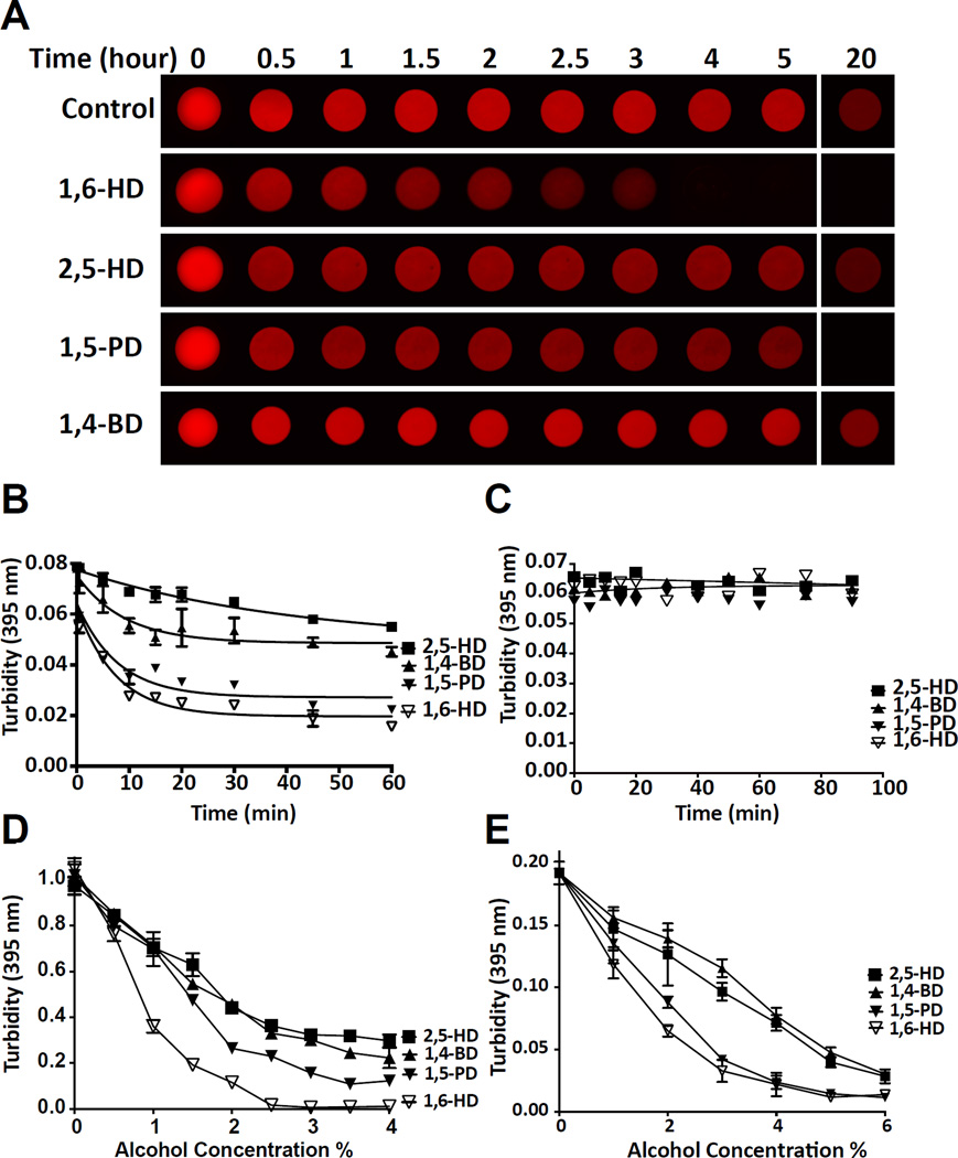
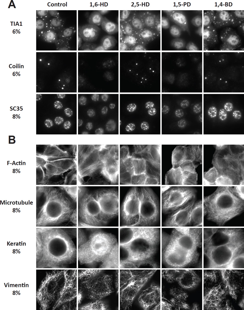
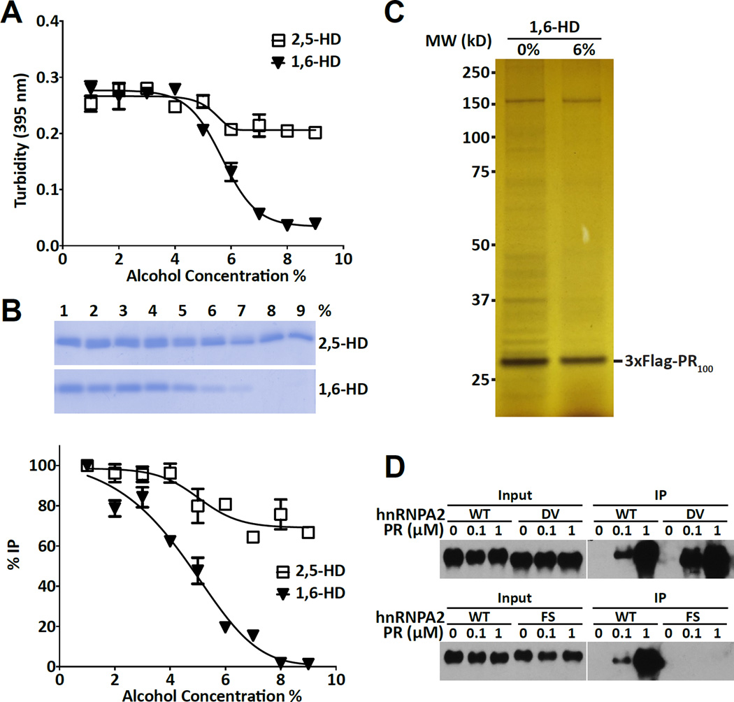
Keywords: 1,6-hexanediol; C9orf72; amyloid-like polymers; cellular puncta not invested by surrounding membranes; intermediate filaments; labile; low complexity sequence polymers; toxic PRn and GRn poly-dipeptides.


Figures

Figure 1
Figure 1. Intracellular targets of the PRn poly-dipeptide
(A) Silver-stained SDS-PAGE gel of HeLa cell proteins crosslinked to PR20BAH (Experimental Procedures). Left lane shows proteins from cells exposed to both PR20BAH probe and UV light, right lane shows proteins from cells exposed to PR20BAH probe but not UV illumination. Proteins were extracted from the gel and identified by shotgun mass spectrometry (See also Figure S1, Table S1). (B) Silver-stained SDS-PAGE gel of immunoprecipitated proteins bound by the 3xFlag:PR100 protein conditionally expressed in U2OS cells. 3xFlag:PR100 expression was either induced with doxycycline (+) or not (−). Proteins were extracted from the gel and identified by mass spectrometry (See also Table S2). (C) Venn diagram showing overlap of 120 proteins commonly identified both by photo-crosslinking to the PR20-BAH probe (PR-Xlink) and immunoprecipitation with the conditionally inducible 3xFlag:PR100 protein (PR-IP). Table below Venn diagram lists the number of proteins associated with low complexity sequences (LCS) in the entire human proteome as compared with the lists of proteins identified to interact with either or both of the PRn probes (See also Table S3). (D) Coomassie stained SDS-PAGE gel of GFP:hnRNPA1 fusion proteins co-precipitated with the PR20-HA probe. Input and immunoprecipitated PR20HA signals are shown below commassie stained gel as dot blots.
Figure 2
Figure 2. Melting of hydrogels, LC domain polymers and liquid-like droplets by aliphatic alcohols
(A) mCherry:FUS hydrogel droplets were incubated with 15% levels of indicated aliphatic alcohols for varying intervals from 30 min to 20 hr (See also Video S1). (B) Melting of mCherry:FUS polymers in suspension with 15% of indicated aliphatic alcohols as measured by light scattering at 395 nm (n=3, data are presented as means ± SD. Note that error bars for most of the data points are smaller than the symbols. See also Figure S2). (C) Exposure of Aβ amyloid fibers to 15% levels of indicated aliphatic alcohols. No change in turbidity (395nm) was observed for up to 90 min. (D) Liquid-like droplets were formed by mixing an MBP:PTB:hnRNPA2 triple fusion protein with TEV protease and a synthetic RNA substrate (Experimental Procedures), followed by exposure to 0.5–4% levels of the indicated aliphatic alcohols (n=2, data are presented as means ± SD). (E) FUS liquid-like droplets were exposed to 0.5–6% levels of the indicated aliphatic alcohols (n=2, data are presented as means ± SD).
Figure 3
Figure 3. Effects of aliphatic alcohols on RNA granules, Cajal bodies, nuclear speckles and cytoskeletal filaments
(A) HeLa cells were exposed to indicated aliphatic alcohols for 5 min, fixed and stained with antibodies diagnostic of stress granules (TIA1), Cajal bodies (coilin) or nuclear speckles (SC35). (B) Hela cells were exposed to indicated aliphatic alcohols for 5 min, fixed and stained with either dye-labeled phalloidin or antibodies to tubulin, keratin or vimentin.
Figure 4
Figure 4. Effects of aliphatic alcohols on interactions between the PRn poly-dipeptide and its cellular targets
(A) Turbidity of hnRNPA2 low complexity polymers in response to indicated concentrations of either 1,6-hexanediol or 2,5-hexanediol as measured by light scattering at 395 nm (n=2, data are presented as means ± SD). (B) Coomassie stained SDS-PAGE gel of immunoprecipitatated samples of the low complexity domain of hnRNPA2 by the PR20:HA probe as a function of exposure to between 1% and 9% of the indicated aliphatic alcohol. Band intensities were quantified from SDS-PAGE gel (top) by image J, and then plotted (bottom) (n=2, data are presented as means ± SD). (C) Silver stained SDS-PAGE gel of immunoprecipitated proteins from U2OS cells expressing the 3xFlag:PR100 protein. Left lane shows proteins co-precipitated in the absence of aliphatic alcohols, right lane shows proteins co-precipitated in the presence of 6% 1,6-hexanediol (See also Figure S3). (D) Western blots showing native hnRNPA2 protein or variants bearing single amino acid substitutions (DV = aspartic acid residue 290 changed to valine, or FS = phenylalanine residue 291 changed to serine) as input to immunoprecipitation reactions (left panels), or as immuno-precipitated in response to varying concentrations of the PR20:HA probe (right panels) (See also Figure S4).
Figure 5
Figure 5. Binding of GFP:PR20 to mCherry:IF head domain hydrogel, and sensitivity of vimentin intermediate filaments to aliphatic alcohols
(A) Hydrogel droplets containing fusion proteins made from mCherry linked to the low complexity head domains of vimentin (left) or neurofilament light isoform (right) were incubated with either GFP (top images) or GFP:PR20 (lower images) and visualized by confocal microscopy (Experimental Procedures) (See also Figure S5). (B) Turbidity measurements as determined by light scattering at 395 nm of polymers formed from an mCherry:vimentin head domain fusion protein in response to 15% of indicated aliphatic alcohols for indicated times (n=3), data are presented as means ± SD. Note that error bars for most of the data points are smaller than the symbols (See also Figure S6A). (C) Electron microscope images of vimentin intermediate filament proteins exposed to indicated aliphatic alcohols for indicated times, scale bar = 1 µm (See also Figure S6B). (D) Quantitation of lengths of intermediate filaments as a function of exposure to indicated aliphatic alcohols for indicated times.
Figure 6
Figure 6. GFP:PR20 and GFP:FUS bind periodically to vimentin intermediate filaments
(A) Electron microscope images of vimentin intermediate filaments exposed to GFP (left), GFP:PR20 (middle) or GFP:FUS (right). Scale bar = 500 nm (See also Figure S6 C–D). (B) Representative image showing the puncta-to-puncta distance of GFP:PR20 bound to a single vimentin intermediate filament. Scale bar = 50 nm. (C) Quantification of puncta-to-puncta distances of GFP:PR20 bound to vimentin intermediate filaments (n=1000). The curve was fitted by Gaussian distribution. (D) Representative images of different sized puncta (15~25 nm or >30 nm) of GFP:PR20 bound to vimentin intermediate filaments. (E) Quantification of frequency to have puncta of GFP:PR20 puncta within 60 nm of small or large puncta. (F) Quantification of puncta-to-puncta distance of GFP-PR20 bound to vimentin intermediate filaments as a function of puncta size. p<0.001 (n=200)

References

    1. ACD/Structure Elucidator. version 15.01. Toronto, ON, Canada: Advanced Chemistry Development, Inc.; 2015. http://www.acdlabs.com/
    1. Ash PE, Bieniek KF, Gendron TF, Caulfield T, Lin WL, Dejesus-Hernandez M, van Blitterswijk MM, Jansen-West K, Paul JW, 3rd, Rademakers R, et al. Unconventional translation of C9ORF72 GGGGCC expansion generates insoluble polypeptides specific to c9FTD/ALS. Neuron. 2013;77:639–646. - PMC - PubMed
    1. Astbury WT, Dickinson S, Bailey K. The X-ray interpretation of denaturation and the structure of the seed globulins. Biochem J. 1935;29:2351–2360. 2351. - PMC - PubMed
    1. Bergeron-Sandoval LP, Safaee N, Michnick SW. Mechanisms and Consequences of Macromolecular Phase Separation. Cell. 2016;165:1067–1079. - PubMed
    1. Brownlees J, Ackerley S, Grierson AJ, Jacobsen NJ, Shea K, Anderton BH, Leigh PN, Shaw CE, Miller CC. Charcot-Marie-Tooth disease neurofilament mutations disrupt neurofilament assembly and axonal transport. Hum Mol Genet. 2002;11:2837–2844. - PubMed

Publication types