. 2016 Dec 20;7(51):84359-84374.
doi: 10.18632/oncotarget.12711.

Inducible expression of cancer-testis antigens in human prostate cancer

Affiliations

Inducible expression of cancer-testis antigens in human prostate cancer

Erika Heninger et al. Oncotarget. .

Abstract

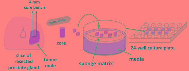
Immune tolerance to self-antigens can limit robust anti-tumor immune responses in the use of tumor vaccines. Expression of novel tumor associated antigens can improve immune recognition and lysis of tumor cells. The cancer-testis antigen (CTA) family of proteins has been hypothesized to be an ideal class of antigens due to tumor-restricted expression, a subset of which have been found to induce antibody responses in patients with prostate disease. We demonstrate that CTA expression is highly inducible in five different Prostate Cancer (PC) cell lines using a hypomethylating agent 5-Aza-2'-deoxycytidine (5AZA) and/or a histone deacetylase inhibitor LBH589. These CTAs include NY-ESO1, multiple members of the MAGE and SSX families and NY-SAR35. A subset of CTAs is synergistically induced by the combination of 5AZA and LBH589. We developed an ex vivo organ culture using human PC biopsies for ex vivo drug treatments to evaluate these agents in clinical samples. These assays found significant induction of SSX2 in 9/9 distinct patient samples and NY-SAR35 in 7/9 samples. Further, we identify expression of SSX2 in circulating tumor cells (CTC) from patients with advanced PC. These results indicate that epigenetic modifying agents can induce expression of a broad range of neoantigens in human PC and may serve as a useful adjunctive therapy with novel tumor vaccines and checkpoint inhibitors.

Keywords: cancer testis antigen; epigenetics; methylation; prostate cancer; tumor immunotherapy.


Conflict of interest statement

CONFLICTS OF INTEREST

Joshua M. Lang holds equity in Salus Discovery LLC, which has licensed some of the technology described in the manuscript. All issues regarding this conflict of interest are managed by the University of Wisconsin and a COI management plan is in place.

Figures

Figure 1
Figure 1. Quantitative analysis of baseline expression of cancer/testis antigen mRNA in 5 PC cell lines and in the RWPE-1 normal epithelial cell line
RNA was evaluated by qRT-PCR for expression relative to an internal control transcript (P0). qRT-PCR was performed using primers specific for each gene, conducted in triplicate, and repeated in an independent experiment. Expression relative to P0 was color scaled from red (highest) to black (middle) to green (lowest). Grey indicates that no relevant expression was detected.
Figure 2
Figure 2. Quantitative analysis of relative expression of CTA mRNA in androgen-receptor expressing PC cell lines and in RWPE-1 normal epithelial cells in response to EMA treatment
RNA was evaluated by qRT-PCR for expression relative to an internal control transcript (P0) following EMA treatment of RWPE-1, LNCaP, LAPC4, and 22rv1 cells. qRT-PCR was performed using primers specific for each gene, conducted in triplicate, and repeated in an independent experiment. Expression relative to P0 was color scaled from red (highest) to black (middle; 50th percentile) to green (lowest) for each CTA separately, across all cell lines, thus similar colors between different rows cannot be directly compared. Grey indicates that no relevant expression was detected.
Figure 3
Figure 3. Quantitative analysis of relative expression of CTA mRNA in androgen-independent PC cell lines treated with EMAs
RNA was evaluated by qRT-PCR for expression relative to an internal control transcript (P0) following EMA treatment of DU-145 and PC3 cells. qRT-PCR was performed using primers specific for each gene, conducted in triplicate, and repeated in an independent experiment. Expression relative to P0 was color scaled from red (highest) to black (middle; 50th percentile) to green (lowest) for each CTA separately, across all cell lines, thus similar colors between different rows cannot be directly compared. Grey indicates that no relevant expression was detected.
Figure 4
Figure 4. Ex vivo human prostate tissue drug culture
A novel preclinical model developed to test the effect of EMA agents on prostate tumor tissue collected from patients undergoing radical prostatectomy. Resected prostate gland slices are subject to gross pathology examination. 4 mm cores are punched from visible tumor nodes and tissue is sliced into ~1 mm3 pieces. Surgical gelatin sponge is soaked in supplemented Ham's media and placed in 24-well tissue cultures wells. Tissue slices are carefully layered on the surface of saturated sponge matrix. The matrix is fully saturated by but is not submerged in media.
Figure 5
Figure 5. EMA treatment induces CTA expression in ex vivo human prostate tissue in a prostate biopsy drug culture model
Quantitative analysis of relative expression of SSX2, NYSAR35, prostatic acid phosphatase (PAP) and androgen receptor (AR) mRNA in PC tissue biopsies treated with EMAs. RNA was evaluated by qRT-PCR for expression relative to an internal control transcript (P0) following EMA treatment. qRT-PCR was performed using primers specific for each gene and was conducted in triplicate. Error bars represent the mean and SD. A missing bar indicates that there was no detectable signal. Comparison between groups was made with a 1-way ANOVA followed by post hoc analysis with the Tukey test. P < 0.05 compared with vehicle (*), 5AZA 10 μM (^), LBH589 100 nM (+), or 5AZA 10 μM + LBH589 100 nM (□) treatment.
Figure 6
Figure 6. Quantitative analysis of expression of SSX2 and prostate-specific marker mRNA in circulating tumor cells
qRT-PCR was performed using primers specific for each gene, and the P0 housekeeping gene is included for reference. Expression was color scaled from red (highest) to black (middle) to green (lowest). Grey indicates that no relevant expression was detected.

References

    1. Ciccarese C, Santoni M, Massari F. Chemohormonal Therapy in Hormone-Sensitive Prostate Cancer. N Engl J Med. 2016;374:286. doi: 10.1056/NEJMc1511800#SA1. - DOI - PubMed
    1. Spratt DE, Zumsteg ZS, Feng FY, Tomlins SA. Translational and clinical implications of the genetic landscape of prostate cancer. Nat Rev Clin Oncol. 2016. - DOI - PMC - PubMed
    1. Strauss J, Madan RA. Therapeutic vaccines for prostate cancer: recent advances and future directions. Expert Rev Vaccines. 2016. pp. 1–8. - DOI - PubMed
    1. Asmar R, Yang J, Carvajal RD. Clinical utility of nivolumab in the treatment of advanced melanoma. Ther Clin Risk Manag. 2016;12:313–25. doi: 10.2147/TCRM.S78039. - DOI - PMC - PubMed
    1. Melosky B, Chu Q, Juergens R, Leighl N, McLeod D, Hirsh V. Pointed Progress in Second-Line Advanced Non-Small-Cell Lung Cancer: The Rapidly Evolving Field of Checkpoint Inhibition. J Clin Oncol. 2016;34:1676–88. doi: 10.1200/JCO.2015.63.8049. - DOI - PubMed

MeSH terms