. 2017 Jan 15:48:258-269.
doi: 10.1016/j.actbio.2016.10.027. Epub 2016 Oct 18.

Enzyme-mediated stiffening hydrogels for probing activation of pancreatic stellate cells

Affiliations

Enzyme-mediated stiffening hydrogels for probing activation of pancreatic stellate cells

Hung-Yi Liu et al. Acta Biomater. .

Abstract

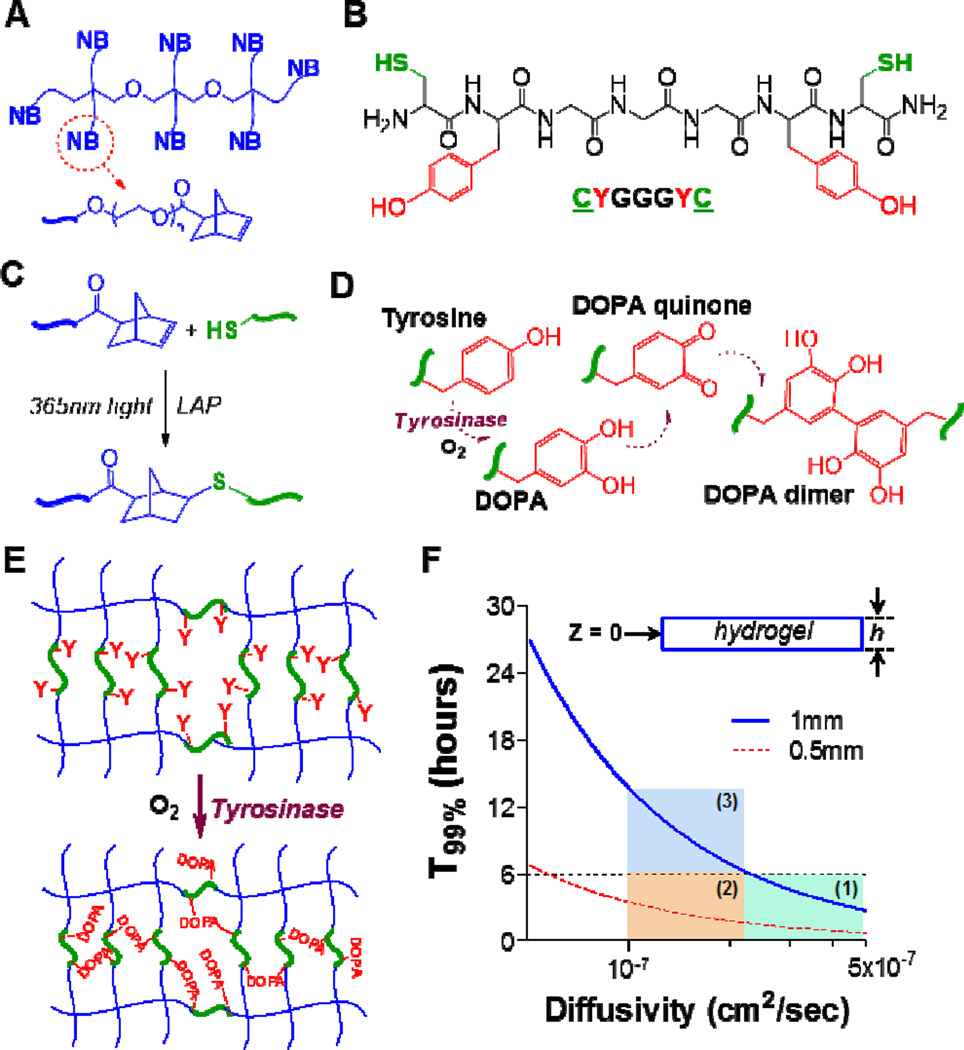
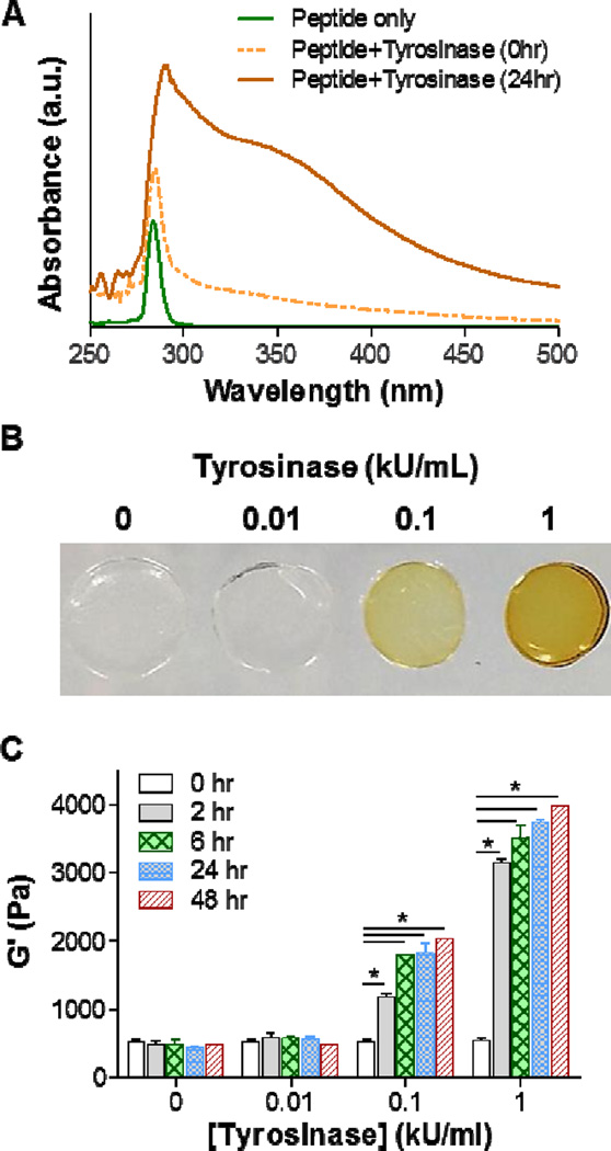
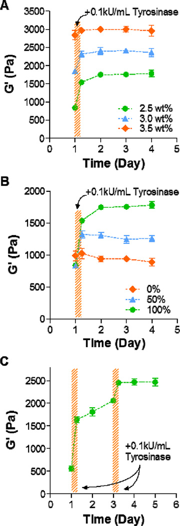
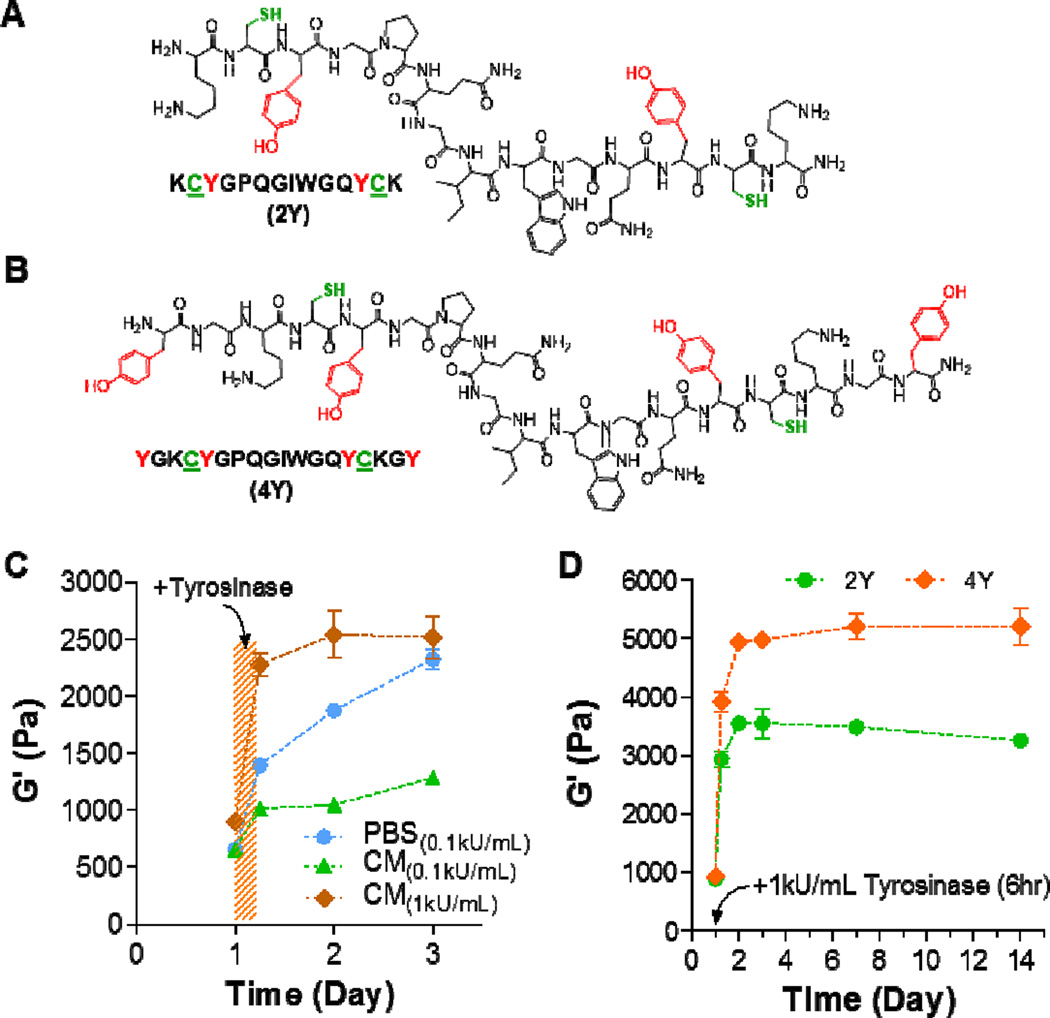
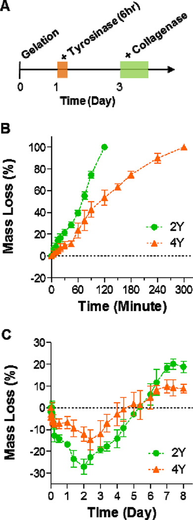
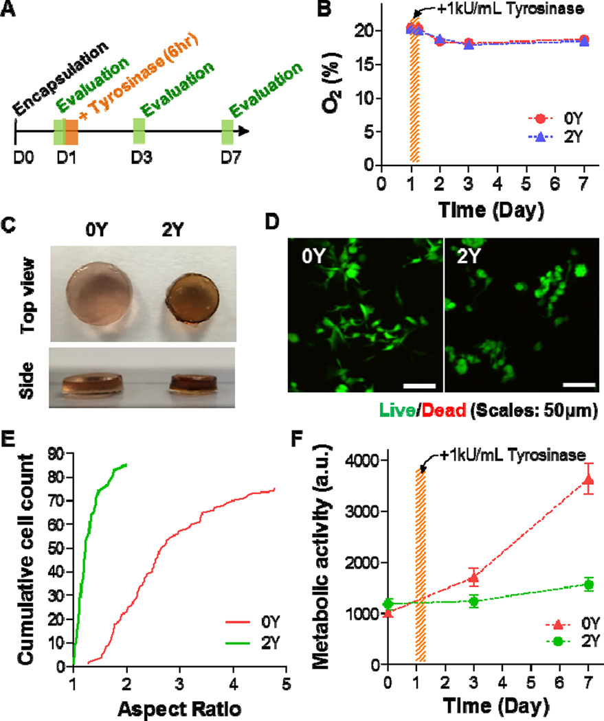
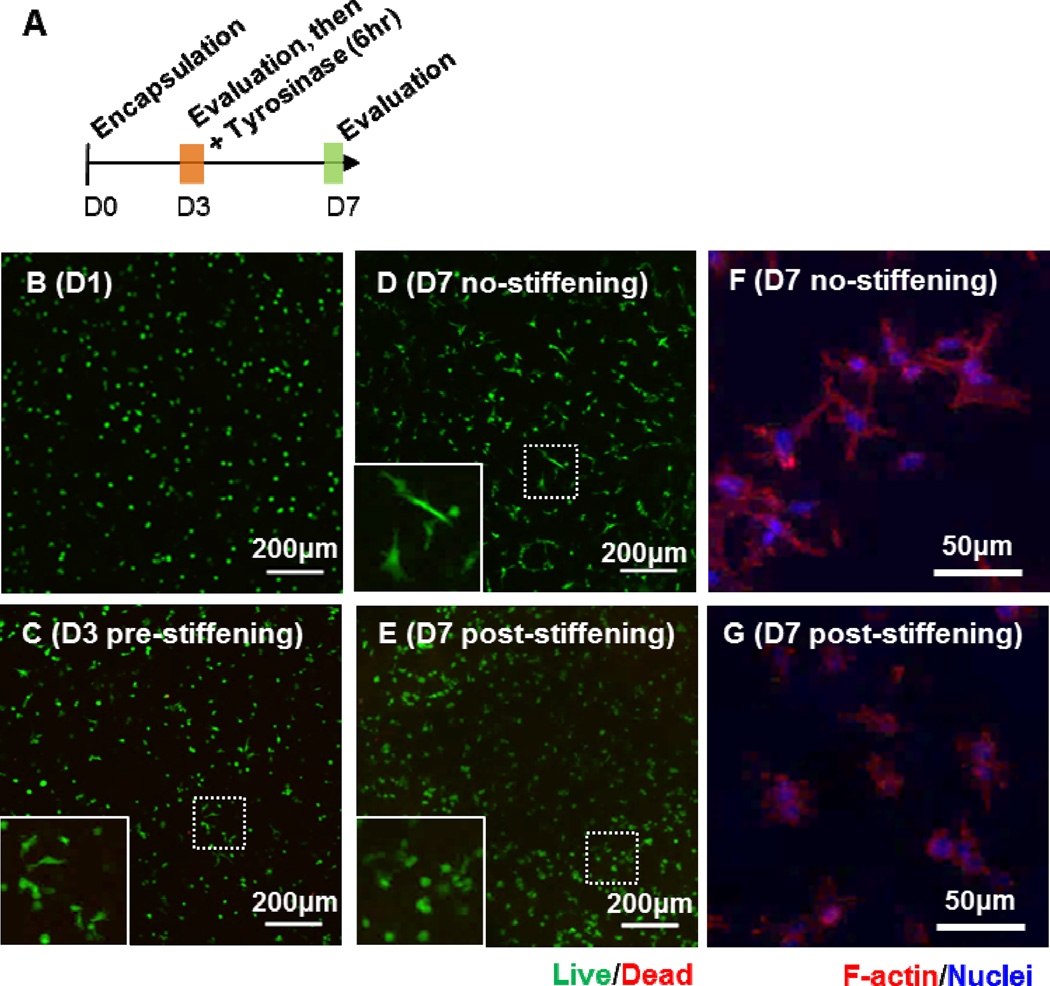
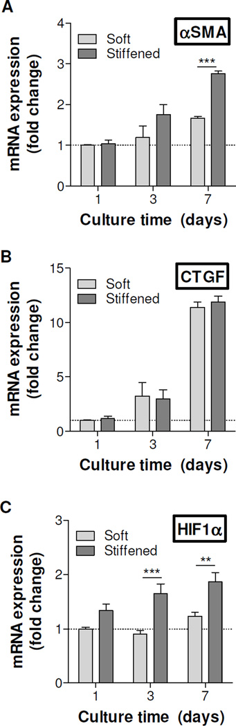
The complex network of biochemical and biophysical cues in the pancreatic desmoplasia not only presents challenges to the fundamental understanding of tumor progression, but also hinders the development of therapeutic strategies against pancreatic cancer. Residing in the desmoplasia, pancreatic stellate cells (PSCs) are the major stromal cells affecting the growth and metastasis of pancreatic cancer cells by means of paracrine effects and extracellular matrix protein deposition. PSCs remain in a quiescent/dormant state until they are 'activated' by various environmental cues. While the mechanisms of PSC activation are increasingly being described in literature, the influence of matrix stiffness on PSC activation is largely unexplored. To test the hypothesis that matrix stiffness affects myofibroblastic activation of PSCs, we have prepared cell-laden hydrogels capable of being dynamically stiffened through an enzymatic reaction. The stiffening of the microenvironment was created by using a peptide linker with additional tyrosine residues, which were susceptible to tyrosinase-mediated crosslinking. Tyrosinase catalyzes the oxidation of tyrosine into dihydroxyphenylalanine (DOPA), DOPA quinone, and finally into DOPA dimer. The formation of DOPA dimer led to additional crosslinks and thus stiffening the cell-laden hydrogel. In addition to systematically studying the various parameters relevant to the enzymatic reaction and hydrogel stiffening, we also designed experiments to probe the influence of dynamic matrix stiffening on cell fate. Protease-sensitive peptides were used to crosslink hydrogels, whereas integrin-binding ligands (e.g., RGD motif) were immobilized in the network to afford cell-matrix interaction. PSC-laden hydrogels were placed in media containing tyrosinase for 6h to achieve in situ gel stiffening. We found that PSCs encapsulated and cultured in a stiffened matrix expressed higher levels of αSMA and hypoxia-inducible factor 1α (HIF-1α), suggestive of a myofibroblastic phenotype. This hydrogel platform offers a facile means of in situ stiffening of cell-laden matrices and should be valuable for probing cell fate process dictated by dynamic matrix stiffness.

Statement of significance: Hydrogels with spatial-temporal controls over crosslinking kinetics (i.e., dynamic hydrogel) are increasingly being developed for studying mechanobiology in 3D. The general principle of designing dynamic hydrogel is to perform cell encapsulation within a hydrogel network that allows for postgelation modification in gel crosslinking density. The enzyme-mediated in situ gel stiffening is innovative because of the specificity and efficiency of enzymatic reaction. Although tyrosinase has been used for hydrogel crosslinking and in situ cell encapsulation, to the best of our knowledge tyrosinase-mediated DOPA formation has not been explored for in situ stiffening of cell-laden hydrogels. Furthermore, the current work provides a gradual matrix stiffening strategy that may more closely mimic the process of tumor development.

Keywords: Hydrogels; Pancreatic cancer; Stellate cells; Tissue stiffening; Tyrosinase.


Figures

Figure 1
Figure 1. Schematics of thiol-norbornene hydrogels susceptible to tyrosinase-mediated in situ gel stiffening
(A) Structure of 8-arm poly(ethylene glycol)-norbornene (PEG8NB). (B) Model bis-cysteine-bis-tyrosine peptide crosslinker CYGGGYC. (C) Light mediated thiol-norbornene photoclick reaction to form PEG8NB-peptide hydrogel. 1 mM LAP was used as the photoinitiator. (D) Tyrosinase-mediated oxidation of tyrosine into DOPA, DOPA quinone, and DOPA dimer. (E) PEG8NB-peptide hydrogel bearing pendant tyrosines for tyrosinase-mediated DOPA dimer formation that leads to increased gel crosslinking density. (F) Prediction of T99% (i.e., Cz=0 ≥ 0.99 × Cz=h) in a disc-shape hydrogel with a thickness of h. Regions 1–3 represent scenarios for which a period of 6-hr tyrosinase incubation are sufficient (regions 1 & 2) or insufficient (region 3) to fulfill the criterion of Cz=0 ≥ 0.99 × Cz=h.
Figure 2
Figure 2. Tyrosinase-mediated in situ stiffening of PEG-based hydrogels
(A) UV/Vis absorbance spectrum of 5 mM CYGGGYC peptide, peptide/tyrosinase (1 kU/mL) mixture before (0hr) and after 24hr incubation. (B) Photographs of PEG-peptide (i.e., 2.5wt% PEG8NB, 5 mM CYGGGYC) hydrogels treated with different concentrations of tyrosinase. (C) Effect of tyrosinase concentration of shear moduli (G’) of the PEG-peptide hydrogels. Crosslinked hydrogels were incubated in PBS for one day prior to 6hr of tyrosinase treatment. Afterward, the gels were transferred to PBS and gel moduli were monitored periodically using oscillatory rheometer. Data represent Mean ± SEM (n = 3). Asterisks indicate p<0.05 (compared with gels at 0hr, i.e., prior to tyrosinase treatment).
Figure 3
Figure 3. Controlling the degree of in situ gel stiffening
(A) Effect of PEG8NB weight percent (2.5, 3.0, and 3.5 wt%) on the degree of gel stiffening. The concentration of CYGGGYC was adjusted such that a stoichiometric ratio of thiol-to-ene moieties was maintained (i.e., 5, 6, and 7 mM peptide, respectively). Hydrogels were allowed to swell for one day post-gelation, followed by tyrosinase treatment for 6 hours (shaded area). (B) Effect of tyrosinase-sensitive peptide content on the degree of in situ gel stiffening. PEG8NB content was fixed at 2.5wt%, whereas tyrosinase-sensitive (i.e., CYGGGYC) and insensitive (i.e., CGGGC) peptide crosslinkers were mixed at different percentages (0, 50, and 100% CYGGGYC). (C) Temporal control in gel stiffening. Swollen hydrogels (2.5wt% PEG8NB and 5 mM CYGGGYC) were treated with tyrosinase at day 1 for 3 hours and again at day 3 for another 3 hours. Data represent Mean ± SEM (n = 3).
Figure 4
Figure 4. In situ stiffening of MMP-sensitive PEG8NB-peptide hydrogels
(A, B) Chemical structures of bis-cysteine MMP-sensitive peptide crosslinker bearing additional two (A) or four (B) tyrosine residues for tyrosinase-mediated gel stiffening. (C) Effect of solution composition on elastic moduli of in situ stiffened PEG-peptide hydrogels. CM: PSC culture media. Gel formulation: 2.5wt% PEG8NB with 2Y peptide. (D) Effect of tyrosine residues (2 or 4 tyrosines) on elastic moduli of in situ stiffened PEG-peptide hydrogels (2.5wt% PEG8NB). Data represent Mean ± SEM (n = 3).
Figure 5
Figure 5. Proteolytic degradation of MMP-sensitive PEG8NB-peptide hydrogels
(A) Timeline of the study. (B, C) Collagenase-mediated mass loss of non-stiffened hydrogels (B) and tyrosinase-stiffened hydrogels (C). All gels were formed with 2.5wt% PEG8NB with 5 mM MMP-sensitive peptide (2Y or 4Y). Data represent Mean ± SEM (n = 3).
Figure 6
Figure 6. Effect of matrix stiffening on pancreatic stellate cell fate in 3D
(A) Timeline of the study. (B) Oxygen contents in PSC culture media with 0Y or 2Y PEG-peptide hydrogels. Tyrosinase-mediated stiffening was conducted on day-1 for 6hr. (C) Photographs of cell-laden hydrogels crosslinked by 0Y or 2Y MMP-sensitive peptide. (D) Representative confocal z-stack images of live/dead stained PSCs encapsulated in 0Y or 2Y MMP-sensitive PEG-peptide hydrogels. (E) Aspect ratio of encapsulated PSCs at day-7 post-encapsulation. (F) Metabolic activity of encapsulated PSCs. All gels were treated with 1 kU/mL tyrosinase at day-1 post-encapsulation. Data represent Mean ± SEM (n = 3).
Figure 7
Figure 7
(A) Timeline of the study. (B–E) Representative confocal z-stack images of live/dead stained PSCs encapsulated in PEG-peptide hydrogels at day-1 (B), day-3 before stiffening (C), day-7 without stiffening (D), and day-7 with stiffening (E). Representative confocal z-stack images of F-actin stained PSCs at day-7 without (F) and with stiffening (G).
Figure 8
Figure 8. Effect of matrix stiffening on gene expression in pancreatic stellate cells
mRNA samples were collected from cell-laden hydrogels before stiffening (day-1), as well as day-2 and day-6 post-stiffening using 1 kU/mL tyrosinase (i.e., day-3, day-7 culture time). Soft: Cell-laden hydrogels crosslinked by regular MMP-sensitive peptide (i.e., 0Y peptide). Stiffened: Cell-laden hydrogels crosslinked by 2Y peptide. GAPDH was used as the housekeeping gene and the expression levels of αSMA (A), CTGF (B), and HIF1α (C) in different samples were normalized to that in day-1 soft gel (1-fold). Experiments were repeated independently for three times with three samples in each group. (Data represent Mean ± SEM). **p<0.01, ***p<0.001.

References

    1. Dupont S, Morsut L, Aragona M, Enzo E, Giulitti S, Cordenonsi M, Zanconato F, Le Digabel J, Forcato M, Bicciato S, Elvassore N, Piccolo S. Role of YAP/TAZ in mechanotransduction. Nature. 2011;474:179–183. - PubMed
    1. Gore J, Korc M. Pancreatic cancer stroma: Friend or foe? Cancer Cell. 2014;25:711–712. - PMC - PubMed
    1. Engler AJ, Sen S, Sweeney HL, Discher DE. Matrix elasticity directs stem cell lineage specification. Cell. 2006;126:677–689. - PubMed
    1. Tibbitt MW, Anseth KS. Hydrogels as extracellular matrix mimics for 3D cell culture. Biotechnol Bioeng. 2009;103:655–663. - PMC - PubMed
    1. Lin C-C, Anseth KS. PEG hydrogels for the controlled release of biomolecules in regenerative medicine. Pharm Res. 2009;26:631–643. - PMC - PubMed

MeSH terms

LinkOut - more resources