. 2016 Dec 9;291(50):25999-26012.
doi: 10.1074/jbc.M116.738856. Epub 2016 Oct 21.

Lysyl Oxidase-like-2 Cross-links Collagen IV of Glomerular Basement Membrane

Affiliations

Lysyl Oxidase-like-2 Cross-links Collagen IV of Glomerular Basement Membrane

Carolina Añazco et al. J Biol Chem. .

Abstract

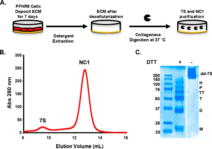
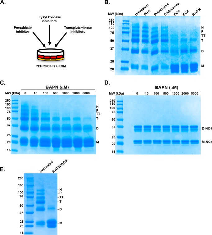
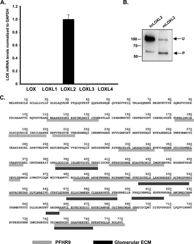
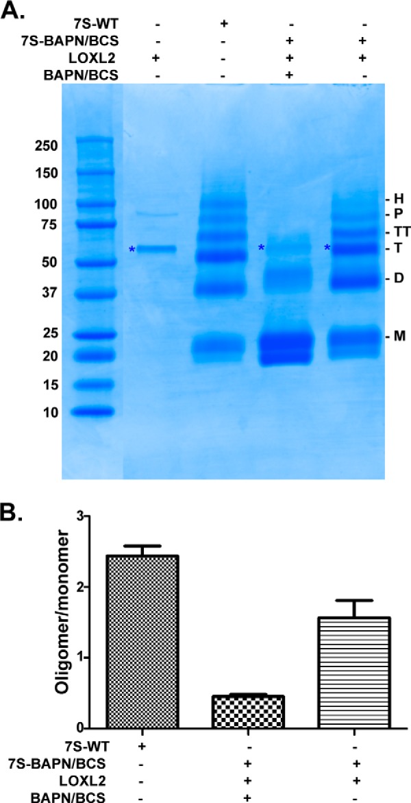
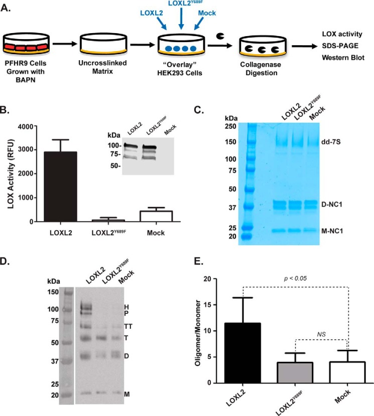
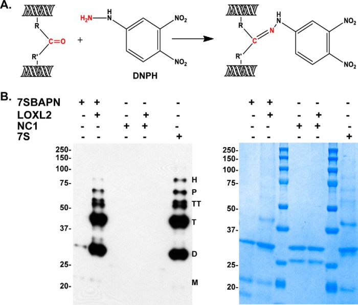
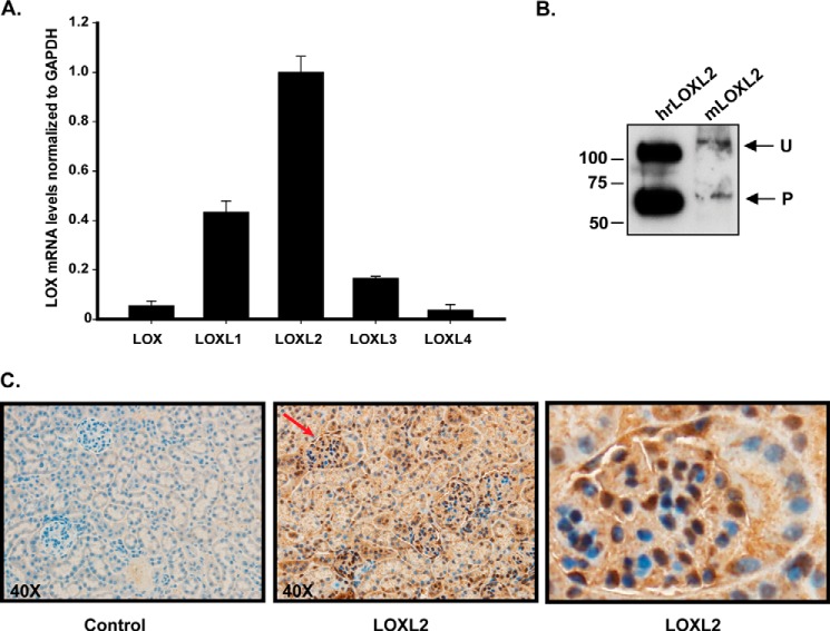
The 7S dodecamer is recognized as an important structural cross-linking domain of collagen IV networks that provide mechanical stability to basement membranes, a specialized form of extracellular matrix essential for the development and maintenance of tissue architecture. Although the 7S dodecamer is stabilized by covalent cross-linking, the molecular mechanism by which such cross-links are formed has not been revealed. Here, we aimed to identify the enzyme(s) that cross-links the 7S dodecamer and characterize its expression in the kidney glomerulus. Pharmacological inhibition of candidate extracellular matrix enzymes revealed that lysyl oxidase activity is required for cross-linking of 7S polypeptides. Among all lysyl oxidase family members, lysyl oxidase-like-2 (LOXL2) was identified as the isoform cross-linking collagen IV in mouse embryonal PFHR-9 cells. Biochemical analyses revealed that LOXL2 readily promoted the formation of lysyl-derived cross-links in the 7S dodecamer but not in the NC1 domain. We also established that LOXL2 is the main lysyl oxidase family member present in the glomerular extracellular matrix. Altogether, we demonstrate that LOXL2 is a novel component of the molecular machinery that forms cross-linked collagen IV networks, which are essential for glomerular basement membrane stability and molecular ultrafiltration function.

Keywords: basement membrane; collagen; extracellular matrix; kidney; lysyl oxidase; protein cross-linking.


Figures

FIGURE 1.
FIGURE 1.
Purification of 7S dodecamers from PFHR-9 cells. A shows a schematic representation of the procedures to purify the 7S dodecamer and NC1 hexamer from PFHR-9 cell matrix. PFHR-9 cells are cultured for 7 days after they reach confluence, allowing the deposition of a very thick ECM. Cells are removed with deoxycholate detergent, and the insoluble ECM is digested with bacterial collagenase. The collagenase-resistant 7S dodecamer and NC1 hexamer are readily solubilized and further purified by ion exchange and gel filtration chromatography. B, a representative gel filtration chromatography profile from which 7S dodecamer and NC1 hexamer are purified. C, SDS-PAGE analysis of purified 7S dodecamer. Molecular mass standard proteins are shown in kDa. Under non-reducing conditions (− DTT), the 7S dodecamer (dd-7S) appears as a single broad band with an apparent molecular weight of ∼250,000. This is in agreement with the combined size of 12 monomeric polypeptide chain of ∼21,000 each, all of which cross-linked together by disulfide and non-disulfide cross-links. Under reducing conditions (+ DTT), the 7S dodecamer dissociates into six major subunits, monomer (M), dimer (D), trimer (T), tetramer (TT), pentamer (P), and hexamer (H). All subunits, except for monomers, are held together by non-disulfide covalent cross-links. Abs, absorbance.
FIGURE 2.
FIGURE 2.
7S subunits are stabilized by LOX-mediated cross-links. A, schematic representation of the experimental strategy to evaluate the effect of enzyme inhibitors and copper chelators on cross-linking of 7S dodecamer. B, PFHR-9 cells were grown in the presence of the indicated enzyme inhibitors for 7 days. 7S dodecamers were purified and analyzed by SDS-PAGE under reducing conditions. The inhibitors and concentrations used were as follows: 5 mm cadaverine and 5 mm putrescine (transglutaminase inhibitors), 1 mm BAPN (lysyl oxidase inhibitor), 50 μm phloroglucinol (PHG) (peroxidasin inhibitor), and 100 μm BCS and 1 mm SCZ (copper chelators). C, SDS-PAGE analysis under reducing conditions of 7S dodecamers purified from PFHR-9 cells grown for 7 days in the presence of increasing concentrations of BAPN (10–5000 μm). D, SDS-PAGE analysis under non-reducing conditions of NC1 hexamers purified from PFHR-9 matrix produced in C. E, SDS-PAGE analysis under reducing conditions of 7S dodecamers purified from untreated (control) cells and cells grown in culture medium containing 1 mm BAPN and 100 μm BCS. M, D, T, TT, P, and H indicate the electrophoretic mobility of monomer, dimer, trimer, tetramer, pentamer, and hexamer 7S subunits, respectively. D-NC1 and M-NC1 indicate the electrophoretic migration of NC1 dimers and NC1 monomers, respectively. The migration of molecular weight (MW) standard proteins is shown on the left.
FIGURE 3.
FIGURE 3.
LOXL2 is the main lysyl oxidase expressed in PFHR-9 matrix. A, the qPCR studies show the detection of LOXL2 as the only LOX isoform expressed in PFHR-9 cells. Error bars represent S.D. from three independent experiments. B, Western blotting detection of LOXL2 protein in a 4 m urea extract from PFHR-9 matrix. Detection of unprocessed (U) and processed (P) forms of LOXL2 from PFHR-9 matrix is similar to the profile observed for human recombinant LOXL2 (hrLOXL2) that was used as a control. C, mass spectrometry identification of mouse LOXL2 (mLOXL2) peptides extracted from PFHR-9 matrix (gray) and glomerular ECM (black).
FIGURE 4.
FIGURE 4.
In vitro cross-linking of 7S dodecamer with LOXL2. A, SDS-PAGE analysis under reducing conditions of in vitro cross-linking reaction of 7S-BAPN/BCS by LOXL2-His6. The asterisk denotes the electrophoretic migration of recombinant LOXL2-His6. M, D, T, TT, P, and H indicate the electrophoretic mobility of monomer, dimer, trimer, tetramer, pentamer, and hexamer 7S subunits, respectively. B, quantitation of signal intensities of cross-linked 7S subunits (dimer, trimer, tetramer, pentamer, and hexamer) relative to 7S monomers (uncross-linked) for each lane from three independent experiments. Error bars represent S.E. Molecular mass standards are indicated in kDa on the left of each gel.
FIGURE 5.
FIGURE 5.
LOXL2 cross-links the 7S dodecamer in a native insoluble basement membrane. A, schematic representation of the overlay assay showing the isolation of PFHR-9 matrix, transient transfection of LOXL2 and LOXL2Y689F of HEK293 cells overlaid on a BAPN-treated substrate matrix, and 7S and NC1 detection after collagenase digestion. B, LOX enzymatic activity in the culture medium of HEK293 cells transfected with either LOXL2 or LOXL2Y689F mutant. Expression of enzymes was confirmed by Western blotting analysis using anti-LOXL2 antibody. Error bars represent S.D. from three independent experiments. C, non-reducing SDS-PAGE analysis of a collagenase digest of PFHR-9 matrices from the overlay assay. 7S-dd, 7S dodecamer; D-NC1, NC1 dimer; M-NC1, NC1 monomer. D, immunoblotting analysis using anti-7S antibody of protein samples from C fractionated under reducing conditions. M, D, T, TT, P, and H indicate the electrophoretic mobility of monomer, dimer, trimer, tetramer, pentamer, and hexamer 7S subunits, respectively. E, quantitation of signal intensities of cross-linked 7S subunits (dimer, trimer, tetramer, pentamer, and hexamer) relative to 7S monomers (uncross-linked) for each lane. Error bars represent S.D. from three independent experiments. Data were analyzed by t test. Statistical significance is shown above the bars. NS, not significant; RFU, relative fluorescence units.
FIGURE 6.
FIGURE 6.
Detection of carbonyl groups in cross-linked 7S subunits. A, schematic representation of the chemical reaction of DNPH with carbonyl groups used for detection of LOX-derived cross-links in 7S subunits. B, immunoblotting detection of carbonyl groups in 7S dodecamers isolated from PFHR-9 cells grown with (7S-BAPN) and without (7S) the lysyl oxidase inhibitor BAPN. 7S-BAPN and NC1 hexamers were incubated overnight at 37 °C with and without purified recombinant LOXL2 to allow for generation of lysyl-derived cross-links. Protein oxidation was detected with the Oxyblot protein oxidation detection kit. M, D, T, TT, P, and H indicate the electrophoretic mobility of monomer, dimer, trimer, tetramer, pentamer, and hexamer 7S subunits, respectively. The panel on the right-hand side shows a replica SDS-polyacrylamide gel stained with Coomassie Blue. Molecular mass standard proteins are shown in kDa on the left of each gel.
FIGURE 7.
FIGURE 7.
LOXL2 is the predominant lysyl oxidase in the mouse glomerulus. A, RT-PCR analyses of mouse glomerulus showing the relative expression of LOX family members. Error bars represent S.D. from three independent experiments. B, detection of LOXL2 in the ECM of mouse glomerulus (mLOXL2). Human recombinant LOXL2 (hrLOXL2) expressed in HEK293 cells is shown for comparison. Unprocessed (U) and processed (P) forms of LOXL2 are indicated. C, immunohistochemical localization of LOXL2 in kidney cortex of adult mouse. The left picture shows a control section of the mouse kidney cortex in which the primary anti-LOXL2 antibody was not included. The section was counterstained with Harris's hematoxylin to demonstrate nuclei (blue). The center picture corresponds to a similar section stained with anti-LOXL2 antibody (dark brown precipitate). The picture shown in the far right is a magnification of a single glomerulus indicated with the red arrow in the middle picture.

References

    1. Hudson B. G., Reeders S. T., and Tryggvason K. (1993) Type IV collagen: structure, gene organization, and role in human diseases. Molecular basis of Goodpasture and Alport syndromes and diffuse leiomyomatosis. J. Biol. Chem. 268, 26033–26036 - PubMed
    1. Miner J. H. (2012) The glomerular basement membrane. Exp. Cell Res. 318, 973–978 - PMC - PubMed
    1. Khoshnoodi J., Pedchenko V., and Hudson B. G. (2008) Mammalian collagen IV. Microsc. Res. Tech. 71, 357–370 - PMC - PubMed
    1. Boutaud A., Borza D.-B., Bondar O., Gunwar S., Netzer K.-O., Singh N., Ninomiya Y., Sado Y., Noelken M. E., and Hudson B. G. (2000) Type IV collagen of the glomerular basement membrane. Evidence that the chain specificity of network assembly is encoded by the noncollagenous NC1 domains. J. Biol. Chem. 275, 30716–30724 - PubMed
    1. Timpl R., Wiedemann H., van Delden V., Furthmayr H., and Kühn K. (1981) A network model for the organization of type IV collagen molecules in basement membranes. Eur. J. Biochem. 120, 203–211 - PubMed

LinkOut - more resources