Expression of Cataract-linked γ-Crystallin Variants in Zebrafish Reveals a Proteostasis Network That Senses Protein Stability
- PMID: 27770023
- PMCID: PMC5207241
- DOI: 10.1074/jbc.M116.749606
Expression of Cataract-linked γ-Crystallin Variants in Zebrafish Reveals a Proteostasis Network That Senses Protein Stability
Abstract
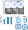
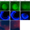
The refractivity and transparency of the ocular lens is dependent on the stability and solubility of the crystallins in the fiber cells. A number of mutations of lens crystallins have been associated with dominant cataracts in humans and mice. Of particular interest were γB- and γD-crystallin mutants linked to dominant cataracts in mouse models. Although thermodynamically destabilized and aggregation-prone, these mutants were found to have weak affinity to the resident chaperone α-crystallin in vitro To better understand the mechanism of the cataract phenotype, we transgenically expressed different γD-crystallin mutants in the zebrafish lens and observed a range of lens defects that arise primarily from the aggregation of the mutant proteins. Unlike mouse models, a strong correlation was observed between the severity and penetrance of the phenotype and the level of destabilization of the mutant. We interpret this result to reflect the presence of a proteostasis network that can "sense" protein stability. In the more destabilized mutants, the capacity of this network is overwhelmed, leading to the observed increase in phenotypic penetrance. Overexpression of αA-crystallin had no significant effects on the penetrance of lens defects, suggesting that its chaperone capacity is not limiting. Although consistent with the prevailing hypothesis that a chaperone network is required for lens transparency, our results suggest that αA-crystallin may not be efficient to inhibit aggregation of lens γ-crystallin. Furthermore, our work implicates additional inputs/factors in this underlying proteostasis network and demonstrates the utility of zebrafish as a platform to delineate mechanisms of cataract.
Keywords: aggregation; cataract; chaperone; crystallin; lens; proteostasis; small heat shock protein (sHsp); zebrafish.
© 2016 by The American Society for Biochemistry and Molecular Biology, Inc.
Figures


Similar articles
-
Transgenic zebrafish models reveal distinct molecular mechanisms for cataract-linked αA-crystallin mutants.PLoS One. 2018 Nov 26;13(11):e0207540. doi: 10.1371/journal.pone.0207540. eCollection 2018. PLoS One. 2018. PMID: 30475834 Free PMC article.
-
AlphaA-crystallin expression prevents gamma-crystallin insolubility and cataract formation in the zebrafish cloche mutant lens.Development. 2006 Jul;133(13):2585-93. doi: 10.1242/dev.02424. Epub 2006 May 25. Development. 2006. PMID: 16728471
-
Loss of the small heat shock protein αA-crystallin does not lead to detectable defects in early zebrafish lens development.Exp Eye Res. 2013 Nov;116:227-33. doi: 10.1016/j.exer.2013.09.007. Epub 2013 Sep 25. Exp Eye Res. 2013. PMID: 24076322 Free PMC article.
-
sHSP in the eye lens: crystallin mutations, cataract and proteostasis.Int J Biochem Cell Biol. 2012 Oct;44(10):1687-97. doi: 10.1016/j.biocel.2012.02.015. Epub 2012 Mar 2. Int J Biochem Cell Biol. 2012. PMID: 22405853 Review.
-
Proteostasis and the Regulation of Intra- and Extracellular Protein Aggregation by ATP-Independent Molecular Chaperones: Lens α-Crystallins and Milk Caseins.Acc Chem Res. 2018 Mar 20;51(3):745-752. doi: 10.1021/acs.accounts.7b00250. Epub 2018 Feb 14. Acc Chem Res. 2018. PMID: 29442498 Review.
Cited by
-
Interaction of α Carboxyl Terminus 1 Peptide With the Connexin 43 Carboxyl Terminus Preserves Left Ventricular Function After Ischemia-Reperfusion Injury.J Am Heart Assoc. 2019 Aug 20;8(16):e012385. doi: 10.1161/JAHA.119.012385. Epub 2019 Aug 19. J Am Heart Assoc. 2019. PMID: 31422747 Free PMC article.
-
Organelle degradation in the lens by PLAAT phospholipases.Nature. 2021 Apr;592(7855):634-638. doi: 10.1038/s41586-021-03439-w. Epub 2021 Apr 14. Nature. 2021. PMID: 33854238
-
Transgenic fluorescent zebrafish lines that have revolutionized biomedical research.Lab Anim Res. 2021 Sep 8;37(1):26. doi: 10.1186/s42826-021-00103-2. Lab Anim Res. 2021. PMID: 34496973 Free PMC article. Review.
-
Model organism data evolving in support of translational medicine.Lab Anim (NY). 2018 Oct;47(10):277-289. doi: 10.1038/s41684-018-0150-4. Epub 2018 Sep 17. Lab Anim (NY). 2018. PMID: 30224793 Free PMC article. Review.
-
The growing world of small heat shock proteins: from structure to functions.Cell Stress Chaperones. 2017 Jul;22(4):601-611. doi: 10.1007/s12192-017-0787-8. Epub 2017 Mar 31. Cell Stress Chaperones. 2017. PMID: 28364346 Free PMC article. Review.
References
-
- Bassnett S. (1997) Fiber cell denucleation in the primate lens. Invest. Ophthalmol. Vis. Sci. 38, 1678–1687 - PubMed
-
- Beebe D. C., Vasiliev O., Guo J., Shui Y. B., and Bassnett S. (2001) Changes in adhesion complexes define stages in the differentiation of lens fiber cells. Invest. Ophthalmol. Vis. Sci. 42, 727–734 - PubMed
-
- Bloemendal H., de Jong W., Jaenicke R., Lubsen N. H., Slingsby C., and Tardieu A. (2004) Ageing and vision: structure, stability and function of lens crystallins. Prog. Biophys. Mol. Biol. 86, 407–485 - PubMed
-
- Delaye M., and Tardieu A. (1983) Short-range order of crystallin proteins accounts for eye lens transparency. Nature 302, 415–417 - PubMed
-
- Tardieu A. (1988) Eye lens proteins and transparency: from light transmission theory to solution X-ray structural analysis. Annu. Rev. Biophys. Biophys. Chem. 17, 47–70 - PubMed
MeSH terms
Substances
Grants and funding
LinkOut - more resources
Full Text Sources
Other Literature Sources
Medical
Molecular Biology Databases

