. 2016 Dec 9;291(50):25837-25852.
doi: 10.1074/jbc.M116.755256. Epub 2016 Oct 21.

Structural and Biochemical Characterization of Spa47 Provides Mechanistic Insight into Type III Secretion System ATPase Activation and Shigella Virulence Regulation

Affiliations

Structural and Biochemical Characterization of Spa47 Provides Mechanistic Insight into Type III Secretion System ATPase Activation and Shigella Virulence Regulation

Jamie L Burgess et al. J Biol Chem. .

Abstract

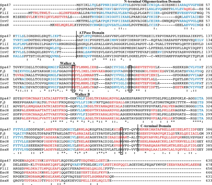
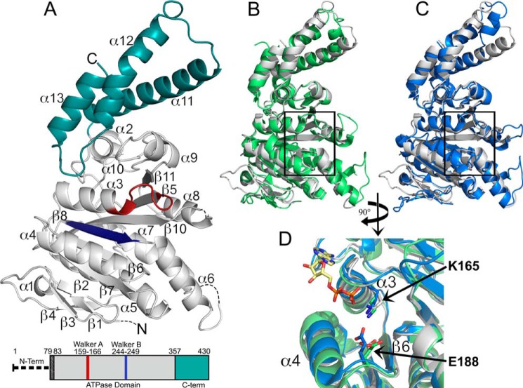
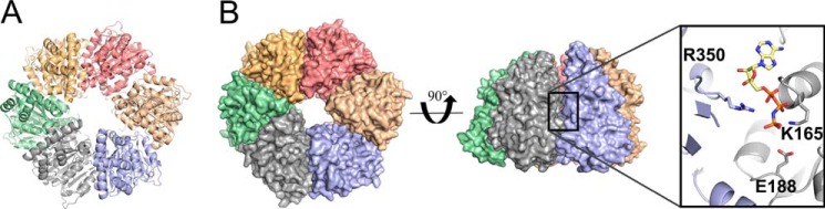
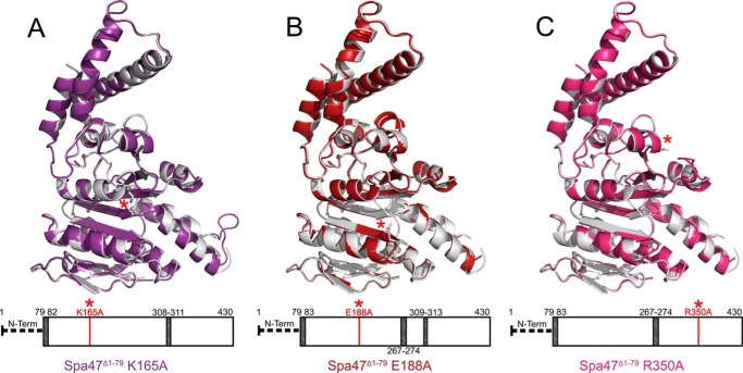
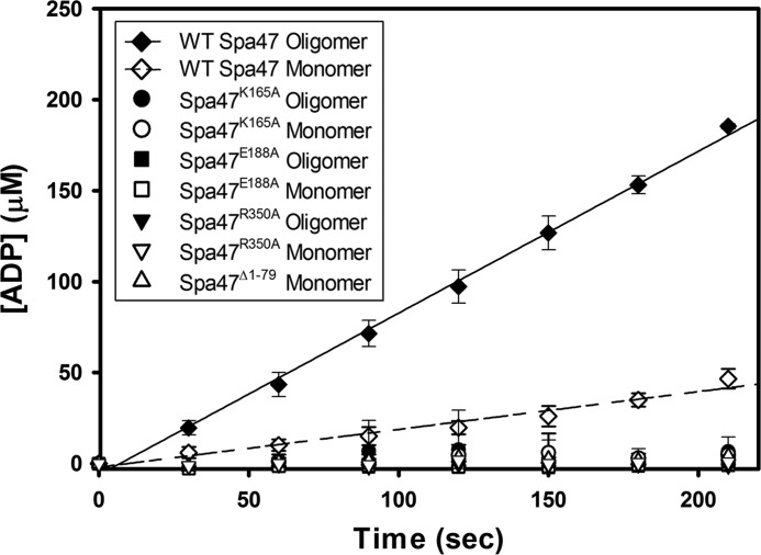
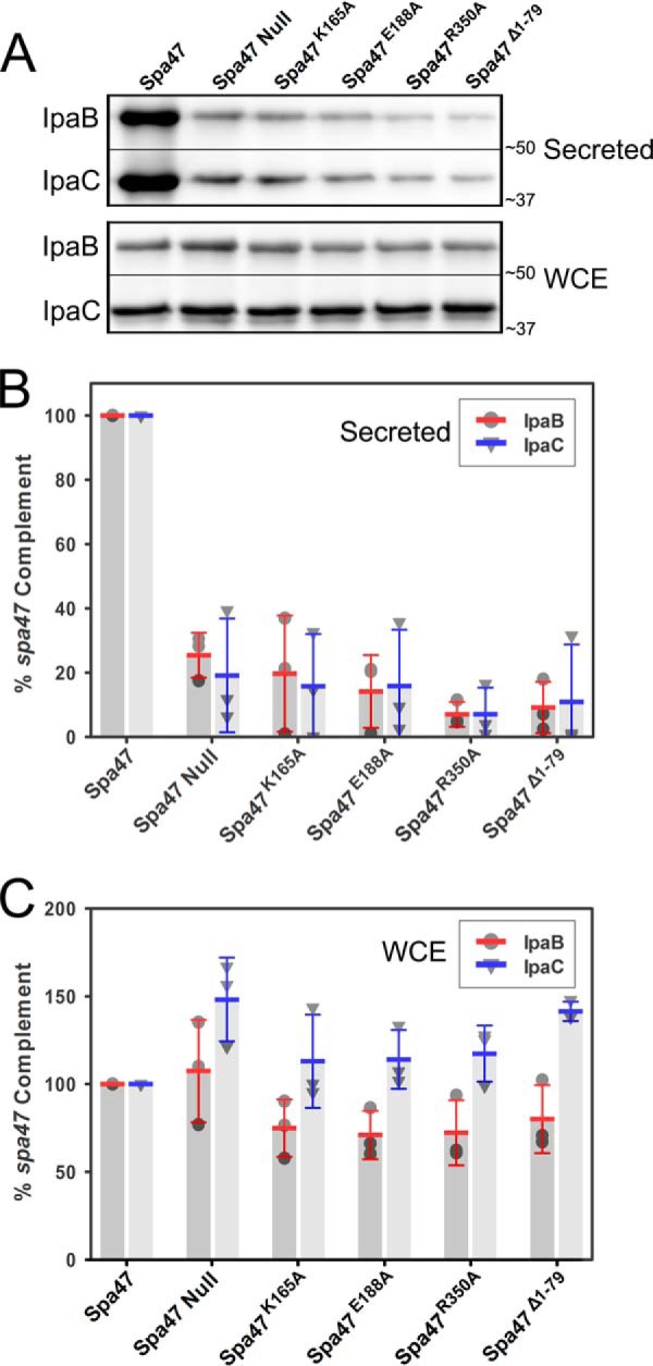
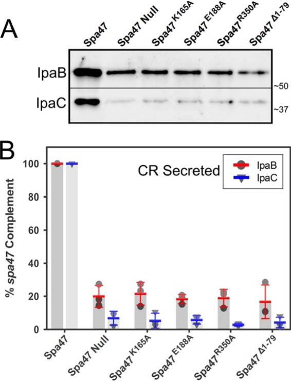
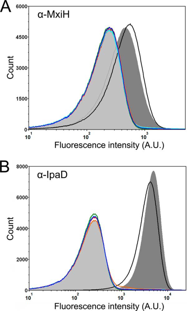
Like many Gram-negative pathogens, Shigella rely on a complex type III secretion system (T3SS) to inject effector proteins into host cells, take over host functions, and ultimately establish infection. Despite these critical roles, the energetics and regulatory mechanisms controlling the T3SS and pathogen virulence remain largely unclear. In this study, we present a series of high resolution crystal structures of Spa47 and use the structures to model an activated Spa47 oligomer, finding that ATP hydrolysis may be supported by specific side chain contributions from adjacent protomers within the complex. Follow-up mutagenesis experiments targeting the predicted active site residues validate the oligomeric model and determined that each of the tested residues are essential for Spa47 ATPase activity, although they are not directly responsible for stable oligomer formation. Although N-terminal domain truncation was necessary for crystal formation, it resulted in strictly monomeric Spa47 that is unable to hydrolyze ATP, despite maintaining the canonical ATPase core structure and active site residues. Coupled with studies of ATPase inactive full-length Spa47 point mutants, we find that Spa47 oligomerization and ATP hydrolysis are needed for complete T3SS apparatus formation, a proper translocator secretion profile, and Shigella virulence. This work represents the first structure-function characterization of Spa47, uniquely complementing the multitude of included Shigella T3SS phenotype assays and providing a more complete understanding of T3SS ATPase-mediated pathogen virulence. Additionally, these findings provide a strong platform for follow-up studies evaluating regulation of Spa47 oligomerization in vivo as a much needed means of treating and perhaps preventing shigellosis.

Keywords: ATPase; Shigella; Spa47; arginine finger; crystal structure; oligomerization; secretion regulation; type III secretion apparatus (T3SA); type III secretion system (T3SS).


Figures

FIGURE 1.
FIGURE 1.
Multiple sequence alignment of Spa47 and related ATPase homologs. Protein sequence alignment of Spa47 (T3SS ATPase, S. flexneri), F1β subunit (ATP synthase, Bos taurus), FliI (Flagellar ATPase, Salmonella typhimurium), EscN (T3SS ATPase, E. coli), InvC (T3SS ATPase, Salmonella gallinrium), and SsaN (T3SS ATPase, S. typhimurium) was performed using the Uniprot multiple sequence alignment tool, Clustal Omega, with single fully conserved residues (*), conservation between groups with strongly similar properties (:), and weakly similar properties (.) identified. Helical and β-sheet regions, as predicted by the PSIPRED structure prediction server, are color-coded red and blue, respectively. Vertical lines separate the predicted N-terminal oligomerization, ATPase, and C-terminal domains. Predicted Walker A (P-loop) and Walker B regions and the fully conserved residues targeted for alanine screening in this study are also identified in the alignment. Uniprot accession numbers used for sequence alignment were Q6XVW8, P00829, P26465, Q7DB71, B5RDL8, P74857 for Spa47, F1 ATPase B subunit, FliI, EscN, InvC, and SsaN, respectively.
FIGURE 2.
FIGURE 2.
2.4 Å X-ray crystal structure of the Shigella T3SS ATPase Spa47. A, crystal structure of Spa47Δ1–79 with coloration corresponding to the predicted domains highlighted in the bar diagram of the Spa47 sequence, where the truncated N-terminal oligomerization domain is represented by a thick dashed line and the black-shaded region corresponds to the N-terminal residues of the construct that are not included in the structure. The gray portion of the Spa47 structure corresponds to the catalytic ATPase domain, the cyan region represents the C-terminal domain, and the Walker A (P-loop) and B motifs are shown in red and blue, respectively. The secondary structure elements are identified, and the dashed lines within the Spa47 structure represent disordered regions with unassigned structure (four N-terminal residues and those connecting α6-β7 and α8-β10). β9 is located directly behind β10 and not visible in this orientation. B and C, alignment of Spa47 (gray) with the catalytic ATPase and C-terminal domains of FliI (green, B) and EscN (blue, C) illustrates the high degree of conservation within the catalytic core of the related ATPases, especially within the nucleotide-binding region (black boxes). D, a closer view of the nucleotide binding site within the aligned structures identifies the conserved active site Lys165 and Glu188 residues positioned near the FliI-bound ADP shown with a yellow carbon skeleton. The PDB codes for the Spa47, FliI, and EscN structures are 5SWJ, 2DPY, and 2OBM, respectively. The crystal structures were rendered using PyMOL.
FIGURE 3.
FIGURE 3.
Hexameric model of activated Spa47. A, Spa47 modeled as an activated homohexamer, based on the F1 ATP synthase structure. Each of the Spa47 monomer subunits are colored independently. B, surface representation of the hexameric model shown in top view and at a 90° rotation. Each of the six nucleotide-binding regions within the hexamer are located at a separate protomer interface with one example outlined. A closer view of the Spa47 active site looking from within the hexamer pore shows the conserved active site Lys165 and Glu188 side chains from one protomer (gray) and an arginine from the adjacent protomer (blue) in relation to the bound ATP homolog (AMPPNP) modeled from its location in the F1 ATP synthase structure.
FIGURE 4.
FIGURE 4.
Structure alignments between wild-type Spa47Δ1–79 and the ATPase inactive alanine point mutants used in this study. The wild-type Spa47Δ1–79 structure is shown in gray and is aligned to the Spa47Δ1–79 K165A (purple), Spa47Δ1–79 E188A (red), and Spa47Δ1–79 R350 (fuchsia) structures shown in A, B, and C, respectively. The bars located below each structure provides a visual representation of the regions included (white) and missing (shaded) in each structure as well as the relative location of each engineered point mutant within the Spa47 sequence and structure (red asterisk). The PDB codes for Spa47Δ1–79, Spa47Δ1–79 K165A, Spa47Δ1–79 E188A, and Spa47Δ1–79 R350A used in the figure are 5SWJ, 5SYP, 5SWL, and 5SYR, respectively.
FIGURE 5.
FIGURE 5.
Analysis of Spa47 oligomeric distribution. A, size exclusion chromatography elution profiles of wild-type Spa47, Spa47K165A, Spa47E188A, and Spa47R350A are all consistent with the formation of monomeric and single oligomeric species. The weak signal observed prior to the early eluting oligomer for the K165A mutant is attributed to nucleic acids and does not result from a change in oligomerization profile. The Spa47Δ1–79 SEC elution profile exhibits a single shifted elution peak, consistent with the reduced molecular mass of the Spa47 N-terminal truncation construct and an essentially exclusive monomeric protein distribution. The void volume and the elution volumes corresponding to commercial protein molecular mass standards are included at the top of the chromatogram. B, representative interference scans of purified Spa47Δ1–79 monitored during SV-AUC. C, representative residuals from fitting the data to a continuous c(s) distribution model as described under “Experimental Procedures.” D, representative sedimentation coefficient distribution (c(s) versus S) showing that Spa47Δ1–79 sediments as an essentially homogeneous species with a sedimentation coefficient of 2.87 ± 0.06 S, corresponding to a calculated molecular mass of 38.9 ± 0.2 kDa and a Spa47Δ1–79 monomer. Reported sedimentation coefficients represent means ± S.D. from three independent measurements.
FIGURE 6.
FIGURE 6.
ATPase activity of engineered Spa47 constructs. Kinetic analysis of ATP hydrolysis by each of the engineered Spa47 constructs used in this study. Wild-type Spa47 monomeric and oligomeric species are ATPase active with an increase in hydrolysis efficiency for the Spa47 oligomer. Isolated monomeric and oligomeric forms of Spa47K165A, Spa47E188A, and Spa47R350A, as well as the monomeric form of the oligomerization-deficient Spa47Δ1–79 construct were all inactive, resulting in background levels of ATP hydrolysis. Each data point represents the means ± S.D. of three independent measurements from two separate protein preparations.
FIGURE 7.
FIGURE 7.
Immunoblot analysis of uninduced translocator secretion profiles of engineered Spa47 mutant Shigella strains. A, the levels of IpaB and IpaC secreted (“leaked”) into overnight Shigella culture media were compared for Shigella strains expressing engineered ATPase inactive Spa47 mutants, as well as positive and negative controls expressing wild-type Spa47 and a spa47 null strain, respectively. The secreted proteins were detected by Western blot and compared with one another using densitometry analysis. B, the protein levels are reported relative to a S. flexneri strain expressing wild-type Spa47 (normalized to 100%) and are reported for each strain. C, the levels of IpaB and IpaC isolated from the bacterial whole cell extract (WCE) were also visualized and are shown relative to the protein levels isolated from the control strain expressing wild-type Spa47. The reported values represent the means ± S.D. from three independent analyses.
FIGURE 8.
FIGURE 8.
Immunoblot analysis of Congo red-induced T3SS secretion profiles for engineered Spa47 mutant Shigella strains. The levels of IpaB and IpaC actively secreted following Congo red induction were compared for Shigella strains expressing engineered ATPase inactive Spa47 mutants, as well as positive and negative controls expressing wild-type Spa47 and a spa47 null strain, respectively. A, actively secreted IpaB and IpaC were detected by Western blot and compared using densitometry analysis. B, the protein levels are reported relative to a S. flexneri strain expressing wild-type Spa47 (normalized to 100%) and are reported for each strain. The reported values represent the means ± S.D. from three independent analyses.
FIGURE 9.
FIGURE 9.
Flow cytometry detection of surface localized Shigella T3SS components. A, fluorescence intensity histograms depicting the levels of surface localization of MxiH for the S. flexneri 2457T strain (black), mxiH null strain (orange), spa47 null S. flexneri strain (light gray shading), and the spa47 null strain complemented with wild-type Spa47 (dark gray shading), Spa47K165A (red), Spa47E188A (blue), Spa47R350A (cyan), and Spa47Δ1–79 (green). B, fluorescence intensity histograms depicting the levels of surface localization of the T3SS tip protein IpaD for the same strains shown in A. The histograms include 500,000 individual intensity measurements per condition and are representative of independent triplicate analysis. The decreased levels of the MxiH and IpaD for all of the ATPase inactive Spa47 mutants are consistent with the in vitro experiments, suggesting that Spa47-catalyzed ATP hydrolysis drives/supports T3SA needle formation and Shigella virulence.

References

    1. Schroeder G. N., and Hilbi H. (2008) Molecular pathogenesis of Shigella spp.: controlling host cell signaling, invasion, and death by type III secretion. Clin. Microbiol. Rev. 21, 134–156 - PMC - PubMed
    1. Portaliou A. G., Tsolis K. C., Loos M. S., Zorzini V., and Economou A. (2016) Type III secretion: building and operating a remarkable nanomachine. Trends Biochem. Sci. 41, 175–189 - PubMed
    1. Galán J. E., Lara-Tejero M., Marlovits T. C., and Wagner S. (2014) Bacterial type III secretion systems: specialized nanomachines for protein delivery into target cells. Annu. Rev. Microbiol. 68, 415–438 - PMC - PubMed
    1. Cossart P., and Sansonetti P. J. (2004) Bacterial invasion: the paradigms of enteroinvasive pathogens. Science 304, 242–248 - PubMed
    1. World Health Organization (Updated April 2013) Diarrhoeal Disease, Fact Sheet no. 330, http://www.who.int/mediacentre/factsheets/fs330/en/

MeSH terms

Substances

LinkOut - more resources