. 2016 Dec;6(3):207-219.
doi: 10.1016/j.ijpddr.2016.10.003. Epub 2016 Oct 12.

Effects of a novel β-lapachone derivative on Trypanosoma cruzi: Parasite death involving apoptosis, autophagy and necrosis

Affiliations

Effects of a novel β-lapachone derivative on Trypanosoma cruzi: Parasite death involving apoptosis, autophagy and necrosis

Danielle Oliveira Dos Anjos et al. Int J Parasitol Drugs Drug Resist. 2016 Dec.

Abstract

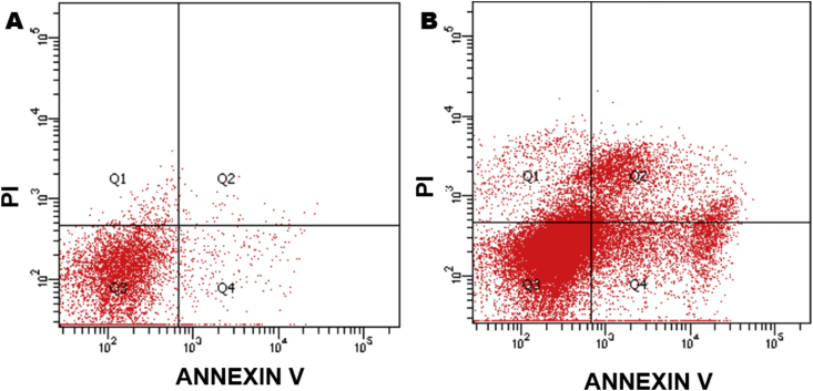
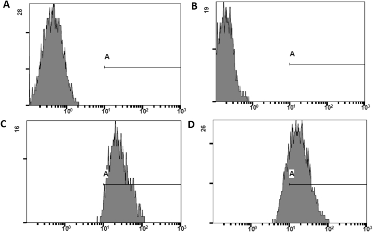
Natural products comprise valuable sources for new antiparasitic drugs. Here we tested the effects of a novel β-lapachone derivative on Trypanosoma cruzi parasite survival and proliferation and used microscopy and cytometry techniques to approach the mechanism(s) underlying parasite death. The selectivity index determination indicate that the compound trypanocidal activity was over ten-fold more cytotoxic to epimastigotes than to macrophages or splenocytes. Scanning electron microscopy analysis revealed that the R72 β-lapachone derivative affected the T. cruzi morphology and surface topography. General plasma membrane waving and blebbing particularly on the cytostome region were observed in the R72-treated parasites. Transmission electron microscopy observations confirmed the surface damage at the cytostome opening vicinity. We also observed ultrastructural evidence of the autophagic mechanism termed macroautophagy. Some of the autophagosomes involved large portions of the parasite cytoplasm and their fusion/confluence may lead to necrotic parasite death. The remarkably enhanced frequency of autophagy triggering was confirmed by quantitating monodansylcadaverine labeling. Some cells displayed evidence of chromatin pycnosis and nuclear fragmentation were detected. This latter phenomenon was also indicated by DAPI staining of R72-treated cells. The apoptotis induction was suggested to take place in circa one-third of the parasites assessed by annexin V labeling measured by flow cytometry. TUNEL staining corroborated the apoptosis induction. Propidium iodide labeling indicate that at least 10% of the R72-treated parasites suffered necrosis within 24 h. The present data indicate that the β-lapachone derivative R72 selectively triggers T. cruzi cell death, involving both apoptosis and autophagy-induced necrosis.

Keywords: Chagas disease; Chemotherapy; Natural products; Trypanosoma cruzi; β–lapachone derivative.


Figures

Image 1
Graphical abstract
Fig. 1
Fig. 1
Decoupled 31P Nuclear magnetic resonance spectrum of N’[(6Z)- 3,4- dihydro - 2,2- dimethyl - 5- oxo- 2H- naphthol [1,2-b] pyran - 6 (5H)- ilidene], ester di-sec-butylic phosphorohydrazidic acid (R72). The R72 synthesis of was performed by adding equimolar amounts of phosphorohydrazidic acid, bis(1-methylpropyl) ester, and β−lapachone in ethyl alcohol, with catalytic amounts of concentrated hydrochloric acid. In the detail: the molecular R72 structure.
Fig. 2
Fig. 2
Inhibitory effect of the β-lapachone derivative R72 upon T. cruzi in vitro proliferation. Epimastigote forms were cultured in the presence of different R72 concentrations and counted under phase microscopy after 96 h. The compound significantly (*p < 0.05; **p < 0.001) impaired the parasite proliferation producing a 19 μM IC50value (A). Inhibitory effect of the β-lapachone derivative R72 upon murine macrophage (B) and splenocyte (C) viability assessed by the MTT method, produced CC50 values of 243 μM and 212 μM, respectively (*p < 0.05). The selectivity indexes (CC50/IC50) determined for these cell types were 12.78 and 11.15, respectively. These data are mean of at least three independent experiments performed in triplicates. Statistical analysis was performed using ANOVA and Tukey post-test.
Fig. 3
Fig. 3
Scanning electron microscopy (SEM) of untreated (A) and DMSO-treated (B) controls, where the normal cytostome opening is evident (B, arrow). R72-treated parasites displayed ruffled plasma membrane and blebbing (*) was observed particularly in the cytostome opening area (C, arrow).
Fig. 4
Fig. 4
Contrary to the regular cytostomes in DMSO-treated parasites observed by TEM (A, arrow), R72-treated parasites presented debris-associated damaged cytostome opening (B, arrow), but the cytopharinx microtubules apparently remained intact (B, *). DMSO-treated parasites showed no alteration as compared to untreated cells. Some R72-treated parasites displayed large kinetoplasts (C - K) with altered kDNA compacting pattern (arrowhead) as well as supernumerary basal bodies (Fig. 4C arrowheads). Bars – 500 nm.
Fig. 5
Fig. 5
ROS production accessed by flow cytometry of parasites incubated with the cell permeant probe reagent 2′,7’ –dichlorofluorescein diacetate (DCFDA). Untreated parasite controls displayed circa 0.9% stained cells (A), whereas parasites incubated with 19 μM R72 for 1 h (B), 3 h (C) and 24 h (D) presented, respectively 93.6%, 53.7% and 41.1% DCFDA-positive cells.
Fig. 6
Fig. 6
R72-treated parasites eventually presented pycnotic nuclei (A - N, *) and some TEM images displayed nuclear protrusions (B arrows) and compartments of similar content suggesting budding of nuclear fragments (B, arrowhead). Fluorescence microscopy using the DNA probe DAPI revealed the normal nucleus and kinetoplast labeling of both untreated and DMSO-treated control cells (C arrows arrowheads) and showed evidence of nuclear fragmentation in some R72-treated parasites (D, arrowhead).
Fig. 7
Fig. 7
Cell death mechanism(s) in R72-treated parasites evaluation by flow cytometry, in cells coincubated with PI and annexin V probes. Untreated controls displayed 91.2% of double negative cells (A), whereas cultures incubated with 19 μM R72 for 24 h (B) displayed 16.8% PI-negative, but annexin V-positive, indicating cells undergoing apoptosis as well as 12.2% PI-positive and AV-positive cells, corresponding to late apoptosis/secondary necrosis. DMSO-treated controls were not labeled.
Fig. 8
Fig. 8
DNA fragmentation detection employing the TUNEL method. 107T. cruzi epimastigotes incubated or not with 19 μM R72 for 24 h were analyzed by flow cytometry. Negative Control (A) assayed in the absence of rTdT (recombinant terminal deoxynucleotidyl transferase) and DMSO-treated parasites (B) displayed similar patterns, with no stained cells in the gate, whereas positive control, employing DNAse (C) and R72-treated parasites (D) showed TUNEL staining on 77.6% and 93.3% cells, respectively.
Fig. 9
Fig. 9
Detection of autophagic process by the probe monodansylcadaverine (MDC). Untreated parasites were poorly and diffusely labelled (A), whereas R72-treated cells displayed numerous and often closely apposed strongly labelled compartments (B). Insets show individual MDC-labelled cells. DMSO-treated parasites showed no alteration as compared to untreated cells (not shown). Quantitation of cells displaying monodansylcadaverine-labeled compartments by fluorescence microscopy (C). The frequency of parasites presenting MDC+ compartments was determined by counting of over 260 cells per group. About 36% of untreated control parasites presented MDC punctate labelling, whereas nearly 83% of parasites grown with 19 μM R72 showed. MDC-stained autophagosomes. Chi-square with Yates correction (1 degree of freedom), equals 7018.369 with P < 0.0001 (two-tailed, ***).
Fig. 10
Fig. 10
Autophagic vacuoles (AV) presenting mitochondrial portion (A–M), membrane profiles (B, arrow) and ribosome-like particles (B, *) were observed in R72-treated parasites. Note that cytoplasmic intact mitochondria may be still observed in the cytoplasm (A, arrow). Compartments lined by double membranes were often observed enveloping condensed mitochondrion fragment (C, thick arrow) and eventually the autophagosome fusion events (C, black arrowheads) formed huge compartments, containing membrane remnants (C, *), which could be associated to parasite cell surface continuity solution (thin arrow). Note condensed mitochondria displaying dilated cristae (C, white arrowheads).

References

    1. Aguiar C., Batista A.M., Pavan T.B., Almeida E.A., Guariento M.E., Wanderley J.S., Costa Serological profiles and evaluation of parasitaemia by PCR and blood culture in individuals chronically infected by Trypanosoma cruzi treated with benzonidazole. Trop. Med. Int. Health. 2012;17:368–373. - PubMed
    1. Almeida M.T., Ramalho-Santos J., Oliveira C.R., de Lima M.C. Parameters affecting fusion between liposomes and synaptosomes. Role of proteins, lipid peroxidation, pH and temperature. J. Membr. Biol. 1994;142:217–222. - PubMed
    1. Alvarez V.E., Kosec G., Sant'Anna C., Turk V., Cazzulo J.J., Turk B. Autophagy is involved in nutritional stress response and differentiation in Trypanosoma cruzi. J. Biol. Chem. 2008;283:3454–3464. - PubMed
    1. Araújo A.J., De Souza A.A., Da Silva Júnior E.N., Marinho-Filho J.D., de Moura M.A., Rocha D.D., Vasconcellos M.C., Costa C.O., Pessoa C., Moraes De, Ferreira V.S., De Abreu F.C., Pinto A.V., Montenegro R.C., Costa-Lotufo L.V., Goulart M.O. Growth inhibitory effects of 3'-nitro-3-phenylamino nor-beta-lapachone against HL-60, a redox-dependent mechanism. Toxicol. In Vitro. 2012;26:585–589. - PubMed
    1. Azeredo C.M., Santos T.G., Maia B.H., Soares M.J. In vitro biological evaluation of eight different essential oils against Trypanosoma cruzi, with emphasis on Cinnamomum verum essential oil. BMC Complem. Altern. Med. 2014;14:309. - PMC - PubMed

MeSH terms

LinkOut - more resources